The Transporter Associated With Antigen Processing (TAP): Structural
Integrity, Expression, Function, and Its Clinical Relevance
Ulrike Ritz and Barbara Seliger
Johannes Gutenberg-University III, Department of Internal Medicine, Mainz, Germany
Accepted December 28, 2000.
Abstract
Background: The transporter associated with antigen processing (TAP), a member of the family of ABC trans-porters, plays a crucial role in the processing and presenta-tion of the major histocompatibility complex (MHC) class I restricted antigens. TAP transports peptides from the cy-tosol into the endoplasmic reticulum, thereby selecting peptides matching in length and sequence to respective MHC class I molecules. Upon loading on MHC class I molecules, the trimeric MHC class I/2-microglobulin/
peptide complex is then transported to the cell surface and presented to CD8 cytotoxic T cells. Abnormalities in
Address correspondence and reprint requests to: Barbara Seliger, PhD, Johannes Gutenberg University, Department of Internal Medicine, Langenbeckstrasse 1, 55101 Mainz, Germany. Phone:
49-6131-176760; Fax: 49-6131-176678; E-mail: B.Seliger@ 3-med.klinik.uni-mainz.de
MHC class I surface expression have been found in a num-ber of different malignancies, including tumors of distinct histology, viral infections, and autoimmune diseases, and therefore represent an important mechanism of malignant or virus-infected cells to escape proper immune response. In many cases, this downregulation has been attributed to impaired TAP expression, which could be due to struc-tural alterations or dysregulation. This review summarizes the physiology and pathophysiology of TAP, thereby fo-cusing on its function in immune responses and its role in human diseases.
Introduction
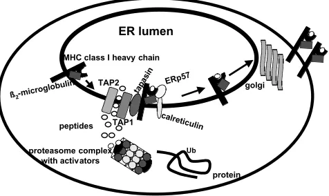
The MHC class I molecules present mainly cytoso-lic peptides to MHC class I restricted CD8 cyto-toxic T lymphocytes (CTL). Most of these peptides are generated by degradation of intracellular pro-teins, which is directed by both constitutive and interferon (IFN)-inducible proteasomal subunits. The yielded peptides are then transported by TAP from the cytosol into the lumen of the endoplasmic reticulum (ER), where they are loaded onto a dimer consisting of MHC class I heavy chain (HC) and 2 -microglobulin (2-m). Both steps—the assembly of these molecules and the formation of the “large TAP complex”—are stabilized by various ER-resident chaperones, such as calnexin, binding protein (BiP), calreticulin, the oxidoreductase ERp57, and the transmembrane protein tapasin (1–3). Tapasin further retains empty MHC class I molecules in the ER and facilitates their peptide loading (4,5). The stable MHC class I HC/2-m/peptide complex is then transported via the trans-golgi to the cell
surface for recognition by CD8T cells (recently re-viewed in 6, 7; Fig. 1).
The Superfamily of ABC Transporters
TAP belongs to the superfamily of ATP-binding cassette (ABC) transporters. All ABC transporters possess two hydrophobic N-terminal domains cross-ing the membrane six to eight times, thereby form-ing a putative translocation pore and two highly conserved C-terminal cytosolic adenosine triphos-phate (ATP)-binding cassettes (Fig. 2). For their proper function, energy in the form of nucleoside triphosphates (NTP) is required. ABC transporters translocate a diverse set of substances/molecules, such as ions, sugars, polysaccharides, amino acids, and oligo- and polypeptides across cell membranes. Each transporter is specialized for the transport of a unique substrate or substrate class and plays an important role in different (patho)physiologic processes (8).
which is mutated in congenital hyperinsulinism (11,12); the P-glycoproteins MDR1 and MDR3, which transport a series of hydrophobic drugs and phospholipids (13–15); and the peptide transporter TAP. TAP consists of two subunits, TAP1 and TAP2, and is the only ABC transporter with a unique func-tion in the immune system. Regarding sequence and topology, the ABC transporters possess signifi-cant homologies that are especially pronounced for the P-glycoproteins and TAP (8,16).
Structure and Function of TAP Molecules
Genomic Organization of TAP The genomic struc-ture of TAP has been well defined in different species, such as human, mouse, and rat (16–18). The genes of the human TAP1 and TAP2 subunits, which are located in the MHC class II locus of the chromosome
587 and 492 bp, respectively. TAP is evolutionary highly conserved with an approximately 35% ho-mology between TAP1 and TAP2 in all species. Thus, the peptide transporter seems to have evolved from a common ancestor gene by duplication prior to the development of the adaptive immune system in vertebrates.
Topology of TAP Heterologous coexpression of TAP1 and TAP2 in insect cells or yeast suggest that the functional peptide transporter is a heterodimer consisting of the two subunits TAP1 and TAP2. This model is further supported by the lack of peptide translocation in TAP1- or TAP2-negative (tumor) cells and TAP1-/- mice, as well as by restoration of TAP function upon TAP gene transfer (19–23). How-ever, despite the sequence differences, TAP subunits of different species are capable of forming a func-tional TAP heterodimer (24,25).
Using a number of different experimental ap-proaches, the localization as well as the topology of TAP have been determined. Immune electron mi-croscopy and confocal laser scanning mimi-croscopy have demonstrated an intracellular distribution of both TAP subunits in the ER (Fig. 3; 26,27), which is consistent with the localization, where MHC class I molecules are loaded with peptides (28).
Hydrophobicity analysis and sequence align-ments of TAP proteins with other ABC transporters revealed that both TAP molecules span the ER mem-brane 6–10 times, containing a highly hydrophobic N-terminal domain linked to the cytosolic nucleotide binding domains (NBT) including the conserved Walker A and Walker B motifs for NTP-binding and hydrolysis (29,30).
Hypothetical TAP Models Using photocross-linking experiments with peptides, potential peptide-binding domains in both TAP subunits have been identified. Such analyses revealed similar binding regions for TAP1 and TAP2 composed of the cytosolic loops between the transmembrane (TM) regions-4 and TM-5 and a carboxy-terminal stretch of about 15 amino acids following TM-6. This topology model suggested that these regions are exposed to the cy-tosol (31).
Recently, Voss et al. (32) developed another topology model for TAP employing a series of C-terminal deletions for TAP1 and TAP2 subunits. As shown in Figure 4a, TAP1 and TAP2 consist of 8 and 7 transmembrane domains, respectively. Thus, the N-and terminal domain of TAP1 as well as the C-terminal domain of TAP2 is located in the cytosol, whereas the N-terminus of TAP2 is located in the ER. The pore-forming domains of the TAP subunits are aligned in a head-head/tail-tail orientation (33; Fig. 4b), which is followed by a cytosolic potential
golgi
proteasome complex with activators
peptides TAP1
Ub
protein MHC class I heavy chain
ß2-microglobulin tapasin
calreticulin
ERp57
TAP2
Fig. 1. The MHC class I antigen processing and
presenta-tion pathway.The classical MHC class I antigen processing and
presentation pathway is divided into four major steps: (1) the generation of antigenic peptides by proteasomal degradation, (2) the ATP-dependent peptide transport from the cytosol in the ER via the TAP heterodimer, (3) the assembly of MHC class I HC, 2-m and peptides, which is assisted by various chaperones (e.g., Erp57, tapasin and calreticulin), and (4) the export and presentation of the trimeric MHC/peptide complex.
cytosol
ATP-binding domains substrate-binding domains
Fig. 2. Topology of typical ABC transporters.The schematic
are controversially discussed and require further in-vestigations.
Peptide Selectivity of TAP During the last decade, the substrate specificity of TAP has been well stu-died by a number of groups employing a peptide translocation assay that traps transported peptides in the ER via glycosylation (35). Information is available about the length and sequence preferences of the transported peptides (36–39). Peptides with a length of 8–16 aa are preferentially transported by TAP, although longer peptides (40 aa) are also transported, but with a lower efficiency (40). In contrast to the peptide transporter assay, the use of peptide-binding domain and subsequently by the
C-terminal domain containing the NTP-binding sites. Deletions of 20 or fewer amino acids (aa) in the potential peptide-binding domain of the TAP1 subunit defined regions between amino acids 366–405 that are essential for proper peptide trans-porter function, whereas deletions of other regions (aa 345–365 and aa 465–487) did not influence TAP activity (34). Although many different approaches have been employed to characterize the structure, or-ganization, and functional domains in TAP, the topology of TAP, the domains responsible for pep-tide binding and for peppep-tide transport, as well as the interaction between the TAP1 and TAP2 subunits
Fig. 3. ER localization of TAP.(A and B) TAP-negative cells were stained either with an anti-calnexin antibody (red color, A) or the
anti-TAP1 monoclonal antibody mAb148.3 (B). (C) TAP overexpressing cells were stained with the anti-TAP1 monoclonal antibody mAb 148.3 (green color). TAP staining is comparable to calnexin staining, suggesting that TAP1 expression is localized in the ER.
ER-lumen
N
N
C C
ATP-binding site ATP-binding site
cytosol TAP1 TAP2
pore forming domains
peptide binding domains
ATP-binding sites
A
TAP2 TAP1
ER-membrane
N N
C C
ATP-binding sites
B
peptide binding domains
Fig. 4. The putative TAP model by Voss et al. (32).(A) Membrane topology of TAP. The light gray regions represent the
termined by screening combinatorial peptide li-braries and determined the influence of a single peptide residue independent of a given sequence context on the affinity of TAP (41). These experi-ments suggest that the carboxy-terminal aa and the first three amino-terminal residues play a key role in peptide selectivity of TAP. Murine TAP alleles se-lectively transport peptides with a hydrophobic C-terminus, whereas human TAP complexes transport both peptides with a hydrophobic C-terminus and peptides with a basic C-terminus (37,39,42,43). The selectivity of TAP is important because it mostly transports peptides matching to MHC class I mole-cules.
Mechanism of Peptide Transport Peptide transport by TAP is a multistep process (44). First, peptides associate with TAP in an ATP-independent manner (45,46). This step determines the peptide selectivity and is followed by a slow isomerization of the TAP complex. The structural reorganization of the mole-cule triggers peptide translocation across the mem-brane, which is ATP dependent (47).
Peptides with a low affinity to MHC class I mole-cules are transported into the ER, but are not rapidly loaded onto MHC class I molecules (48). Thus, they are available for export out of the ER into the cytosol via the Sec61 channel (49).
Biochemical Modifications of TAP TAP1 and TAP2 are mainly nonglycosylated, although three putative glycosylation sites, two facing the cytosol and one placed in the ER, have been identified (26). Recently, it has been demonstrated that the TAP subunits are phosphorylated under physiologic conditions. They form a high molecular complex with tapasin, MHC class I heavy chain, and a putative phosphatase, which dephosphorylates the complex. This dephos-phorylation process is essential for proper TAP func-tion, because phosphorylated TAP is not capable of transporting peptides into the ER, suggesting that an altered phosphorylation status of TAP may represent an immune escape mechanism (50).
Modulation of TAP Expression
Promoters of the TAP Genes The potential promo-ters of both TAP genes contain no TATA motifs in the 5’ flanking sequence, but putative GC-rich elements containing SP1-binding sites either 128 or 70 nu-cleotides upstream of the translation start codon for TAP1 and TAP2, respectively (51). These are at least essential for proper activity of the TAP1 promoter (52). The human TAP1 promoter is a bidirectional promoter consisting of 593 bp, which also coordinately regulates the low-molecular-weight protein (LMP)2. It contains an IFN- and a p53 response element, as well as a NF-B–binding site
promoter, the human TAP2 promoter still has to be characterized in detail.
Regulation of TAP Expression by Cytokines With a few exceptions, most cell types constitutively express MHC class I surface molecules. However, the MHC class I expression can be strongly enhanced by vari-ous cytokines, such as IFN-, IFN-, TNF- or gra-nulocyte-macrophage colony stimulating factor (GM-CSF). Analogous to MHC class I molecules, TAP1 and TAP2 subunits are upregulated by IFNs and TNF-, but with distinct time kinetics and to a differ-ent extdiffer-ent (55–57). TAP1 and TAP2 transcription is rapidly upregulated after IFN- treatment consecu-tively increasing over 48 hours to 10- to 20-fold, whereas the IFN-–mediated induction of the human leukocyte antigen (HLA) class I heavy chain tran-scription occurs more slowly and later, increasing only 10-fold in 48 hours. Although most cytokines enhance TAP expression, interleukin (IL)-10 causes a downregulation of both transcription and translation of the peptide transporter, thereby abrogating its function (58, 59). The IL-10–mediated downregula-tion of TAP leads to reduced cell surface expression of MHC class I antigens (59). The inhibitory effect of IL-10 may have in vivo relevance, because various tu-mors secrete IL-10. Results with murine IL-10 trans-duced cells suggest that IL-10 production decreases TAP function, which is accompanied by low levels of MHC class I surface expression and reduced CTL-specific lysis, but enhances sensitivity to natural killer cell–mediated cytotoxicity (58).
TAP Polymorphism, Functional Significance, and Association With Autoimmune Disease
Several alleles of human TAP1 and TAP2 have been identified in different populations, but their influ-ence on TAP activity and/or peptide-binding speci-ficity varies (60,61). A functional polymorphism was found for a human TAP2 splice variant (TAP2iso) that consists of an altered C-terminus. When compared to the wild-type TAP1/TAP2, the TAP1-TAP2iso heterodimer exhibited a distinct pep-tide selectivity, transporting peppep-tides with hy-drophobic C-terminal residues 30 times more effi-ciently (62), thereby creating a new pattern of presented peptides/antigens. To the best of our knowledge, other functional TAP polymorphisms have only been detected in the rat (38,63).
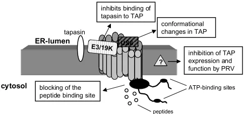
au-process that requires the presentation of viral pep-tide fragments in the context of MHC class I mole-cules on the cell surface. Persistent viruses, such as herpes simplex virus (HSV), human cytomegalovirus (HCMV), adenoviruses (Ad), and human papilloma virus (HPV) have been forced to develop a series of strategies to evade immune defense by interfering with various steps of the antigen processing and representation pathway. Some directly affect TAP expression and/or function, resulting in defects in MHC class I cell surface expression and immune re-sponses (88; summarized in Fig. 5).
The immediate early protein ICP47 encoded by the HSV type 1 (HSV-1) is responsible for the downregulation of MHC class I surface expression in human fibroblasts, which is accompanied by a lack of CTL response. ICP47 binds with high affin-ity to the peptide-binding site of TAP, thereby desta-bilizing TAP and inhibiting the TAP-mediated pep-tide transport into the ER (89–91). The interaction of human ICP47 and the TAP complex is species spe-cific because ICP47 has a 100-fold higher affinity for human than for murine TAP (92). The identification of the critical amino acids of ICP47 responsible for inhibition of TAP function represents the basis for the development of therapeutic drugs. These may be applicable in novel vaccination strategies against HSV owing to restoration of CTL-mediated recogni-tion of virus-infected cells.
The HCMV encodes the ER-resident glycopro-tein gpUS6 that inhibits the TAP-mediated peptide transport due to its binding to the ER luminal part of the TAP complex (93–95). This results in a con-formational change of TAP. Moreover, HCMV in-hibits dephosphorylation of TAP, which results in lack of peptide transport and impaired MHC class I surface expression (50). Furthermore, another mem-ber of the herpes virus family, the pseudorabies virus (PRV), interferes and inhibits the porcine pep-thors failed to detect any correlation (68–70).
Simi-lar discrepancies between association of type I dia-betes, multiple sclerosis, and Grave’s disease and rejection of renal transplants with TAP polymor-phism have been found by various groups (71–77). However, the deficiencies of HLA class I presenta-tion in lymphoid cells from individuals with type I diabetes was corrected by TAP transfection (78).
Human Diseases Associated With Dysfunction of TAP: Clinical Relevance?
Any effect that impairs the TAP-mediated peptide translocation into the ER results in reduced MHC class I surface expression. Thus, cells with a defective MHC class I antigen processing and presentation pathway can be identified by measuring the MHC class I surface expression, which can be either down-regulated or totally lost. The molecular defects under-lying the abnormalities of MHC class I and TAP ex-pression in human diseases as well as the role of TAP deficiencies for immune response have only been ad-dressed to a limited extent. However, this information is important; it might contribute both to our under-standing of the basis of immune escape mechanisms in diseases as well as to the design or optimization of the treatment of patients with TAP defects.
Genetic Diseases Bare lymphocyte syndrome (BLS) is characterized by a severe downregulation of MHC class I or class II antigens. BLS is divided into two subtypes. In type I BLS, the defect is confined to the MHC class I molecules, whereas in type II BLS, MHC class II cell surface expression is downregu-lated (79). Only a small number of patients with type I BLS have been identified worldwide, which could be differentiated into three disease subsets de-pending on their clinical and immunologic appear-ance. For example, one group of BLS patients (10 patients from 7 families) exhibits defective TAP ex-pression and has similar clinical manifestations, in-cluding bacterial and viral infections (sinusitis, chronic bronchitis) and necrotizing granulomatous skin lesions (80). In these BLS patients, structural al-terations have been found in both TAP1 and TAP2 subunits (81–84). The mutations lead to the genera-tion of a premature stop codon resulting in nonfunc-tional TAP proteins (81,85). Interestingly, mutations in the TAP2 subunit result in a more severe disease than mutations in the TAP1 subunit (80).
The lymphocyte repertoire of peripheral blood displayed low numbers of CD8 T cells, but an expansion of NK and T cells in most BLS patients (86). The NK and T cells may be involved both in the pathogenesis of granulomatous skin lesions and in antiviral immune responses that do not require TAP-dependent antigen processing (80,83).
Viral Infections and Viral Persistence: Transcriptional and Posttranscriptional Regulation CD8CTL efficiently recognize and eliminate virus-infected cells (87), a
ICP47 US6
conformational changes in TAP
cytosol
ER-lumen
ATP-binding sites
peptides ?
inhibition of TAP expression and function by PRV
blocking of the peptide binding site
E3/19K
inhibits binding of tapasin to TAP
tapasin
Fig. 5. Viral interference with TAP function.The major
have been shown to affect TAP function (97,98). This could be either due to transcriptional downreg-ulation of TAP expression or binding of the aden-oviral protein E3gp19K to TAP, thereby inhibiting complex formation with tapasin, further preventing MHC class I/TAP association (98). In contrast to hu-man Ad virus, infection of mouse cells with murine Ad virus does not influence MHC class I surface ex-pression (99).
It has been known for a long time that cervical cancer and respiratory papillomatosis are diseases of viral origin caused by the infection of different types of HPV. MHC class I surface expression was variably downregulated in both diseases, which was associ-ated with decreased TAP1 expression (100,101). In addition, the level of TAP1 and MHC class I antigen expression correlated inversely with disease recur-rence (100,102). The E7 early proteins of HPV types 6 and 18, essential for the maintenance of respiratory papillomas (RRP) and cervical cancer, respectively, repress the bidirectional LMP2/TAP1 promoter, which could be responsible for low MHC class I sur-face expression in these diseases (103). These find-ings suggest that viruses may evade T-cell recognition and killing of infected cells by decreasing MHC class I surface expression through modulation of TAP. However, it is speculated that additional viruses that influence the antigen-processing pathway, including the peptide transporter activity, will be identified.
Impaired TAP Expression and Function Tumors of Distinct Histology: Sequence Alterations and Dysregulation Analysis of both surgically removed lesions and cell lines derived from tumors of distinct histology has
been restricted to the analysis of TAP1 expression because of the limited availability of anti-TAP2 an-tibodies. In comparison to autologous normal tis-sues, TAP1 downregulation or loss was found in all tumor types analyzed with a frequency ranging from 10–84% (23). Similar results were obtained by analysis of tumor cell lines (Table 1). The clinical impact of TAP deficiencies by malignant cells varies among tumors. The frequency of TAP1 downregula-tion was more pronounced in metastatic than in primary lesions in breast, cervical, colon, small cell lung carcinomas (SCLC), and melanomas, whereas deficient TAP expression of renal cell carcinomas (RCC) is not correlated with tumor grading and staging (104–108). In melanoma, impaired TAP ex-pression is also associated with poor prognosis and reduced survival rate (107). Heterogeneity in terms of quantity and quality of TAP downregulation among various types of tumors (lesions/cell lines) has been detected, which might reflect differences in the patient populations selected for analysis, im-munobiology of the tumor types, the proliferative status of the tumor cells, and the sensitivity of the techniques employed (109).
Although impaired TAP expression is frequently found in tumors of distinct origin, the underlying molecular mechanisms of these TAP deficiencies have not yet been defined. Consequently, until recently, tu-mor lesions/cell lines have not been monitored for TAP mutations. In one lung cell carcinoma cell line, a new TAP1 allele has been identified that results in expression of a nonfunctional TAP protein (110). In addition, a bp deletion in the TAP1 subunit of a melanoma cell line also results in TAP dysfunction
Table 1. TAP downregulation in lesions and cell lines derived from human tumors of distinct histology
Downregulation of Expression (%)
Primary Lesions Cell Lines
Tumor Type Samples (n) TAP1 TAP2 Samples (n) TAP1 TAP2
Breast carcinoma 63 13–44 21 ND ND ND
SCLC 93 38 ND 6 100 100
Colon carcinoma 81 14 ND 5 32 23
RCC 70 52 ND 19 79 74
Cervical carcinoma 76 49 ND 7 0 0
Melanoma 53 31 16 9 33 57
ND, not determined.
as to the development of innovative therapeutic ap-proaches for autoimmune diseases.
Acknowledgement
We thank Dr. Dennis Strand, I. Medical Clinic, Jo-hannes-Gutenberg-University, Mainz for his help re-garding the immunfluorescence analysis and Mrs. I. Schmidt for help in preparing the manuscript.
References
1. Ortmann B, Copeman J, Lehner PJ, et al. (1997) A critical role for tapasin in the assembly and function of multimeric MHC class I-TAP complexes. Science277:1306–1309. 2. Spee P, Neefjes J. (1997) TAP-translocated peptides
specifi-cally bind proteins in the ER, including gp96, protein disul-fide isomerase and calreticulin. Eur. J. Immunol. 27:
1441–2449.
3. Suh W, Mitchell E, Yang Y, Peterson P, Waneck G, Williams DB. (1996) MHC class I molecules form ternary complexes with calnexin and TAP and undergo peptide regulated in-teraction with TAP via their extracellular domains. J. Exp. Med.184:337–348.
4. Sadasivan B, Lehner PJ, Ortmann B, Spies T, Cresswell P. (1996) Roles for calreticulin and a novel glycoprotein, tapsin, in the interaction of MHC class I molecules with TAP. Immunity5:103–114.
5. Grandea AG, Golovina TN, Hamilton SE, et al. (2000) Im-paired assembly yet normal trafficking of MHC class I mol-ecules in tapasin mutant mice. Immunity13:213–222. 6. Pamer E, Cresswell P. (1998) Mechanism of MHC class
I-resticted antigen processing. Annu. Rev. Immunol. 16:
323–358.
7. van Endert PM. (1999) Genes regulating MHC class I pro-cessing of antigen. Curr. Opin. Immunol.11:82–88.
8. Higgins CF. (1992) ABC transporters: from microorganisms to man. Annu. Rev. Cell Biol.8:67–113.
9. Anderson MP, Gregory RJ, Thompson S, et al. (1991) Demonstration that CFTR is a chloride channel by alteration of its anion selectivity. Science98:202–205.
10. Riordan JR, Chang XB. (1992) CFTR, a channel with the structure of a transporter. Biochim. Biophys. Acta 1101:
221–222.
11. Aguilar-Bryan L, Clement JP, Nelson DA. (1998) Sulfony-lurea receptors and ATP-sensitive potassium ion channels. Methods Enzymol.292:732–744.
12. Meissner T, Brune W, Mayatepek E. (1997) Persistent hy-perinsulinaemic hypoglycaemia of infancy: therapy, clinical outcome and mutational analysis. Eur. J. Pediatr. 156:
754–757.
13. Gottesman MM, Pastan I. (1993) Biochemistry of multidrug resistance mediated by the multidrug transporter. Annu. Rev. Biochem.62:385–427.
14. Saeki T, Ueda K, Tanigawara Y, Hori R, Komano T. (1993) Human P-glycoprotein transports cyclosporin A and FK506. J. Biol. Chem.268:6077–6080.
15. Saeki T, Ueda K, Tanigawara Y, Hori R, Komano T. (1993) P-glycoprotein-mediated transcellular transport of MDR-reversing agents. FEBS Lett.324:99–102.
16. Powis SH, Mockridge I, Kelly A, et al. (1992) Polymor-phism in a second ABC transporter gene located within the class II region of the human major histocompatibility com-plex. Proc. Natl. Acad. Sci. USA89:1463–1467.
17. Spies T, Bresnahan M, Bahram S, Arnold D, Blanck G, Mellins E. (1990) A gene in the human major histocompat-ibility complex class II region controlling the class I antigen presentation pathway. Nature348:744–747.
18. Trowsdale J, Hanson I, Mockridge I, Beck S, Townsend A, Kelly A. (1990) Sequence encoded in the class II region of the MHC related to the ABC superfamily of transporters. Na-ture348:741–743.
accompanied by abnormalities of MHC class I sur-face expression (Seliger et al. submitted). Transfec-tion of these melanoma cells with wild-type TAP1 restores peptide transport as well as MHC class I surface expression. In contrast, a large number of RCC lesions with deficient TAP expression demon-strated neither mutations nor deletions in TAP (Seliger et al., submitted). Because correction of re-duced TAP expression can be achieved in many tu-mor cells of different origin by cytokine treatment, TAP abnormalities mainly appear to be due to de-fects in regulatory mechanisms rather than structural alterations (23,111,112). This hypothesis is further supported by recent data from Zhu et al. (54) demonstrating that p53 as well as DNA damaging agents enhance TAP1 expression. Because more than 50% of human tumors exhibit mutations of the p53 tumor suppressor gene, it is anticipated that these p53 mutations abrogate immune surveillance. Thus, dysfunctional p53 is not capable to induce TAP1 upon genotoxic stress (54).
The impact of TAP abnormalities on the recogni-tion of tumor cells by CD8 CTL has only been investigated in a few cases utilizing tumor cell lines, surrogate antigens, and HLA class I–restricted antigen-specific CTL (112). A correlation between the degree of TAP downregulation, impaired MHC class I surface expression, and the recognition of tumor cells by tumor antigen-specific MHC class I–restricted cells has been found (23,113). These results further sug-gest a negative impact of deficient TAP expression on the outcome of T-cell–based immunotherapy, although CD8 CTL that specifically recognize TAP-negative cells have been identified (114).
Conclusions and Unanswered
Questions
transporter. Nature351:323–324.
21. Powis SJ, Townsend AR, Deverson EV, Bastin J, Butcher GW, Howard JC. (1991) Restoration of antigenic presenta-tion to the mutant cell line RMA-S by an MHC-linked trans-porter. Nature354:528–531.
22. van Kaer L, Ashton-Rickardt PG, Ploegh HL, Tonegawa S. (1992) TAP1 mutant mice are deficient in antigen presenta-tion, surface class I molecules, and CD48T cells. Cell71:
1205–1209.
23. Seliger B, Maeurer MJ, Ferrone S. (2000) Antigen process-ing machinery break-down and tumor growth. Immunol. To-day21:455–464.
24. Armandola E, Momburg F, Nijenhuis M, Bulbuc N, Früh K, Ha¨mmerling GJ. (1996) A point mutation in the human transporter associated with antigen processing (TAP2) alters peptide transport specificity. Eur. J. Immunol. 26: 1748– 1755.
25. Yewdell JW, Esquivel F, Arnold D, Spies T, Eisenlohr LC, Bennink JR. (1993). Presentation of numerous viral pep-tides to mouse MHC class I-restricted T lymphocytes is me-diated by the human MHC-encoded transporter or a hybrid mouse-human transporter. J. Exp. Med.177:1785–1789. 26. Meyer T, van Endert PM, Uebel S, Ehring B, Tampé R.
(1994) Functional expression and purification of the ABC transporter complex associated with antigen processing (TAP) in insect cells. FEBS Letters351:443–447.
27. Russ G, Esquivel F, Yewdell JW, Cresswell P, Spies T, Ben-nink JR. (1995) Assembly, intracellular location and nu-cleotide binding properties of the human peptide trans-porters TAP1 and TAP2 expressed by recombinant vaccinia virus. J. Biol. Chem.270:21312–21318.
28. Kleijmeer MJ, Kelly Y, Geute HJ, Slot JW, Townsend A, Trowsdale J. (1992) Location of MHC-encoded transporters in the endoplasmic reticulum. Nature357:342–344. 29. Walker JE, Saraste M, Runswick MJ, Gay NJ. (1982)
Dis-tantly related sequences in the alpha and beta subunits of ATP synthase, myosin, kinases and other ATP-requiring en-zymes and a common nucleotide binding fold. EMBO J. 1:
945–951.
30. Monaco JJ, Cho S, Attaya M. (1990) Transport protein genes in the murine MHC: possible implications for antigen pro-cessing. Science250:1723–1726.
31. Nijenhuis M, Hämmerling GJ. (1996) Multiple regions of the transporter associated with antigen processing (TAP) con-tribute to its peptide binding site. J. Immunol.157:5467–5472. 32. Voss JC, Spee P, Momburg F, Neefjes JJ. (1999) Membrane topology and dimerization of the two subunits of the trans-porter associated with antigen processing reveal a three do-main structure. J. Immunol.163:6679–6685.
33. Vos JC, Reits EA, Wojcik-Jacobs E, Neefjes JJ. (2000) Subunit interactions visualized by post-translational translocation and ER-mobility indicate a head-head/tail-tail orientation for the pore of the ABC transporter TAP. Curr. Biol.13:1–7.
34. Ritz U, Momburg F, Huber C, Pircher HP, Seliger B. (2001) Identification of domains in the human peptide transporter subunit TAP1 required for TAP function. Int. Immunol. 13:
31–41.
35. Neefjes JJ, Momburg F, Ha¨mmerling GJ. (1993) Selectivity and ATP-dependent translocation of peptides by the MHC-encoded transporter. Science261:769–771.
36. Heemels MT, Ploegh HL. (1994) Substrate specificity of al-lelic variants of the TAP peptide transporter. Immunity 1:
775–779.
37. Momburg F, Roelse J, Ha¨mmerling G, Neefjes J. (1994) Pep-tide size selection by the major histocompatibility complex-encoded peptide transporter. J. Exp. Med.179:1613–1618. 38. Momburg F, Armandola E, Post M, Ha¨mmerling GJ. (1996)
Residues in TAP2 peptide transporters controlling substrate specificity. J. Immunol.156:1756–1759.
40. Koopmann JO, Post M, Neefjes JJ, Hämmerling GJ, Mom-burg F. (1996) Translocation of long peptides by transporters associated with antigen processing (TAP). Eur. J. Immunol.
26:1720–1724.
41. Uebel S, Meyer TH, Kraas W, Kienle S, Jung G, Wiesmüller KH, Tampé R. (1995) Requirement for peptide binding to the human transporter associated with antigen processing revealed by peptide scans and complex peptide libraries. J. Biol. Chem.270:18512–18516.
42. Neisig A, Wubbolts R, Zang X, Melief C, Neefjes JJ. (1996) Allele-specific differences in the interaction of MHC class I molecules with transporter associated with antigen process-ing. J. Immunol. 156:3196–3400.
43. Uebel S, Kraas W, Kienle S, Wiesmüller KH, Jung G, Tampé R. (1997) Recognition principle of the TAP transporters dis-closed by combinatorial peptide libraries. Proc. Natl. Acad. Sci. USA94:8976–8982.
44. van Endert PM, Tampé R, Meyer TH, Tisch R, Bach FJ, McDevitt HO. (1994) A sequential model for peptide bind-ing and transport by the transporters associated with anti-gen processing. Immunity1:4591–500.
45. van Endert PM, Riganelli D, Greco G, Fleischauer K, Sette A, Bach JF. (1995) The peptide-binding motif of the human transporter associated with antigen processing. J. Exp. Med.
182:1883–1895.
46. Androlewicz M, Cresswell P. (1994) Human transporters as-sociated with antigen processing possess a promiscuous peptide-binding site. Immunity1:7–14.
47. Knittler MR, Alberts P, Deverson EV, Howard JC. (1999) Nucleotide binding by TAP mediates association with pep-tide and release of assembled MHC class I molecules. Curr. Biol.9:999–1008.
48. Androlewicz M, Ortmann B, van Endert P, Spies T, Cress-well P. (1994) Characteristics of peptide and major histo-compatibility complex class I/2-microglobulin binding to
the transporter associated with antigen processing (TAP1 and TAP2). Proc. Natl. Acad. Sci. USA91:12716–12720. 49. Koopmann JO, Albring J, Hu¨ter E, et al. (2000) Export of
antigenic peptides from the endoplasmic reticulum inter-sects with retrograde protein translocation through the Sec61p channel. Immunity13:1–20.
50. Li Y, Salter-Cid L, Vitiello A, et al. (2000) Regulation of transporter associated with antigen processing by phospho-rylation. J. Biol. Chem.275:24130–24135.
51. Beck S, Kelly A, Radley E, Kurshid F, Alderton RP, Trows-dale J. (1992) DNA sequence analysis of 66 kb of the human MHC class II region encoding a cluster of genes for antigen processing. J. Mol. Biol.228:433–441.
52. Wright KL, White LC, Kelly A, Beck S, Trowsdale J, Ting JP. (1995) Coordinate regulation of the human TAP1 and LMP2 genes from a shared bidirectional promoter. J. Exp. Med.181:
1459–1471.
53. Chatterjee-Kishore M, Kishore R, Hicklin DJ, Marincola FM, Ferrone S. (1998) Different requirement for signal transducer and activator of transcription 1 and interferon regulatory factor 1 in the regulation of low molecular mass polypeptide 2 and TAP1 gene expression. J. Biol. Chem.273:
16177–16183.
54. Zhu K, Wang J, Zhu J, Jiang J, Shou J, Chen X. (1999) p53 induces TAP1 and enhances the transport of MHC class I peptides. Oncogene18:7740–7747.
55. Epperson DE, Arnold D, Spies T, Cresswell P, Pober JS, Johnson DR. (1993) Cytokines increase transporter in anti-gen processing-1 expression more rapidly than HLA class I expression in endothelial cells. J. Immunol. 149: 3297– 3301.
75. Ma L, Penfornis A, Wang X, et al. (1997) Evaluation of TAP1 polymorphism with insulin dependent diabetes mellitus in Finnish diabetic patients. Hum. Immunol.53:159–166. 76. Rau H, Nicolay A, Usadel KH, Finke R, Donner H, Walfish
PG, Badenhoop K. (1997) Polymorphism of TAP1 and TAP2 genes in Graves disease. Tissue Antigens49:16–22.
77. Vandevyver C, Stinissen P, Cassiman JJ, Raus J. (1994) TAP1 and TAP2 transporter gene polymorphisms in multi-ple sclerosis: no evidence for disease association with TAP. J. Neuroimmunol.54:35–40.
78. Wang F, Li X, Annis B, Faustman DL. (1995) TAP1 and TAP2 gene therapy selectively restores conformationally depen-dent HLA class I expression in type I diabetic cells. Hum. Gene Ther.6:1005–1017.
79. Touraine JL. (1981) The bare lymphocyte syndrome. Lancet.
1:319–321.
80. Gadola SD, Moins-Teisserenc HT, Trowsdale J, Gross WL, Cerundolo V. (2000) TAP deficiency syndrome. Clin. Exp. Immunol.212:173–178.
81. de la Salle H, Hanau D, Fricker D, et al. (1994) Homozygous human TAP transporter mutation in HLA class I deficiency. Science265:237–241.
82. de la Salle H, Zimmer J, Fricker D, et al. (1999) HLA class I deficiencies due to mutations in subunit 1 of the peptide transporter TAP1. J. Clin. Invest.103:R9–R13.
83. de la Salle H, Houssaint E, Peyrat MA, et al. (1997) Human peptide transporter deficiency. J. Immunol. 158: 4555– 4563.
84. Teisserenc H, Schmitt W, Blake N, et al. (1997) A case of pri-mary immunodeficiency due to a defect of the major histo-compatibility gene complex class I processing and presenta-tion pathway. Immunol. Lett.57:183–187.
85. Furukawa H, Murata S, Yabe N, et al. (1999) Splice acceptor site mutation of the transporter associated with antigen pro-cessing 1 gene in human bare Iymphocyte syndrome. J. Clin. Invest.103:755–758.
86. Zimmer J, Donato L, Hanau D, Cazenave JP, Tongio MM, Moretta A, De la Salle H. (1998) Activity and phenotype of natural killer cells in peptide transporter (TAP) deficient pa-tients (type I bare lymphocyte syndrome). J. Exp. Med.187:
117–122.
87. Zinkernagel RM, Doherty PC. (1979) MHC-restricted cyto-toxic T cells: studies on the biological role of polymorphic major transplantation antigens determining T-cell restric-tion—specificity, function and responsiveness. Adv. Immunol.
27:51–177.
88. Ploegh HL. (1998) Viral strategies of immune evasion. Sci-ence280:248–253.
89. Früh K, Ahn K, Djaballah H, Sampe P, van Endert PM, Tampé R. (1995) A viral inhibitor of peptide transporter for antigen presentation. Nature375:415–418.
90. Hill A, Jugovic P, York I, Russ G, Bennink J, Yewdell J. (1995) Herpes simplex virus turns off TAP to evade host im-munity. Nature375:411–415.
91. Lacaille VG, Androlewicz MJ. (1998) Herpes simplex virus inhibitor ICP47 destabilizes the transporter associated with antigen processing (TAP) heterodimer. J. Biol. Chem. 273:
17386–17390.
92. Tomazin R, van Schoot NE, Goldsmith K, Jugovic P, Sempe P, Fruh K, Johnson DC. (1998) Herpes simplex virus type 2 ICP47 inhibits human TAP but not mouse TAP. J. Virol.72:
2560–2563.
93. Ahn K, Gruhler A, Galocha B, Jones TR, Wiertz EJHJ, Ploegh HL. (1997) The ER-luminal domain of the HCMV Glycoprotein US6 inhibits peptide translocation by TAP. Im-munity6:613–621.
94. Gilbert MJ, Riddell SR, Plachter B, Greenberg PD. (1996) Cytomegalovirus selectively blocks antigen processing and presentation of its immediate-early gene product. Nature
383:720–722.
95. Hengel H, Koopmann JO, Flohr T, Muranyi W, Goulmy E, Hämmerling GJ. (1997) A viral ER-glycoprotein inactivates the MHC-encoded peptide transporter. Immunity6:623–632. 57. Seliger B, Hammers S, Höhne A, Zeidler R, Knuth A,
Ger-harz CD, Huber C. (1997) IFN-–mediated coordinated tran-scriptional regulation of the human TAP-1 and LMP-2 genes in human renal cell carcinoma. Clin. Cancer Res.3:573–578. 58. Petersson M, Charo J, Salazar-Onfray F, et al. (1998)
Consti-tutive IL-10 production accounts for the high NK sensitivity, low MHC class I expression, and poor transporter associated with antigen processing (TAP)-1/2 function in the prototype NK target YAC-1. J. Immunol.161:2099–2105.
59. Zeidler R, Eissner G, Meissner P, Uebel S, Tampé R, Lazis S, Hammerschmidt W. (1997) Downregulation of TAP1 in B lymphocytes by cellular and EpsteBarr virus-encoded in-terleukin-10. Blood90:2390–2397.
60. Obst R, Armandola EA, Nijenhuis M, Momburg F, Hämmerling GJ. (1995) TAP polymorphism does not influence transport of peptide variants in mice and humans. Eur. J. Immunol.25:2170–2176.
61. Rueda-Faucz FR, Macagnan-Probst C, Petzl-Erler ML. (2000) Polymorphism of LMP2, TAP1, LMP7 and TAP2 in Brazilian amerindians and caucasoids: implications for the evolution of allelic and haplotypic diversity. Eur. J. Immunogenet.27:5–16.
62. Yan G, Shi L, Faustmann D. (1999) Novel splicing of the hu-man MHC-encoded peptide transporter confers unique properties. J. Immunol.162:852–856.
63. Deverson EV, Leong L, Seelig A, Coadwell WJ, Tredgett EM, Butcher GW, Howard JC. (1998) Functional analysis by site-directed mutagenesis of the complex polymorphism in rat transporter associated with antigen processing. J. Immunol.
160:2767–2779.
64. Faustman D, Li XP, Lin HY, Fu YE, Eisenbarth G, Avruch J, Guo J. (1991) Linkage of faulty major histocompatibility com-plex class I to autoimmune diabetes. Science254:1756–1761. 65. Fu Y, Yan G, Shi L, Faustman D. (1998) Antigen processing
and autoimmunity. Evaluation of mRNA abundance and function of HLA-linked genes. Ann. N.Y. Acad. Sci. 842:
138–155.
66. van Endert PM, Lopez MT, Patel SD, Monaco JJ, McDevitt HO. (1992) Genomic polymorphism, recombination, and linkage equilibrium in human major histocompatibility complex-encoded antigen-processing genes. Proc. Natl. Acad. Sci. USA89:11594–11597.
67. Hohler T, Weimann A, Schneider PM, et al. (1996) TAP-polymorphism in juvenile onset psoriasis and psoriatic arthritis. Hum. Immunol.51:49–54.
68. Singal DP, Ye M, Qiu X, D’Souza M. (1994) Polymorphisms in the TAP-2 gene and their association with rheumatoid arthritis. Clin. Exp. Rheumatol.12:29–33.
69. Vandevyver C, Geusens P, Cassiman JJ, Raus J. (1995) Pep-tide transporter genes (TAP) polymorphisms and genetic susceptibility to rheumatoid arthritis. Br. J. Rheumatol. 34:
207–214.
70. Vejbaesya S, Luangtrakool P, Luangtrakool K, Sermduang-prateep C, Pasivisutt L. (2000) Analysis of TAP and HLA-DM polymorphism in Thai rheumatoid arthritis. Hum. Im-munol.61:309–313.
71. Cucca F, Congia M, Trowsdale J, Powis SH. (1994) Insulin-dependent diabetes mellitus and the major histocompatibil-ity complex peptide transporters TAP1 and TAP2: no associ-ation in a populassoci-ation with a high disease incidence. Tissue Antigens44:234–240.
72. Chevrier D, Giral M, Braud V, Bourbigot B, Muller JY, Bignon JD, Soulillou JP. (1995) Effects of MHC-encoded TAP1 and TAP2 gene polymorphism and matching on kid-ney graft rejection. Transplant.60:292–296.
73. Kobayashi T, Yokoyama I, Inoko H, et al. (2000) Significance of transporter associated with antigen processing gene poly-morphism in living renal transplantation. Hum. Immunol.61:
670–674.
97. Rotem-Yehudar R, Groettrup M, Soza A, Kloetzel PM, Ehrlich R. (1996) LMPO-associated proteolytic activities and TAP-dependent peptide transport for Class I MHC molecules are suppressed in cell lines transformed by the highly oncogenic Adenovirus 12. J. Exp. Med.183:499–514. 98. Bennett EM, Bennink JR, Yewdell JW, Brodsky FM. (1999) Adenovirus E19 has two mechanisms for affecting class I MHC expression. J. Immunol.162:5049–5055.
99. Kring SC, Spindler KR. (1996) Lack of effect of mouse ade-novirus type 1 infection on cell surface expression of major histocompatibility complex class I antigens. J. Virol. 70:
5495–5502.
100. Vambutas A, Bonagura VA, Steinberg BM. (2000) Altered expression of TAP1 and MHC class I in laryngeal papillo-matosis: correlation of TAP1 with disease. Clin. Diagn. Lab. Immunol.7:79–85.
101. Keating PJ, Cromme FV, Duggan-Keen M, et al. (1995) Fre-quency of downregulation of individual HLA-A and -B alle-les in cervical carcinomas in relation to TAP1 expression. Br. J. Cancer.72:405–411.
102. Cromme FV, van Bommel PFJ, Walboomers JMM, et al. (1994) Differences in MHC and TAP1 expression in cervical cancer lymph node metastases as compared with the pri-mary tumors. Br. J. Cancer69:1176–1181.
103. Georgopoulos NT, Proffitt JL, Blair, GE. (2000) Transcrip-tional regulation of the major histocompatibility complex (MHC) class I heavy chain, TAP1 and LMP2 genes by the human papillomavirus (HPV) type 6b, 16 and 18 E7 onco-proteins. Oncogene19:4930–4935.
104. Seliger B, Harders C, Lohmann S, Momburg F, Urlinger S, Tampé R, Huber C. (1998) Down-regulation of the MHC class I antigen processing machinery after oncogenic trans-formation of murine fibroblasts. Eur. J. Immunol.28:122–133.
106. Kaklamanis L, Townsend A, Doussis-Ananostopoulou IA, Mortensen N, Harris AL, Gatter KC. (1994) Loss of MHC-encoded transporter associated with antigen processing (TAP) in colorectal cancer. Am. J. Pathol.145:505–509. 107. Kageshita T, Hirai S, Ono T, Hicklin DJ, Ferrone S. (1999)
Down-regulation of HLA class I antigen-processing mole-cules in malignant melanoma: association with disease pro-gression. Am. J. Pathol.154:745–754.
108. Vitale M, Rezzani R, Zauli G, Grigolato P, Cadeio M, Hick-lin DJ, Ferrone S. (1998). HLA class I antigen and TAP downregulation in high grade primary breast carcinoma le-sions. Cancer Res.58:737–742.
109. Alpan RS, Zhang M, Pardee AB (1996) Cell cycle-dependent expression of TAP1, TAP2 and HLA-B27 messenger RNAs in a human breast cancer cell line. Cancer Res.56:4358–4361. 110. Chen HL, Gabrilovich D, Tampe R, Girgis KR, Nadaf S,
Car-bone DP. (1996) A functionally defective allele of TAP1 re-sults in loss of MHC class I antigen presentation in a human lung cancer. Nat. Genet.13:210–213.
111. Kallfelz M, Jung D, Hilmes C, Knuth A, Jaeger E, Huber C, Seliger B. (1999) Induction of immunogenicity of a human renal-cell carcinoma cell line by TAP-gene transfer. Int. J. Cancer.81:125–129.
112. Seliger B, Höhne A, Jung D, et al. (1997) Expression and function of the peptide transporter in escape variants of hu-man rental cell carcinomas. Exp. Haematol.25:608–614. 113. Restifo N, Esquivel F, Kawakami Y, Yewdell JW, Mulé JJ,
Rosenberg SA. (1993) Identification of human cancers defi-cient in antigen processing. J. Exp. Med.177:265–272. 114. Wolpert EZ, Petersson M, Chambers BJ, et al. (1997)
Gener-ation of CD8T cells specific for transporter associated with antigen processing deficient cells. Proc. Natl. Acad. Sci. USA94: