Page 36 www.ijiras.com | Email: contact@ijiras.com
Prevalence Of Diabetic Retinopathy In Patients With Diabetes
Mellitus
Dr. Mayur Kulkarni
Department of Ophthalmology, MIMSR Medical College, Latur, Maharashtra, India
I. INTRODUCTION
Diabetic retinopathy is a potentially blinding complication of diabetes mellitus. Reasons for loss of vision are diabetic maculopathy and complications of proliferative diabetic retinopathy (PDR) such as vitreous haemorrhage, tractional retinal detachment, and neovascular glaucoma. By 2030 developing countries will face an increase by 69% and industrialized countries by 20% of the number of patients with diabetes compared to 2010.
Diabetic retinopathy is the most common microvascular complication of diabetes mellitus and affects between 3%-4% of people, while the relative risk for developing diabetic retinopathy is higher in type 1 diabetes compared to type 2. Diabetes mellitus is responsible for about 15% of all cases of
legal blindness (best corrected visual acuity less than 0.02). It is the main cause of blindness within the working-age population in industrialized nations.
While retinal changes are rarely seen in patients with type 1 diabetes before adolescence, about one third of patients have signs of diabetic retinopathy at time of initial diagnosis of diabetes mellitus. The risk of PDR is higher in type 1 diabetes than in type 2, while diabetic macular edema is more commonly found in type 2 diabetes (prevalence after 15 years of disease: type 1 vs type 2 = 15% vs 25%).
The prevalence of diabetic retinopathy in India varies from study to study. Raman R et al reported the prevalence of 18% in diabetic population, whereas Agrawal RP et al reported a prevalence of 28.9% in type 2 diabetic population. Although various population and hospital based studies have
Abstract:
Background: Various population and hospital based studies have been conducted till now to estimate the prevalence of diabetic retinopathy in urban areas, but there is paucity in the literature regarding the prevalence of diabetic retinopathy in rural area. With above context, a rural hospital based cross sectional study is designed to find out prevalence of diabetic retinopathy in rural area.
Aim: To determine the Prevalence of diabetic retinopathy among diabetics visiting rural hospital.
Setting & Design Rural Hospital based Prospective, Cross-sectional, observational study is done at tertiary eye care centre.
Material & Method: All diabetic patients who came to the OPD during this period were considered for the study. Every patient underwent a complete ocular examination including visual acuity, anterior segment and fundus examination by an attending Ophthalmologist and grading of diabetic retinopathy noted down.
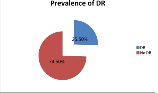
Results: Prevalence of diabetic retinopathy was 25.5% (51/200). Most of the cases were of non-proliferative DR (21.50%) and 4% patients were having proliferative diabetic retinopathy
Conclusion: In our study, there is no general awareness of diabetic retinopathy amongst majority of diabetic patients. Therefore, there is a very much need for awareness and also provision of access to screening services to the patient.
Page 37 www.ijiras.com | Email: contact@ijiras.com been conducted to estimate the prevalence of diabetic
retinopathy in urban areas, there is paucity in the literature regarding the prevalence of diabetic retinopathy in rural area. With above context, a rural hospital based cross sectional study is designed to find out prevalence of diabetic retinopathy in rural area.
Hence the present study was done at our tertiary care centre to assess the knowledge and attitude of the general population residing in rural areas regarding diabetes mellitus and diabetic retinopathy with an aim to create awareness and understand the shortcomings in such programs.
II. MATERIALS AND METHODS
This is Prospective, cross-sectional, observational study done at tertiary care centre in rural area during usual clinical practice from 1 January 2016 to 30 June 2017.The study was approved by the Institutional Ethics Committee and followed the tenets of Declaration of Helsinki. A total of 200 patients were included.
Inclusion criteria in this study were a) Patients of all age groups having diabetes mellitus. b) Newly diagnosed diabetic patients. c)Includes both male and females. Exclusion Criteria were - a) Patients who do not have diabetes mellitus. b) Patients having dense cataract and hazy media, whose fundi cannot be examined. c) Patients with history of exposure to radiation, hypertensive retinopathy without diabetes, sickle cell disease and pheochromocytoma as these conditions can mimic fundus features with diabetic retinopathy. d) Noncompliant patients. e) Immunocompromised patients. f) Patients having severe ocular infections.
CASE REPORT FORMS / PROFORMA: Standardized format of case report forms (CRF) (proforma) was used to document the patient‟s data during the course of the study. The investigator ensured that all data are entered promptly, legibly, completely and accurately. The CRF along with their respective lab reports were recorded.
All diabetic patients who came to the OPD during this period were considered for the study. Informed consent was taken. Detailed clinical history was taken and ocular examination was done. After exclusion criteria were applied as mentioned above, the selected subjects were given a case report form which was approved by the ethics committee. The case report form was filled after a thorough interview.
All participants were asked about their age, sex, address, religion, educational attainment, Risk factors for diabetic retinopathy and vision-threatening diabetic retinopathy (haemoglobin A1c, duration of diabetes, insulin use, systolic and diastolic blood pressure, addiction history and history of end organ diseases) were examined. Haemoglobin A1c, duration of diabetes, and systolic and diastolic blood pressure were used as continuous variables. Prior history of CVD was ascertained by self-report of coronary heart disease, angina, myocardial infarction, stroke, or congestive heart failure. We considered adherence to treatment indirectly as subjects taking >90% of the prescribed doses from the last visit.
Every patient underwent a complete ocular examination including visual acuity, anterior segment evaluation and fundus by an attending Ophthalmologist. Examination of
posterior segment was done with the indirect ophthalmoscopy and slit lamp (78D or 90D) direct ophthalmoscopy and grading of diabetic retinopathy if any were noted according to the ETDRS guidelines.
III. RESULTS STATISTICAL ANALYSIS
Quantitative data is presented with the help of Mean and Standard deviation. Qualitative data is presented with the help of frequency and percentage table. Association among the study groups is assessed with the help of Chi-Square test. „p‟ value less than 0.05 is taken as significant.
Pearson's chi-squared test
Where Χ2 = Pearson's cumulative test statistic. Oi = an observed frequency;
Ei = an expected frequency, asserted by the null hypothesis;
n = the number of cells in the table.
Results were graphically represented where deemed necessary.
Appropriate statistical software, including but not restricted to MS Excel, SPSS ver. 20 will be used for statistical analysis. Graphical representation will be done in MS Excel 2010.
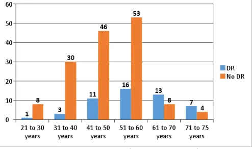
A rural hospital based study was done at our tertiary care centre to assess the prevalence of diabetic retinopathy in patients with diabetes mellitus. Mean age of the diabetics in our study was 50.47±11.601 years. Most of the diabetics were of age group 51 to 60 years (34.5%) followed by 61 to 70 years (28.5%). (Figure 1.) Diabetics of age group 21 to 30 years and 71 to 75 years were less in number. Significant association was found between age and DR.
Figure 1: Association between Age and DR
68.5% patients had no knowledge that diabetes affects vision in complicated cases. (Table 1)
Knowledge that diabetes affect vision
No of patients Percent
No 137 68.50%
Yes 63 31.50%
Page 38 www.ijiras.com | Email: contact@ijiras.com Most of the patients were male by gender (52%) and rest
(48%)were females. No significant association was found between gender and DR. Almost equal number of males are suffering from DR as compared to affected females.
59.50% were hypertensive in our study which was prevalence of hypertensive with diabetes in our study. Significant association was found that most of the hypertensive patients in our study suffered from DR (Table 2)
HT status DR No DR
Hypertension 35 84
No hypertension 16 65
Chi-square 12.513 P value< 0.05
Table 2: Distribution of subjects according to hypertension status
Recent cases i.e. diabetes less than 10-year duration was present in only 28.50% cases but most of the diabetics were not recent cases, they had DM from >10-year duration 51.5%. Most of the cases were of non-proliferative DR and were having >10 years‟ duration of diabetes mellitus (68.5%) and 17.6% had proliferative diabetic retinopathy with diabetes duration of more than 10 years (Table 3). 37.2% cases had moderate NPDR with a diabetes duration of >10 years. There was significant association found between prevalence of diabetic retinopathy and duration of diabetes.
Severity of DR
Not Recent(>10
years)
Recent(<10 years)
Unkno wn Mild
NPDR(14)
9(17.6%) 2(3.9%) 3(5.8%)
Moderate NPDR(21)
19(37.2%) 0(0%) 2(3.9%)
Severe NPDR(7)
7(13.7%) 0(0%) 0(0%)
Proliferative DR(9)
9(17.6%) 0(0%) 0(0%)
Total(51) 44(86.2%) 2(3.9%) 5(9.8%) Chi-square 30.254 P value <0.05
Table 3: Prevalence of DR relative to duration of diabetes
According to severity, prevalence of diabetic retinopathy was 25.5% (51/200) (Figure 2). Patients having mild NPDR, moderate NPDR, severe NPDR and Clinically significant macular oedema were 7%,10.5%,3.5% and 0.5% respectively and 4% patients were having PDR (Table 4).
Figure 2: Prevalence of diabetic retinopathy amongst diabetics
Diabetic retinopathy No of patients Percent
Mild NPDR 14 7%
Moderate NPDR 21 10.5%
Severe NPDR 7 3.50%
Proliferative DR 8 4%
Clinically significant macular edema
1 0.5%
No DR 149 74.5%
Table 4: Prevalence of Diabetic retinopathy amongst diabetics
IV. DISCUSSION
A rural hospital based study was done at our tertiary care centre to assess the prevalence of diabetic retinopathy in patients with diabetes mellitus.
Significant association was found between age and DR. Most of the diabetics were in the age group of 51 to 60 years (34.5%) followed by 61 to 70 years (28.5%). Diabetics of age group 21 to 30 years and 71 to 75 years were less in number. Most of the patients were male by gender 52% and rest 48% were females.
Shetgar AC et al study assessing the awareness of diabetic retinopathy in individuals with type 2 diabetes mellitus found study group comprised of 80 males and 70 females. Patients were aged between 40 and 83 years with the mean age being 61.5 years. All individuals were diagnosed with type 2 diabetes mellitus. Sheth NR et al10 study assessing the awareness about diabetes mellitus and diabetic retinopathy in patients with diabetes mellitus had 42% females and 58% males. Mean age was 58.5 years and age range was 40 to 71 years.
68.5% patients in our study had no knowledge that diabetes affects vision in complicated cases. Shetgar AC et al study assessing the awareness of diabetic retinopathy in individuals with type 2 diabetes mellitus observed of the 150 patients, 68 (45.3%) were aware of Diabetic Retinopathy compared to 82(54.6%) who were not.
Kiran D et al study assessing the awareness and knowledge of diabetic retinopathy among diabetic patients reported 37.4% had heard about the eye complications of diabetes and 62.6% never heard about it. Out of 37.4% only 24.9% knew the relationship between diabetes and diabetic eye disease; 29.5 % had heard that vision can be affected due to high blood sugar levels.
Page 39 www.ijiras.com | Email: contact@ijiras.com is on anti-VEGF for prevention of systemic complications, the
chances of the diabetic hypertensive patient suffering from DR may reduce upto a certain extent.
There was significant association found between prevalence of diabetic retinopathy and duration of diabetes. Alam M et al study to know the effect of duration of diabetes on severity of diabetic retinopathy reported in group-A 185 (86.04%) patients had normal fundi and 24 (11.16%) had NPDR and 6(2.79%) had PDR. In Group-B 39 (33.33%) had normal fundi, 46 (39.31%) had NPDR and 32 (27.35%) had PDR. In group C no patient had normal fundi, 15 (39.47%) had NPDR and 23 (60.52%) had PDR.
Shrote AP et al cross-sectional study found Retinopathy was prevalent in type 2 diabetes compared with those of type 1 (84% vs. 2% for NPDR; and 14% vs. 0% for PDR), majority fortunately being mild to moderate NPDR cases and a few diabetics unfortunately diagnosed to have PDR.
Most of the cases of DR in our study were having diabetes for >10 years duration and significant association was found. Most of the cases were of non-proliferative DR and were having >10 years‟ duration of diabetes mellitus (68.5%) and 17.6% had proliferative diabetic retinopathy with diabetes duration of more than 10 years. 37.2% cases had moderate NPDR with a diabetes duration of >10 years. Baba D et al study estimating the prevalence of diabetic retinopathy and to determine the correlation of systemic risk factors with retinopathy observed a strong association between the duration of diabetes and the onset of retinopathy. 56.3% acquired retinopathy in the first 5 years after diagnosis.
According to severity prevalence of diabetic retinopathy was 25.5% (51/200). Jenchitr W et al study to determine the prevalence and severity of diabetic retinopathy reported prevalence of the background or nonproliferative diabetic retinopathy (BDR or NPDR) was 18.9% and proliferative diabetic retinopathy (PDR) was 3% in all age groups. For the relationship of the duration of diabetes, it showed that the longer the duration of diabetes the higher the prevalence of diabetic retinopathy. In NPDR, the retinopathy varied from 13.11 to 22.91% in persons having diabetes for less than 10 years and up to 42.86% in those with diabetes for up to 20 years. In the PDR group, the prevalence was 2.15 to 2.42% in persons with diabetes for less than 10 years and up to 10.20% for those with diabetes for up to 20 years.
V. CONCLUSION
In our study, there is no general awareness of diabetic retinopathy amongst majority of diabetic patients. Therefore, there is a very much need for awareness and also provision of access to screening services to the patient. Comprehensive and aggressive awareness is very much required to educate diabetic patients on diabetic retinopathy. Creation of awareness is therefore very necessary as one of the first steps in any program which aims at reducing Diabetic Retinopathy. This can only be achieved if people with diabetes have the proper information, awareness and tools to make changes based on best practice.
REFERENCES
[1] Shaw J., Sicree R., & Zimmet, P. (2010). Global estimates of the prevalence of diabetes for 2010 and 2030. Diabetes Research and Clinical Practice,87(1), 4-14.
[2] Prokofyeva E., & Zrenner E. (2012). Epidemiology of Major Eye Diseases Leading to Blindness in Europe: A Literature Review. Ophthalmic Research, 47(4).
[3] Hammes H. (2013). Optimal treatment of diabetic retinopathy. Therapeutic Advances in Endocrinology and Metabolism, 4(2), 61-71.
[4] Struck H. (2006). Nationale Versorgungsleitlinie Typ-2-Diabetes – Prävention und Therapie von Netzhautkomplikationen. Klinische Monatsblätter Für Augenheilkunde, 223(S 3).
[5] Giani G, Janka H, Hauner H, Standl E, Schiel R, Neu (2004),A Epidemiologie und Verlauf des Diabetes mellitus in Deutschland. Evidenzbasierte Leitlinie DDG-Aktualisierung , 5, 1–12.
[6] Nentwich, M., & Ulbig, M. (2010). Diabetische Retinopathie. Der Diabetologe, 6(6), 491-502.
[7] Raman, R., Rani, P. K., Rachepalle, S. R., Gnanamoorthy, P., Uthra, S., Kumaramanickavel, G., & Sharma, T. (2009). Prevalence of Diabetic Retinopathy in India. Ophthalmology, 116(2), 311-318.
[8] Agrawal R, Ranka M, Beniwal R, Gothwal S, Jain G, Kochar D, et al. (2003) Prevalence of diabetic retinopathy in type 2 diabetes in relation to risk factors: Hospital based study. Int J Diab Dev Countries, 23 ,16–19.
[9] Shetgar, A. C., Patil, B., Salagar, M. C., & Nanditha, A. M. (2015). Assessment of awareness of diabetic retinopathy among diabetics: A Clinical Survey. Indian Journal of Clinical and Experimental Ophthalmology, 1(4), 260.
[10] Sheth, N. R., Sareshwala, N. A., Chaudhary, S. G., & Matai, H. D. (2017). Awareness about diabetes mellitus and diabetic retinopathy in patients with diabetes mellitus. International Journal of Research in Medical Sciences, 5(8), 3570.
[11] 11. Kiran, D., & Mendonca, D. N. (2016). “Assessment of Awareness of Diabetic Retinopathy among The Diabetics.”. IOSR Journal of Humanities and Social Science, 21(08), 61-64.
[12] 12. Shrote, A. P. (2015). Clinical Evaluation of Correlation Between Diabetic Retinopathy with Modifiable, Non-Modifiable and Other Independent Risk Factors in Tertiary Set-up in Central Rural India. Journal Of Clinical And Diagnostic Research
[13] 13. Alam M, Ihsanullah M, Saeed R, Saleem M. (2011) Effect of duration of diabetes on severity of retinopathy. Gomal Journal of Medical Sciences. 9(2).
[14] 14. Baba D, Muthukrishnan V, Bhaskaran S, Kumar PS, Poovitha R. (2015) Prevalence of Diabetic Retinopathy and Correlation with Systemic Risk Factors in Type 2 Diabetes Mellitus in a Tertiary Care Hospital. Sch. J. App. Med. Sci,, 3(7C) ,2659-2664
Page 40 www.ijiras.com | Email: contact@ijiras.com in relation to duration of diabetes mellitus in community