ISMB TUMS
The Epidemiology of Tuberculosis in Tabriz, Iran: A Five Year Retrospective Study
Mehrdad Asghari
1, Siamak Heidarzadeh
2, Ziba Vaise Malekshahi
3, Maryam
Hemmatzadeh
4, Mohammad Javad Razzaghe Karimi
4, Hamideh Ashrafi
5, Fatemeh
Fallah
6, Raheleh Sadat Sajadi Nia
7*1
Students Research Committee, Faculty of Paramedicine, Tabriz University of Medical Sciences, Tabriz, IR Iran 2Department of Pathobiology, School of Public Health, Tehran University of Medical Sciences, Tehran, IR Iran 3
Department of biology, Faculty of Science, Shahed University, Tehran, IR Iran 4
Department of Laboratory Sciences, Faculty of Paramedicine, Tabriz University of Medical Sciences, Tabriz, IR Iran 5
Tuberculosis and Lung Disease Research Center, Tabriz University of Medical Sciences, Tabriz, IR Iran 6
Department of Microbiology, Shahid Beheshti University of Medical Sciences, Tehran, IR Iran 7
Shahid Beheshti University of Medical Sciences, Tehran, IR Iran
ARTICLE INFO ABSTRACT
Article type:
Original Article Background: Tuberculosis is one of the most important causes of morbidityand mortality worldwide. Approximately 10-20% of tuberculosis is
Extra-pulmonary tuberculosis (EPTB), which is much higher (50%) in patients suffering from immunity defects such as AIDS. EPTB diagnosis is difficult mostly because of various clinical manifestations and aggressive procedures needed for its diagnosis. The main goal of this study was to determine the prevalence of EPTB in the north west of Iran and also to investigate the different clinical characteristics of the studied population, the various clinical manifestations and organ involvement of EPTB, as well.
Methods: This study was carried out retrospectively using the data from
Tabriz Tuberculosis and Lung Disease Research Center from 2007 through 2011. Questionnaires were designed to extract relevant information to describe characteristics of EPTB affected population and also various clinical manifestations and organ involvement of the disease among the patients.
Results: The study included 203 EPTB cases notified from 2007 through
2011 including, 91 (44.83%) males and 112 (55.17%) females. The mean age of the patients was 46.55 ± 18.3. The main extra pulmonary involvements of the studied population were lymphadenitis (9.35%), pleural (7.39%) and spinal (5.42%) among males and lymphadenitis (17.24%), ocular (7.88%), pleural (6.40%) and spinal (5.91%) among females, respectively.
Conclusion: Since EPTB diagnosis is a challenging and time sparing attempt
even by the expert physicians, there is a need to perform further researches in order to identify the main clinical manifestations and organ involvement of EPTB in patients.
Article history: Received: 31 Jul 2012 Revised: 16 Aug 2012 Accepted: 25 Aug 2012
Keywords:
Tuberculosis, Pulmonary
Epidemiology
Tabriz
Please cite this paper as: Asghari M, Heidarzadeh S, Vaise Malekshahi Z, Hemmatzadeh M, Razzaghe
Karimi MJ, Ashrafi H, Fallah F, Sajadi Nia RS. The Epidemiology of Tuberculosis in Tabriz, Iran: A Five Year Retrospective Study. J Med Bacteriol. 2012; 1 (1, 2): pp. 23-30.
Asghari M et al. The Epidemiologyof Tuberculosis in Tabriz, Iran …
24 J Med Bacteriol. Vol. 1, No. 1, 2 (2012): pp. 23-30 jmb.tums.ac.ir
Introduction
Tuberculosis is one of the most important
causes of morbidity and mortality worldwide (1).
Approximately 2 billion people have latent
infection, 8 million are affected with active
disease and 2-3 million die due to tuberculosis
annually (2-4). The disease frequently involves
pulmonary system however it can disseminate
through hematogenous or lymphogenous
circulation and thus may involve any other
organ, which is then called Extra Pulmonary
Tuberculosis (EPTB) (4-6).
Approximately 10-20% of tuberculosis is
EPTB, that is much higher (50%) in patients
suffering from immunity defects such as AIDS
(7). The most common sites of EPBT
involvement are reported to be lymph nodes and
pleura and mean age of involvement is under 35
years old (8, 9). EPTB incidence is higher in
developing countries, however number of
patients with EPTB has increased in developed
countries during the last two decades as well
which can be as a result of increased mobility of
people, resistance against antibiotics and
growing number of patients with suppressed
immune system (10). EPTB diagnosis is difficult
mostly because of various clinical manifestations
and aggressive procedures needed for its
diagnosis (11).
The main goal of this study was to determine
the prevalence of EPTB in the north west of Iran
and also to investigate the different clinical
characteristics of the studied population, the
various clinical manifestations and organ
involvement of EPTB, as well.
Methods
This study was carried out retrospectively using
the data from Tabriz Tuberculosis and Lung
Disease Research Center obtained during 2007
through 2011.
From the questionnaires which were designed
to extract relevant information to describe
characteristics of EPTB affected population,
personal data including age, sex, weight, place of
residence, nationality, date of EPTB diagnosis,
diagnostic criteria, TB type (i.e. “new case”,
where a first time TB diagnosis is made and
reported, or “re-treated cases”),involved organs,
rate of End Stage Renal Disease (ESRD) due to
EPTB, family history of TB, history of BCG
vaccination and referral center were collected.
We followed standard methods for EPTB
diagnosis, which has been designed to identify
tuberculosis such as: staining (acid-fast) of
samples obtained from skin, bone marrow,
Cerebrospinal fluid (CSF), sputum with
Ziehl-Neelsen method and culturing in solid egg
medium (Lowenstein-Jensen), measuring levels
of Niacin-Nitrate reduction, semiquantitative
catalase , urease, arylsulfatase, tellurite reduction
in the specimens, and evaluation of tuberculin
test. Specimen processing for the recovery of
acid-fast bacilli involves a number of complex
procedures, each of which must be carried out
with precision. Specimens from sterile site can
be inoculated directly to media (small volume),
other specimens required decontamination and
concentration. All incomplete questionnaires
were rechecked and full filled. Data analysis was
carried out using SPSS software (version 16;
(frequencies and means) were used to describe
the demographic characteristics.
Ethics
The protocol for this study was reviewed and
granted by the Research Deputy of Tabriz
University of Medical Sciences and was found to
be public health surveillance and not a human
subject research.
Results
The study included 203 EPTB cases notified
from 2007 through 2011 including, 91 (44.83%)
males and 112 (55.17%) females.
The most common EPTB manifestations were
night sweating (33.99%), chronic fever lasting
more than 2 weeks (33.50%) and weight loss
(28.80%), respectively (Figure 1) (Table 4, 5).
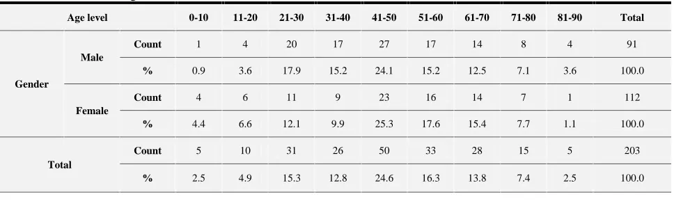
The mean age of the patients was 46.55 ± 18.3
(46.38 ± 18.79 among males and 46.68 ± 17.79
among females) (Table 1). The main extra
pulmonary involvements of the studied
population were lymphadenitis (9.35%), pleural
(7.39%) and spinal (5.42%) among males and
lymphadenitis (17.24%), ocular (7.88%), pleural
(6.40%) and spinal (5.91%) among females,
respectively (Table 2, 3). Genital TB, TB
peritonitis and pericarditis were more dominant
among females, however epididymal and also
bladder TB were found only in males.
It should be noted that the most useful method
for EPTB diagnosis in this study was imaging,
by which 129 cases were identified (Figure 2),
and the most prevalent type of TB among the
studied group were new cases (Figure 3).
Figure 1. EPTB manifestations among the patients
referred to Tabriz Tuberculosis and Lung Disease Research Center from 2007 through 2011
Figure 2. Diagnostic methods for EPTB in patients
referred to Tabriz Tuberculosis and Lung Disease Research Center from 2007 through 2011
Figure 3. Types of TB in patients referred to Tabriz
Tuberculosis and Lung Disease Research Center from 2007 through 2011
(frequencies and means) were used to describe
the demographic characteristics.
Ethics
The protocol for this study was reviewed and
granted by the Research Deputy of Tabriz
University of Medical Sciences and was found to
be public health surveillance and not a human
subject research.
Results
The study included 203 EPTB cases notified
from 2007 through 2011 including, 91 (44.83%)
males and 112 (55.17%) females.
The most common EPTB manifestations were
night sweating (33.99%), chronic fever lasting
more than 2 weeks (33.50%) and weight loss
(28.80%), respectively (Figure 1) (Table 4, 5).
The mean age of the patients was 46.55 ± 18.3
(46.38 ± 18.79 among males and 46.68 ± 17.79
among females) (Table 1). The main extra
pulmonary involvements of the studied
population were lymphadenitis (9.35%), pleural
(7.39%) and spinal (5.42%) among males and
lymphadenitis (17.24%), ocular (7.88%), pleural
(6.40%) and spinal (5.91%) among females,
respectively (Table 2, 3). Genital TB, TB
peritonitis and pericarditis were more dominant
among females, however epididymal and also
bladder TB were found only in males.
It should be noted that the most useful method
for EPTB diagnosis in this study was imaging,
by which 129 cases were identified (Figure 2),
and the most prevalent type of TB among the
studied group were new cases (Figure 3).
Figure 1. EPTB manifestations among the patients
referred to Tabriz Tuberculosis and Lung Disease Research Center from 2007 through 2011
Figure 2. Diagnostic methods for EPTB in patients
referred to Tabriz Tuberculosis and Lung Disease Research Center from 2007 through 2011
Figure 3. Types of TB in patients referred to Tabriz
Tuberculosis and Lung Disease Research Center from 2007 through 2011
(frequencies and means) were used to describe
the demographic characteristics.
Ethics
The protocol for this study was reviewed and
granted by the Research Deputy of Tabriz
University of Medical Sciences and was found to
be public health surveillance and not a human
subject research.
Results
The study included 203 EPTB cases notified
from 2007 through 2011 including, 91 (44.83%)
males and 112 (55.17%) females.
The most common EPTB manifestations were
night sweating (33.99%), chronic fever lasting
more than 2 weeks (33.50%) and weight loss
(28.80%), respectively (Figure 1) (Table 4, 5).
The mean age of the patients was 46.55 ± 18.3
(46.38 ± 18.79 among males and 46.68 ± 17.79
among females) (Table 1). The main extra
pulmonary involvements of the studied
population were lymphadenitis (9.35%), pleural
(7.39%) and spinal (5.42%) among males and
lymphadenitis (17.24%), ocular (7.88%), pleural
(6.40%) and spinal (5.91%) among females,
respectively (Table 2, 3). Genital TB, TB
peritonitis and pericarditis were more dominant
among females, however epididymal and also
bladder TB were found only in males.
It should be noted that the most useful method
for EPTB diagnosis in this study was imaging,
by which 129 cases were identified (Figure 2),
and the most prevalent type of TB among the
studied group were new cases (Figure 3).
Figure 1. EPTB manifestations among the patients
referred to Tabriz Tuberculosis and Lung Disease Research Center from 2007 through 2011
Figure 2. Diagnostic methods for EPTB in patients
referred to Tabriz Tuberculosis and Lung Disease Research Center from 2007 through 2011
Figure 3. Types of TB in patients referred to Tabriz
Asghari M et al. The Epidemiologyof Tuberculosis in Tabriz, Iran …
26 J Med Bacteriol. Vol. 1, No. 1, 2 (2012): pp. 23-30 jmb.tums.ac.ir
Table 1. Age and sex distribution of the patients with EPTB referred to Tabriz Tuberculosis and Lung Disease Research
Center from 2007 through 2011
Age level 0-10 11-20 21-30 31-40 41-50 51-60 61-70 71-80 81-90 Total
Gender
Male
Count 1 4 20 17 27 17 14 8 4 91
% 0.9 3.6 17.9 15.2 24.1 15.2 12.5 7.1 3.6 100.0
Female
Count 4 6 11 9 23 16 14 7 1 112
% 4.4 6.6 12.1 9.9 25.3 17.6 15.4 7.7 1.1 100.0
Total
Count 5 10 31 26 50 33 28 15 5 203
% 2.5 4.9 15.3 12.8 24.6 16.3 13.8 7.4 2.5 100.0
Table 2. Clinical characteristics and different organ involvements of the patients with EPTB referred to Tabriz Tuberculosis
and Lung Disease Research Center from 2007 through 2011
M
ili
a
ry
tu
b
er
cu
lo
si
s
B
la
d
d
er
tu
b
er
cu
lo
si
s
A
b
d
o
m
in
a
l
tu
b
er
cu
lo
si
s
R
en
a
l
tu
b
er
cu
lo
si
s
B
o
n
e
tu
b
er
cu
lo
si
s
Ga
st
ro
in
te
st
in
a
l
tu
b
er
cu
lo
si
s
T
h
y
ro
id
tu
b
er
cu
lo
si
s
M
en
in
g
es
tu
b
er
cu
lo
si
s
D
er
m
a
l
tu
b
er
cu
lo
si
s
L
y
m
p
h
a
d
en
it
is
Radiology % 100 66.7 0 25 70 100 0 100 90.9 63.3
Pathology % 0 0 50 25 50 100 0 0 100 83.7
Microbiology % 50 100 50 100 40 100 100 100 36.4 26.5
PPD 0 26±12.72 9.5±0.7 16.75±13.14 14±6.14 0 0 0 15.28±10.41 19.29±2.28
ESR 45 6.5±2.12 40.5±14.84 33.25±29.21 62.25±32.27 69±53.74 0 0 22.22±30.57 22.72±3.54
Family history 1 0 1 2 1 0 0 0 3 7
Vaccine history 0 1 0 0 2 1 0 0 3 8
AIDS 0 0 0 0 0 0 0 0 0 1
Cancer 0 1 0 0 0 0 0 0 0 1
Diabetes 0 0 0 0 0 0 0 0 0 2
New disease 1 4 3 5 10 3 1 1 12 52
Recurrent 1 0 0 0 1 0 0 0 1 1
Discussion
Extra-pulmonary tuberculosis is caused by the
spread of TB bacilli from a pulmonary focus
(12). Spreading is carried out mainly through
hematogenous dissemination or lymphogenous
spread from a primary, usually a pulmonary,
focus. This hematogenous spread may happen
years before the onset of progressive
tuberculosis (6, 13).
Although some damages caused by tuberculosis
and extra-pulmonary tuberculosis heal up in
most patients, but granuloma containing TB
bacilli usually stay alive for decades (12). The
organism gains access to the blood stream via
the lymphohematogenous route and may then
affect any organ (6, 14). The next failures related
to damages could reactivate the extra-pulmonary
disease once again (12). Since there is no
particular sign of infection, therefore reactivation
of the disease may not be recognized clinically
(12).
There are many reports of unidentified
extra-pulmonary tuberculosis that could be
distinguished only by necrosis (12). Diagnosis is
often difficult, but successful outcomes are
possible when made at an early stage (15).
In many different studies the role of various
factors on the incidence of pulmonary and
Table 3. Clinical characteristics and different organ involvements of the patients with EPTB referred to Tabriz Tuberculosis
and Lung Disease Research Center from 2007 through2011
P le u ra l tu b er cu lo si s Oc u la r tu b er cu lo si s S p in a l tu b er cu lo si s Ge n it a l tu b er cu lo si s P er ic a rd ia l tu b er cu lo si s L iv er tu b er cu lo si s E p id id y m a l tu b er cu lo si s P er it o n eu m tu b er cu lo si s
Radiology % 100 93.8 95 55.6 100 100 50 67.7
Pathology % 40 6.3 50 44.4 50 50 50 50
Microbiology % 40 25 15 66.7 25 50 50 50
PPD 15.13±7.08 29.92±14.18 23.15±3.67 24.44±13.42 26.25±36.83 0 13.75±11.05 16±5.35
ESR 31.95±26.8 28.05±31 42.88±33.27 31.30 14.33±7.02 - 23±29 32.33±14.64
Family history 3 2 1 2 0 0 2 1
Vaccine history 1 0 2 1 0 0 1 0
AIDS 0 0 0 0 0 0 0 0
Cancer 0 0 0 0 0 0 0 0
Diabetes 2 0 1 0 1 0 1 1
New disease 26 27 23 11 5 2 5 6
Recurrent 2 0 0 0 0 0 0 0
Table 4. Clinical manifestations of EPTB in patients referred to Tabriz Tuberculosis and Lung Disease Research Center from
2007 through 2011
M ili a ry tu b er cu lo si s B la d d er tu b er cu lo si s A b d o m in a l tu b er cu lo si s R en a l tu b er cu lo si s B o n e tu b er cu lo si s Ga st ro in te st in a l tu b er cu lo si s T h y ro id tu b er cu lo si s M en in g es tu b er cu lo si s D er m a l tu b er cu lo si s L y m p h a d en it is
Age mean 63±24 62±12.19 54.33±18.50 41.8±18.47 39.36±30.62 31.33±14.74 29±0 59±0 41.15±15.45 43.61±18.57
Frequency 2 4 3 5 11 3 1 1 13 54
G Male 2 4 1 2 7 2 0 1 5 19
Female 0 0 2 3 4 1 1 0 8 35
Fever 2 2 2 0 2 0 0 0 2 20
Weight loss 1 1 3 2 4 1 0 0 1 15
Sweating 2 1 3 1 3 0 0 0 3 18
Chess pain 0 0 0 0 2 1 0 0 0 7
Shortness
of breath 0 0 0 1 2 0 0 0 0 8
Backache 0 0 1 1 3 1 0 0 3 5
Cough (more than
2 week)
1 0 1 2 1 0 0 0 1 12
Phlegm
(mucus) 0 0 0 1 0 0 0 0 2 9
G = Gender
Table 5. Clinical manifestations of EPTB in patients referred to Tabriz Tuberculosis and Lung Disease Research Center
from 2007 through 2011
P le u ra l tu b er cu lo si s Oc u la r tu b er cu lo si s S p in a l tu b er cu lo si s Ge n it a l tu b er cu lo si s P er ic a rd ia l tu b er cu lo si s L iv er tu b er cu lo si s E p id id y m a l tu b er cu lo si s P er it o n eu m tu b er cu lo si s
Age mean 48.5±19.4 53.11±11.9 54.6±17.4 35.6±5.6 60.4±14.4 46±5.6 50.4±17.2 52.6±9.6
Frequency 28 27 23 11 5 2 5 6
Gender Male 15 10 11 2 4 0 5 1
Female 13 16 12 9 1 2 0 5
Fever 22 1 10 2 2 0 0 2
Weight loss 12 1 7 2 3 1 0 3
Sweating 16 4 12 2 2 0 0 3
Chess pain 12 0 2 2 2 1 0 3
Shortness of breath 13 0 6 1 3 0 0 0
Backache 9 1 11 2 2 0 0 0
Cough (more than 2 week) 17 1 3 2 1 0 0 2
Asghari M et al. The Epidemiologyof Tuberculosis in Tabriz, Iran …
28 J Med Bacteriol. Vol. 1, No. 1, 2 (2012): pp. 23-30 jmb.tums.ac.ir Extra-pulmonary tuberculosis after observance
of HIV infection has become more common
(17). It has been investigated among 50% of the
patients affected simultaneously with
tuberculosis and AIDS (17, 18) and therefore
extra-pulmonary tuberculosis most probably is
an index of immune deficiency (19). Black men
and women have a higher rate of EPTB
incidence (19). Furthermore, extra-pulmonary
tuberculosis is most common among young
patients with active tuberculosis and usually
diagnosed as a cancer inadvertently (20).
More than 3.8 million cases of extra-pulmonary
tuberculosis were reported to WHO in 2001,
90% of them came from developing countries
(21, 22). Among patients with AIDS up to two
thirds of infection was either in combined
involvement of lungs and extra-pulmonary or
extra-pulmonary tuberculosis alone (23). Among
26283 cases with tuberculosis reported to the
Centers for Disease Control and Prevention
(CDC) in the United States until 1991, 18% of
them had extra-pulmonary tuberculosis but
following AIDS epidemic, the cases of
extra-pulmonary tuberculosis increased and thus
30-60% of patients affected with AIDS and
tuberculosis had extra-pulmonary tuberculosis
(24).
Annual average incidence rate of tuberculosis
in Iran is 17.9 in 100000 patients. Tuberculosis
incidence is higher in Balochistan, Khorasan,
Golestan Gilan, Kurdistan, Western Azerbaijan,
Khuzestan, and southern coasts of Iran (16).
During 2001-2006, pulmonary tuberculosis and
extra-pulmonary tuberculosis has had a
decreased rate of incidence especially in Birjand
(-18.6%) and Eastern Azerbaijan (-10.2%) cities
whereas from 2006 through 2008, it has had an
upward move particularly in Sabzevar (+24.9%).
Throughout this period 56629 were affected with
tuberculosis and 23442 of patients were
suffering from extra-pulmonary tuberculosis
(25).
In a study performed by Iran Pasteur Institute in
1982, of the patients suffering from tuberculosis,
22.5% were diagnosed with EPTB and the most
common organs involved were reported to be
lymph nodes (39%) joints and bones (18%),
meninges (16%), urinary tract (12%),
endometrial tissue (5%). These findings were
repeated in another study conducted by the same
institute in 1997 (16).
In our study it was shown that the prevalence of
EPTB among women (which may be due to
having contact with TB affected animals in the
farms) is higher than men which is in contrast
with the researches by Tam et al. in Hong Kong
and Lado Lado et al. in Santiago (26, 27).
In our study, EPTB incidence was found more
commonly (38.9%) between 25-40 years of age,
however it was rarely reported in over 65 years
old individuals (13%). The most common sites
of EPTB involvement were lymph nodes
(26.60%), pleural (13.79%), ocular (13.30%) and
spinal (10.0%) which is similar to other studies
performed in Germany and Netherlands,
however in a research by Demiralay in Turkey,
the most common sites of EPTB involvement
were pleural (52.8%) and lymph nodes,
Conclusion
Since EPTB diagnosis is a challenging and time
sparing attempt even by the expert physicians,
there is a need to perform further researches in
order to identify the main clinical manifestations
and organ involvement of EPTB in patients.
Figure 4. Occupation of patients with EPTB referred to
Tabriz Tuberculosis and Lung Disease Research Center from 2007 through 2011
Acknowledgment
Hereby we would like to express our gratitude
to the Research Deputy of Tabriz University of
Medical Sciences for their sincere cooperation
and financial support.
References
1. Chong VH, Lim KS. Gastrointestinal
tuberculosis. Singapore Med J 2009; 50 (6):
638-46.
2. Almaguer-Chavez J, Ocampo-Candiani J, Rendon
A. Current panorama in the diagnosis of
cutaneous tuberculosis. Actas Dermosifiliogr
2009; 100 (7): 562-70.
3. Malikova H, Mikova B. Abdominal tuberculosis
in CT imaging. Cas Lek Cesk 2007; 146 (6):
557-9.
4. Malaviya AN, Kotwal PP. Arthritis associated
with tuberculosis. Best Pract Res Clin Rheumatol
2003; 17 (2): 319-43.
5. Tinsa F, Essaddam L, Fitouri Z, et al.
Extra-pulmonary tuberculosis in children: a study of 41
cases. Tunis Med 2009; 87 (10): 693-8.
6. De Backer AI, Mortele KJ, De Keulenaer BL, et
al. Tuberculosis: epidemiology, manifestations,
and the value of medical imaging in diagnosis.
JBR-BTR 2006; 89 (5): 243-50.
7. Ullah S, Shah SH, Aziz ur R, et al.
Extrapulmonary tuberculosis in Lady Reading
Hospital Peshawar, NWFP, Pakistan: survey of
biopsy results. J Ayub Med Coll Abbottabad
2008; 20 (2): 43-6.
8. Özvaran MK, Baran R, Tor M, et al.
Extrapulmonary tuberculosis in non-human
immunodeficiency virus-infected adults in an
endemic region. Annals of thoracic medicine
2007; 2 (3): 118.
9. Ilgazli A, Boyaci H, Basyigit I, et al.
Extrapulmonary tuberculosis: clinical and
epidemiologic spectrum of 636 cases. Arch Med
Res 2004; 35 (5): 435-41.
10. Galois L, Chary-Valckenaere I, Mainard D, et al..
Tuberculosis of the patella. Arch Orthop Trauma
Surg 2003; 123 (4): 192-4.
11. Mazza-Stalder J, Nicod L, Janssens JP.
Extrapulmonary tuberculosis. Rev Mal Respir
2012; 29 (4): 566-78.
12. Ang EP, Quimosing EM, Alora BD.
Extrapulmonary tuberculosis. Spine 1982; 4 (3):
1-7.
13. Vanhoenacker F, De Backer A, Op de Beeck B,
et al. Imaging of gastrointestinal and abdominal
tuberculosis. European Radiology Supplements
2004; 14 (3): 103-15.
14. Kader E. The CT features of abdominal
tuberculosis in children. Pediatr Radiol 2002; 32
(2): 75-81.
15. Tahar G, Goucha-Louzir R, Rachid LM.
Tuberculosis in children undergoing
Conclusion
Since EPTB diagnosis is a challenging and time
sparing attempt even by the expert physicians,
there is a need to perform further researches in
order to identify the main clinical manifestations
and organ involvement of EPTB in patients.
Figure 4. Occupation of patients with EPTB referred to
Tabriz Tuberculosis and Lung Disease Research Center from 2007 through 2011
Acknowledgment
Hereby we would like to express our gratitude
to the Research Deputy of Tabriz University of
Medical Sciences for their sincere cooperation
and financial support.
References
1. Chong VH, Lim KS. Gastrointestinal
tuberculosis. Singapore Med J 2009; 50 (6):
638-46.
2. Almaguer-Chavez J, Ocampo-Candiani J, Rendon
A. Current panorama in the diagnosis of
cutaneous tuberculosis. Actas Dermosifiliogr
2009; 100 (7): 562-70.
3. Malikova H, Mikova B. Abdominal tuberculosis
in CT imaging. Cas Lek Cesk 2007; 146 (6):
557-9.
4. Malaviya AN, Kotwal PP. Arthritis associated
with tuberculosis. Best Pract Res Clin Rheumatol
2003; 17 (2): 319-43.
5. Tinsa F, Essaddam L, Fitouri Z, et al.
Extra-pulmonary tuberculosis in children: a study of 41
cases. Tunis Med 2009; 87 (10): 693-8.
6. De Backer AI, Mortele KJ, De Keulenaer BL, et
al. Tuberculosis: epidemiology, manifestations,
and the value of medical imaging in diagnosis.
JBR-BTR 2006; 89 (5): 243-50.
7. Ullah S, Shah SH, Aziz ur R, et al.
Extrapulmonary tuberculosis in Lady Reading
Hospital Peshawar, NWFP, Pakistan: survey of
biopsy results. J Ayub Med Coll Abbottabad
2008; 20 (2): 43-6.
8. Özvaran MK, Baran R, Tor M, et al.
Extrapulmonary tuberculosis in non-human
immunodeficiency virus-infected adults in an
endemic region. Annals of thoracic medicine
2007; 2 (3): 118.
9. Ilgazli A, Boyaci H, Basyigit I, et al.
Extrapulmonary tuberculosis: clinical and
epidemiologic spectrum of 636 cases. Arch Med
Res 2004; 35 (5): 435-41.
10. Galois L, Chary-Valckenaere I, Mainard D, et al..
Tuberculosis of the patella. Arch Orthop Trauma
Surg 2003; 123 (4): 192-4.
11. Mazza-Stalder J, Nicod L, Janssens JP.
Extrapulmonary tuberculosis. Rev Mal Respir
2012; 29 (4): 566-78.
12. Ang EP, Quimosing EM, Alora BD.
Extrapulmonary tuberculosis. Spine 1982; 4 (3):
1-7.
13. Vanhoenacker F, De Backer A, Op de Beeck B,
et al. Imaging of gastrointestinal and abdominal
tuberculosis. European Radiology Supplements
2004; 14 (3): 103-15.
14. Kader E. The CT features of abdominal
tuberculosis in children. Pediatr Radiol 2002; 32
(2): 75-81.
15. Tahar G, Goucha-Louzir R, Rachid LM.
Tuberculosis in children undergoing
Conclusion
Since EPTB diagnosis is a challenging and time
sparing attempt even by the expert physicians,
there is a need to perform further researches in
order to identify the main clinical manifestations
and organ involvement of EPTB in patients.
Figure 4. Occupation of patients with EPTB referred to
Tabriz Tuberculosis and Lung Disease Research Center from 2007 through 2011
Acknowledgment
Hereby we would like to express our gratitude
to the Research Deputy of Tabriz University of
Medical Sciences for their sincere cooperation
and financial support.
References
1. Chong VH, Lim KS. Gastrointestinal
tuberculosis. Singapore Med J 2009; 50 (6):
638-46.
2. Almaguer-Chavez J, Ocampo-Candiani J, Rendon
A. Current panorama in the diagnosis of
cutaneous tuberculosis. Actas Dermosifiliogr
2009; 100 (7): 562-70.
3. Malikova H, Mikova B. Abdominal tuberculosis
in CT imaging. Cas Lek Cesk 2007; 146 (6):
557-9.
4. Malaviya AN, Kotwal PP. Arthritis associated
with tuberculosis. Best Pract Res Clin Rheumatol
2003; 17 (2): 319-43.
5. Tinsa F, Essaddam L, Fitouri Z, et al.
Extra-pulmonary tuberculosis in children: a study of 41
cases. Tunis Med 2009; 87 (10): 693-8.
6. De Backer AI, Mortele KJ, De Keulenaer BL, et
al. Tuberculosis: epidemiology, manifestations,
and the value of medical imaging in diagnosis.
JBR-BTR 2006; 89 (5): 243-50.
7. Ullah S, Shah SH, Aziz ur R, et al.
Extrapulmonary tuberculosis in Lady Reading
Hospital Peshawar, NWFP, Pakistan: survey of
biopsy results. J Ayub Med Coll Abbottabad
2008; 20 (2): 43-6.
8. Özvaran MK, Baran R, Tor M, et al.
Extrapulmonary tuberculosis in non-human
immunodeficiency virus-infected adults in an
endemic region. Annals of thoracic medicine
2007; 2 (3): 118.
9. Ilgazli A, Boyaci H, Basyigit I, et al.
Extrapulmonary tuberculosis: clinical and
epidemiologic spectrum of 636 cases. Arch Med
Res 2004; 35 (5): 435-41.
10. Galois L, Chary-Valckenaere I, Mainard D, et al..
Tuberculosis of the patella. Arch Orthop Trauma
Surg 2003; 123 (4): 192-4.
11. Mazza-Stalder J, Nicod L, Janssens JP.
Extrapulmonary tuberculosis. Rev Mal Respir
2012; 29 (4): 566-78.
12. Ang EP, Quimosing EM, Alora BD.
Extrapulmonary tuberculosis. Spine 1982; 4 (3):
1-7.
13. Vanhoenacker F, De Backer A, Op de Beeck B,
et al. Imaging of gastrointestinal and abdominal
tuberculosis. European Radiology Supplements
2004; 14 (3): 103-15.
14. Kader E. The CT features of abdominal
tuberculosis in children. Pediatr Radiol 2002; 32
(2): 75-81.
15. Tahar G, Goucha-Louzir R, Rachid LM.
Asghari M et al. The Epidemiologyof Tuberculosis in Tabriz, Iran …
30 J Med Bacteriol. Vol. 1, No. 1, 2 (2012): pp. 23-30 jmb.tums.ac.ir hemodialysis. Int J Nephrol Renovasc Dis 2010;
3: 47-50.
16. Metanat M, Salehi MB, Sharifi mood B, et al.
Epidemiology of extra pulmonary tuberculosis in
Zahedan. Tabib-E-Shargh 2006; 7 (4): 275-81.
17. Golden MP, Vikram HR. Extrapulmonary
tuberculosis: an overview. Am Fam Physician
2005; 72 (9): 1761-8.
18. Shafer RW, Kim DS, Weiss JP, et al.
Extrapulmonary tuberculosis in patients with
human immunodeficiency virus infection.
Medicine (Baltimore) 1991; 70 (6): 384-97.
19. Fiske CT, Griffin MR, Erin H, et al. Black race,
sex, and extrapulmonary tuberculosis risk: an
observational study. BMC Infect Dis 2010; 10:
16.
20. Aisenberg GM, Jacobson K, Chemaly RF, et al.
Extrapulmonary tuberculosis active infection
misdiagnosed as cancer: Mycobacterium
tuberculosis disease in patients at a
Comprehensive Cancer Center (2001-2005).
Cancer 2005 Dec 15; 104 (12): 2882-7.
21. Cegielski JP, Chin DP, Espinal MA, et al. The
global tuberculosis situation: progress and
problems in the 20th century, prospects for the
21st century. Infect Dis Clin North Am 2002; 16
(1): 1-58.
22. Attaran A, Barnes KI, Curtis C, et al. WHO, the
Global Fund, and medical malpractice in malaria
treatment. Lancet 2004; 363 (9404): 237-40.
23. Denis MF. Extrapulmonary tuberculosis in
HIV-positive and HIV-negative children in Haiti: A
hospital-based Investigation 2005: 1-60.
24. Yoon HJ, Song YG, Park WI, et al. Clinical
manifestations and diagnosis of extrapulmonary
tuberculosis. Yonsei Med J 2004; 45 (3): 453-61.
25. Arsang SH, Kazemnejad A, Amani F.
Epidemiology of Tuberculosis in Iran (2001-08).
J Gorgan Uni Med Sci 2011; 13 (3): 78-86.
26. Tam CM, Leung CC, Noertjojo K, et al.
Tuberculosis in Hong Kong-patient
characteristics and treatment outcome. Hong
Kong Med J 2003; 9 (2): 83-90.
27. Lado Lado FL, Tunez Bastida V, Golpe Gomez
AL, et al. Extrapulmonary tuberculosis in our
area. Forms of presentation. An Med Interna
2000; 17 (12): 637-41.
28. Demiralay R. Some epidemiological features of
extrapulmonary tuberculosis registered in the
tuberculous struggle dispensaries in Isparta.