Asian Journal of Pharmaceutical Research and Development Vol.1 (4) July– August 2013: 88-97
Asian Journal of Pharmaceutical Research and Development
(An International Peer-Reviewed Journal of Pharmaceutical Research and Development)www.ajprd.com
ISSN 2320-4850
Research Article
VIRTUAL DOCKING STUDIES OF FLAVONOID COMPOUNDS
AGAINST CELL WALL PROTEINS OF
MYCOBACTERIUM
TUBERCULOSIS
D. Gnanslin Sheeba1, V. Subha*2, Dr. K. Suseela Gomathi3 and Dr
T.Citarasu4
1. Biotechnology, Udaya College of Arts and Science, Vellamodi, KK District, Tamil Nadu, India,
2. Bioinformatics, Dept. of Biotechnology, College of Agriculture, Trivandrum, Kerala, India, 3. Santhigiri IGNOU Community College, Trivandrum, Kerala, India,
4. Centre for Marine Science and Technology, Rajackamangalam, KK District, Tamil Nadu, India.
Received: 20 June 2013, Revised and Accepted: 16 July 2013
ABSTRACT
Tuberculosis continues to be a major cause of morbidity and mortality throughout the world. Considering the world–wide TB problems, there is an urgent need to develop relatively inexpensive new drugs to treat this deadly disease. The two main avenues of drug discovery are: identifying new microbial proteins for which to direct drug discovery efforts, and designing innovative drugs that target existing proteins. Natural products isolated from plants have played an important role in discovery of drugs against infectious diseases. In this present study, 50 ligand molecules (basically secondary metabolites, flavonoids) which were commonly present in the plants were docked with the selected Mycobacterium tuberculosis receptors (PDB ID- 1DQY, 1KPI and 1TQ8) using iGEMDOCK. Among them, five compounds had a significant inhibitory activity with the receptors at a very low energy value. This was also found to obey the Lipinski’s Rule of five and showed the drug likeliness and bioavailability. Since it is from a natural source the compound is non toxic and has reduced side effects.
Keywords: Cell wall proteins, docking, flavonoids, iGEMDOCK, Mycobacterium tuberculosis.
INTRODUCTION
ycobacterium tuberculosis is the
etiologic agent of tuberculosis in humans. Tuberculosis (TB), a decimating disease affecting one third of the human population and causing around two million deaths every year according to the World Health Organization [1]. M. tuberculosis belongs to the
genus Mycobacterium and is a slow-growing, gram positive, aerobic rod-shaped, facultative intracellular pathogen which has the ability to survive and multiply inside macrophages [2, 3]. Tuberculosis, a lung infection and is one of the contagious and deadly diseases which have added to the woes of the mankind.
*Correspondence Author V. Subha
Dept: of Biotechnology, College of Agriculture, Vellayani Kerala Agricultural University Trivandrum, India.
E-mail: itsmesubhav@gmail.com +91-9496369166
Although several antibiotics and the ‘Directly Observed Treatment, Short-course’ (DOTS) [4] have been used to effectively reduce the burden of TB, emergence of drug resistant and drug-sensitive TB and co-infection with HIV result in increasing incidence of TB in recent years [5]. Therefore, it is crucial to identify novel targets to develop new approaches and agents for anti-drug-resistant and drug-sensitive M. tuberculosis. To do this,
biochemical pathways specific to the mycobacteria and related organism’s disease cycle must be better understood. Many unique metabolic processes occur during the biosynthesis of mycobacterial cell wall components [6]. One of these attractive targets for the rational design of new antitubercular agents are the mycolic acids and the major components of the cell wall of
Mycobacterium tuberculosis [7].
Mycobacteria have an unusual cell wall in which mycolic acids play a critical role in pathogenesis and persistence. Important characteristics conferred by this structure are resistance to
V.Subha et al www.ajprd.com 89
chemical injury, low permeability to antibiotics, resistance to dehydration, and ability to thrive within the hostile environment of the macrophage phagolysosome [8]. Proteins of the antigen 85 complex are responsible for the high affinity of mycobacteria to fibronectin. Each protein possesses a mycolyltransferase activity required for the biogenesis of trehalose dimycolate (cord factor), a dominant structure necessary for maintaining cell wall integrity [9].
Several studies indicate that functional groups in the acyl chain of mycolic acids are important for pathogenesis and persistence. There are three Mycolic acid cyclopropane synthases (PcaA, CmaA1, and CmaA2) responsible for the site-specific modifications of mycolic acids [10]. Considering the importance of the cell envelope structure for bacterial survival [11,12], many attempts have been made to identify the enzymes involved in the metabolism of such specific compounds which obviously represent attractive targets for the design of new anti mycobacterial drugs.
The discovery of novel drugs to treat diseases is still an important area of pharmaceutical research. Structure-based drug design (SBDD) is one of the most promising ways in this endeavor. It has been
shown for a large number of three dimensional (3D) structures of proteins can be used to design small drug molecules that can bind tightly to the active site of protein [13-15]. In silico methods are
used to analyze the target structures for possible binding sites, generate candidate molecules, check for their drug likeness, dock these molecules with the target, rank them according to their binding affinities, and further optimize the molecules to improve binding characteristics.
MATERIALS AND METHODS
Receptors selected for this study
The three dimensional structure of the target proteins, Antigen 85C complexed with Diethyl phosphate (Figure 1. (A) PDB ID – 1DQY), Mycolic acid cyclopropane synthase CmaA2 complexed with SAH and DDDMAB (Figure.1.(B) PDB ID –1KPI), and a hypothetical protein Rv1636 complexed with MSE (Figure 1.(C) PDB ID –1TQ8) which is present in the cell wall of Mycobacterium tuberculosis were
obtained from Protein Data Bank [16].
A B C
Figure 1: Crystal structure of targets: PDB ID (A) 1DQY (B) 1KPI (C) 1TQ8
Preparation of ligand library
Chemical structures were retrieved from ZINC database [17]. The set of ligand molecules selected for this study were 50 flavonoids compounds from different plant sources. Flavonoids are polyphenolic compounds that are ubiquitous in nature and are categorized, according to chemical structure [18]. Over 6,500 flavonoids have been identified, many of which occur in fruits,
vegetables and beverages [19]. The flavonoids have aroused considerable interest recently because of their potential beneficial effects on human health-they have been reported to have microbial, allergic, platelet, anti-inflammatory, anti-tumor and anti-oxidant activities [20-22]. These phytochemicals were screened in silico for their inhibitory activity
Asian Journal of Pharmaceutical Research and Development Vol.1 (4) July– August 2013:
V.Subha et al www.ajprd.com 90
Myricetin Isorhamnetin Cianidanol Fisetin
Kaempferol Hesperetin Quercetin Pelargonidin Chloride
Luteolin Apigenin Eriodictyol Coumestrol
Figure 2: Structure of screened flavonoid compounds used in this study
Docking and Screening
In order to carry out the docking simulation, we used the iGEMDOCKv [23] as molecular docking
tool. iGEMDOCKv is an integrated Virtual
Screening environment from pre-preparation through post-screening analysis with pharmacological interactions. The docking protocol consisted of 70 generations per ligand and the population size of 200 random individuals. All the docking conformations were performed twice using genetic evolutionary algorithm and the fitness of the docked structures were calculated. The hydrophobic preference and electrostatic preference were set to 1.00. The binding site of the target was identified at a distance 8Å.
The empirical scoring function of iGEMDOCK
was estimated as:
Fitness = vdW + Hbond + Elec.
Here, the vdW term is van der Waal energy. Hbond and Elect terms are hydrogen bonding energy and electro statistic energy, respectively.
The pharmacological scoring function of
iGEMDOCK was estimated as
Epharma=EGEMDOCK+E (E) pharma+2E (H) pharma+0.5E
(V) pharma
Where EGEMDOCK is the docked energy of
GEMDOCK and E (E) pharma, E (H) pharmaand E (V)
pharma are the pharmacological scores of
electrostatic, hydrogen bonding and vdW interactions, respectively.
Based on these profiles and compound structure,
iGEMDOCK infers the pharmacological
interactions and clusters the screening compounds for the post screening analysis. Finally
iGEMDOCK ranks and visualizes the screening
V.Subha et al www.ajprd.com 91
interactions and energy- based scoring functions of
iGEMDOCK.
RESULTS AND DISCUSSION
Docking of small chemical compounds onto the binding site of a receptor and estimating the binding energy of the complex is an important part of Structure Based Drug Discovery (SBDD). In this present study, selected three receptor protein molecules were docked with 50 flavonoid compounds. These ligands were screened for their ability to dock within the receptor molecule and to find those chemical compounds which can inhibit
the activity of the protein. In order to enhance the accuracy of the prediction, the docked pose was ranked by using iGEMDOCK scoring function.
From this virtual screening process, we finally got 13 chemical compounds that bound at different binding pockets of the selected proteins. The top ranked potential leads compounds from each selected protein molecules with their corresponding energy values are listed in Table I. Lesser the energy greater the acceptability of chemical as a drug. Molecules that scored best by
iGEMDOCK scoring functions were identified as
potential leads for tuberculosis drug discovery process.
Table I: Showing iGEMDOCK energy values.
Pharmacological Interactions
The pharmacological interactions derived by
iGEMDOCK are useful for identifying lead
compounds and understanding ligand binding mechanisms for a therapeutic target. In
iGEMDOCK, an interaction conservation is
viewed as a pharmacological preferences and an interaction is considered as the pharmacological interaction if Wj >=0.4 [23]. Here, for the H and V
profile of all the selected target shows that all the residues have pharmacological preferences >=0.4 (Table II). The residues that lie within 8 Å unit
area of ligand that interact with it through their Side chain and Main chain were considered as active site residues.
While mining the pharmacological interactions of three selected target proteins, we observed that for 1KPI and 1DQY, among the 9 predicted pharmacological interactions, 9 of 9 residues agree with the hot spots. For 1TQ8, 8 of 8 residues agree with the hot spots (Table II).These results indicate that the pharmacological interactions from screening compounds are often essential for the ligand binding.
Compound ID Chemical
Name
1DQY 1KPI 1TQ8
S. No.
EGEMDOCK Epharma EGEMDOCK Epharma EGEMDOCK Epharma
1 ZINC03874317 Myricetin -116.3 -159.4 - - - - 2 ZINC00517261 Isorhamnetin - - - - -94.3 -118.9 3 ZINC00119983 Cianidanol - - -115 -165 - - 4 ZINC003911 Fisetin - - - - -104.1 -133 5 ZINC03869768 Kaempferol - - -126.9 -148.4 - - 6 ZINC00039092 Hesperetin -111.5 -152.1 - - -96.4 -123.1 7 ZINC03869685 Quercetin - - -127.8 -136.4 -94 -134.3 8 ZINC00391840 Pelargonidin
chloride - - - - -92 -132.4
Asian Journal of Pharmaceutical Research and Development Vol.1 (4) July– August 2013:
V.Subha et al www.ajprd.com 92 Table II: Pharmacological interactions and consensus interaction ratio.
PDB ID Predicted Pharmacological
Interactions
Consensus interaction ratio*
1DQY Asp 38-H-S (0.55) 0.80
Arg 41-H-M (0.76) 0.90 Ala 42-H-M (0.54) 0.80 Asn 52-H-S (1.00) 1.00 Trp 262-H-S (0.58) 0.80 Leu 40-V-M (0.93) 1.00 Arg 41-V-M (1.00) 1.00 Arg 41-V-S (0.99) 1.00 Trp 262-V-M (0.92) 1.00
1KPI Tyr 24-H-S (1.00) 0.90
Gly 82-H-M (0.93) 0.90 Gln 107-H-S (0.61) 0.70 Tyr 24-V-S (0.89) 0.90 Gly 80-V-M (0.70) 0.80 Leu 103-V-S (0.68) 0.80 Ala 146-V-M(1.00) 1.00 His 149-V-S (0.47) 0.70 Phe 150-V-S(0.70) 0.80
1TQ8 Arg 32- H-M (1.00) 1.00
Ile 82- H-M (0.49) 0.70 Asp 85- H-S (0.51) 0.70 Arg 89- H-S (0.70) 0.80 Gly 26- V-M (0.97) 1.00 Ser 27-V-M (0.68) 0.80 Asp 28-V-M (0.71) 0.80 Arg 89-V-S (1.00) 1.00
*The consensus interaction ratio of the residue i is defined as Aj/A, where Aj is the number of active
compounds interacting to the residue i and A is the total number of active compounds. residue i is
considered as “hot spot” if the consensus interaction ratio >=0.5. [23].
H and V are interaction types. M and S are Main chain and Side chain.
Post Screening Analysis
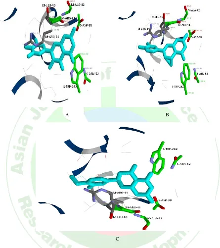
In the post screening analysis PDB ID- 1DQY shows that the compound ID ZINC03874317 (Myricetin), ZINC18185774 (Luteolin) and ZINC00039092 Hesperetin) have better drug activity (Figure 3). In 1KPI, the post screening analysis predicted that the compound ID ZINC03869768 (Kaempferol), ZINC03869685 (Quercetin), ZINC0119983 (Cianidanol) and ZINC00001219 (Coumestrol) have better drug activity with the target protein (Figure 4).
V.Subha et al www.ajprd.com 93
A B
C
Asian Journal of Pharmaceutical Research and Development Vol.1 (4) July– August 2013:
A B
C
D
V.Subha et al www.ajprd.com 95
A B
C
Figure 5: Predicted docking pose of (A) ZINC003911 (B) ZINC0039092 (C) ZINC03869685 lie within the active site of the target protein, PDB ID- 1TQ8. Cyan color represents the corresponding ligand molecule, green and grey color represents the amino acids involved in hydrogen bonding and van der Waals interactions respectively.
Satisfying Lipinski’s rule of five?
No discussion of drug-likeness would be complete without reference to the influential Rule of 5 (Ro5) which is essentially a statement of property distributions for compounds taken into Phase II clinical trials. Lipinski's Rule of Five is a rule of thumb to evaluate drug likeness, or determine if a chemical compound with a certain pharmacological or biological activity has properties that would make it a likely orally active drug in humans. The rule was formulated by Christopher A. Lipinski in 1997, based on the observation that most medication drugs are relatively small and lipophilic molecules[24].
The rule describes molecular properties important for a drug's pharmacokinetics in the human body, including their Absorption, Distribution, Metabolism, and Excretion ("ADME"). The rule is
Asian Journal of Pharmaceutical Research and Development Vol.1 (4) July– August 2013:
Table III*: Molecular properties including Lipinski’s rule of five and drug likeness.
Compound ID Chemical
name
Molecular Formula
M.Wa g/mol
X logPb
TPSAc Hbond
donor
Hbond acceptor
No. of r bd
ZINC03874317 Myricetin C15H10O8 318.24 -1.39 152 5 8 1
ZINC00517261 Isorhamnetin C16H12O7 316.26 1.99 120 4 7 2
ZINC00119983 Cianidanol C15H14O6 290.27 1.37 110 5 6 1
ZINC003911 Fisetin C15H10O6 286.23 1.97 111 4 6 1
ZINC03869768 Kaempferol C15H10O6 286.23 2.17 111 4 6 1
ZINC00039092 Hesperetin C16H14O6 302.27 -1.94 96 3 6 2
ZINC03869685 Quercetin C15H10O7 302.23 1.68 131 5 7 1
ZINC00391840 Pelargonidin
chloride
C15H11O5Cl 306.69 -0.26 92 4 5 1
ZINC18185774 Luteolin C15H10O6 286.24 1.97 111 4 6 1
ZINC03871576 Apigenin C15H10O5 270.24 2.46 91 3 5 1
ZINC00058117 Eriodictyol C15H12O6 288.25 1.63 107 4 6 1
ZINC00001785 Naringenin C15H12O5 272.25 2.12 87 3 5 1
ZINC0001219 Coumesterol C15H8O5 268.22 2.54 84 2 5 0
aMolecular Weight bOctanol/Water partition coefficient cTopological Polar Surface Area
dNo. of rotatable bonds.
*Data collected from Zinc database and PubChem [26].
CONCLUSION
We found in silico drug docking a better
approach to check utility of any chemical as a drug before going through any in vivo or in vitro analysis to shorten out the experiments
and cost cutting. Our study suggest that the flavonoid compounds, Coumestrol, Fisetin, Hesperetin, Myricetin, and Quercetin can be used as a lead molecule against the cell wall proteins of M. tuberculosis for performing in vitro and in vivo study. The set of molecules
identified by us in this study are very likely to serve as potential leads in the search for new drugs against tuberculosis.
REFERENCES
1. www.who.int
2. Vincent J, Hiroshi N. Mycobacterial cell wall: structure and role in natural resistance to antibiotics. FEMS Microbiol Lett. 1994; 123(1-2):11-18. 3. Mamadou D, Philip D. The Envelope Layers of
Mycobacteria with Reference to their Pathogenicity. Adv Microb Physiol. 1998; 39:131-203.
4. Qureshi H, Arif A, Alam E, Qadir N. Integration of informal medical practitioners in DOTS implementation to improve case detection rate. J Pak Med Assoc .2010; 60, 33–37.
6. Pasquato KFM, Ferreira EI. An approach for the rational design of new antitubercular agents. Curr. Drug Targets 2001; 2:427–437.
7. Diacovich L, Mitchell D, Pham H, Gago G, Melgar MM, Khosla C, Gramajo H, Tsai SC. Crystal structure of the beta–subunit of acyl–CoA carboxylase: structure–based engineering of substrate specificity. Biochemistry 2004; 43:14027– 36.
8. Barry CE, Lee RE, Mdluli K, Sampson AE, Schroeder BG, Slayden RA, YuanY. Prog. Lipid Res.1998; 37, 143–179
9. Ronning DR, Klabunde T, Besra GS, Vissa VD, Belisle JT, Sacchettini JC. Crystal structure of the secreted form of antigen 85C reveals potential targets for mycobacterial drugs and vaccines. Nat Struct Biol. 2000; 7(2):141-6.
10. Huang CC, Smith CV, Glickman MS, Jacobs WR, Sacchettini JC. Crystal structure of mycolic acid cyclopropane synthases from Mycobacterium tuberculosis. J Biol Chem 2002; 277: 11559- 11569 .
11. Banerjee A, Dubnau E, Quémard A, Balasubramanian V, Um KS, Wilson T, Collins D, Lisle G, Jacobs WR. inhA, a gene encoding a target for isoniazid and ethionamide in Mycobacterium tuberculosis. Science. 1994; 263: 227–230. 12. Belanger AE, Besra GS, Ford ME, Mikusova K,
Belisle JT, Brennan PJ, Inamine JM. The embAB genes of Mycobacterium avium encode an arabinosyl transferase involved in cell wall arabinan biosynthesis that is the target for the antimycobacterial drug ethambutol. Proc Natl Acad Sci USA.1996; 93: 11919–11924.
13. Lyne PD: Structure-based virtual screening: an overview. Drug Discovery Today. 2002; 7:1047-1055.
14. Tanrikulu Y, Schneider G. Pseudoreceptor models in drug design: bridging ligand- and receptor-based virtual screening. Nature Reviews Drug Discovery. 2008; 7:667-677.
15. Kitchen DB, Decornez H, Furr JR, Bajorath J. Docking and scoring in virtual screening for drug discovery: methods and applications. Nature Reviews Drug Discovery. 2004; 3:935-949. 16. www.rcsb.org/pdb
17. www.zinc.docking.org
18. Middleton EJ. Effect of plant flavonoids on immune and inflammatory cell function. Adv.Exp. Med. Biol. 1998; 439:175–182.
19. Boumendjel A, Pietro AD, Dumontet C, Barron D. Recent Advances in the Discovery of Flavonoids and Analogs with High-Affinity Binding to P-Glycoprotein Responsible for Cancer Cell Multidrug Resistance. Med.Res. Rev. 2002; 22: 512-529.
20. Havsteen B. Flavonoids, a class of natural products of high pharmacological potency. Biochem Pharmacol. 1983; 32:1141–8.
21. Tim Cushnie TP, Andrew JL. Antimicrobial activity of flavonoids. International Journal of Antimicrobial Agents. 2005; 26:343–356.
22. Braca A, Sortino C, Politi M, Morelli I, Mendez J. Antioxidant activity of flavonoids from Licania licaniaeflora. J Ethnopharmacol. 2002; 79(3):379-81.
23. Kai CH, Yen FC, Shen RL, Jinn.MY. iGEMDOCK: a graphical environment of enhancing GEMDOCK using pharmacological interactions and post-screening analysis. BMC Bioinformatics 2011, 12(Suppl 1):S33.
24. Lipinski CA, Lombardo.F, Dominy BW, Feeney P.J. Experimental and computational approaches to estimate solubility and permeability in drug discovery and development settings. Adv Drug Del Rev. 2001; 46: 3–26.
25. Oprea TI, Davis AM, Teague SJ, Leeson PD. Is There a Difference between Leads and Drugs? A Historical Perspective. J. Chem. Inf. Comput. Sci. 2001; 200141 (5): 1308–1315.