Asian Journal of Pharmaceutical Research and Development
(An International Peer-Reviewed Journal of Pharmaceutical Research and Development)
www.ajprd.com
ISSN 2320-4850
Review Article
A BRIEF REVIEW ON DENGU
Saquib Tanweer
*, Mohd. Shahid Khan, M.P. Khinchi, Ashiya Ansari,
Shubham Gautam
Department of Pharmacology, Kota College of Pharmacy, Kota, Rajasthan, India.
ABSTRACT
Globally important arboviral infection transmitted by the Aedes genus of mosquito.Endemic in more than 120 countries, particularly the Southeast Asian and Western Pacific regions, the Caribbean, Latin America, and some regions in the US, Africa, and Middle East.Severe dengue fever is characterised by marked thrombocytopenia, severe haemorrhage, plasma leakage leading to shock or fluid accumulation with respiratory distress, and severe organ impairment. Confirmatory tests include viral antigen or nucleic acid detection and serology. Difficult to distinguish clinically from Zika and chikungunya virus infections without diagnostic testing. A tetravalent vaccine has been approved in Mexico, making it the first vaccine to be licensed in the world for the prevention of dengue. Dengue viruses (DENVs) cause the most common arthropod-borne viral disease in man with 50–100 million infections per year. Because of the lack of a vaccine and antiviral drugs, the sole measure of control is limiting the Aedes mosquito vectors. DENV infection can be asymptomatic or a self-limited, acute febrile disease ranging in severity. The classical form of dengue fever (DF) is characterized by high fever, headache, stomach ache, rash, myalgia, and arthralgia. Severe dengue, dengue hemorrhagic fever (DHF), and dengue shock syndrome (DSS) are accompanied by thrombocytopenia, vascular leakage, and hypotension. DSS, which can be fatal, is characterized by systemic shock. Despite intensive research, the underlying mechanisms causing severe dengue is still not well understood partly due to the lack of appropriate animal models of infection and disease. However, even though it is clear that both viral and host factors play important roles in the course of infection, a fundamental knowledge gap still remains to be filled regarding host cell tropism, crucial host immune response mechanisms, and viral markers for virulence.
Keywords: dengue virus, dengue fever, dengue hemorrhagic fever, dengue shock syndrome, flavivirus, vector-borne virus, arbovirus
INTRODUCTION
engue fever is a mosquito-borne tropical disease caused by the dengue virus. Symptoms typically begin three to fourteen days after infection. This may include a high fever, headache, vomiting, muscle and joint paints, and a characteristic skin rash. Recovery generally takes two to seven days. In a small proportion of cases, the disease develops into the life-threatening dengue hemorrhagic fever, resulting in bleeding, low levels of blood platelets and blood plasma leakage,
*Corresponding author: Saquib Tanweer
Department of pharmacology, Kota college of Pharmacy, Kota
Sp-1 RIICO Industrial Area, Ranpur, Kota Mob. 9549756755
Email- [email protected]
or into dengue shock syndrome,
where dangerously low blood
pressure occurs. Dengue is spread by several species of mosquito of the Aedes type, principally A. aegypti. The virus has five different types infection with one type usually gives lifelong immunity to that type, but only short-term immunity to the others. Subsequent infection with a different type increases the risk of severe complications. A number of tests are available to confirm the diagnosis including detecting antibodies to the virus or it’s RNA.
Dengue causes severe flu-like symptoms, such as:
A high temperature (fever) of 40C (104F) or over
Severe headache
Muscle and joint pain
facial flushing and skin rash
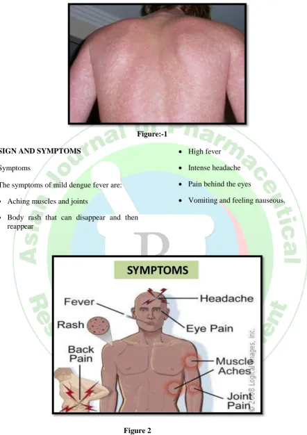
Figure:-1
SIGN AND SYMPTOMS
Symptoms
The symptoms of mild dengue fever are:
Aching muscles and joints
Body rash that can disappear and then reappear
High fever
Intense headache
Pain behind the eyes
Vomiting and feeling nauseous.
Figure 2
CAUSES
Virology
A transmission electron microscopy image showing dengue virus. A TEM micrograph showing dengue virus virions (the cluster of dark dots near the center).
fever virus. Most are transmitted by arthropods (mosquitoes or ticks), and are therefore also referred to as arboviruses (arthropod-borne viruses).
The dengue virus genome (genetic material) contains about 11,000 nucleotide bases, which code for the three different types of protein molecules (C, prM and E) that form the virus particle and seven other types of
protein molecules (NS1, NS2a, NS2b, NS3, NS4a, NS4b, NS5) that are found in infected host cells only and are required for replication of the virus. There are five strains of the virus, called serotypes, of which the first four are referred to as 1, DENV-2, DENV-3 and DENV-4. The fifth type was announced in 2013. The distinctions between the serotypes are based on their antigenicity.
Figure 3 Transmission
Close-up photograph of an Aedesaegypti mosquito biting human skin. The mosquito Aedesaegypti feeding on a human host. Dengue virus is primarily transmitted by Aedes mosquitoes, particularly A. aegypti. These mosquitoes usually live between the latitudes of 35° North and 35° South below an elevation of 1,000 metres (3,300 ft). They typically bite during the early morning and in the evening, but they may bite and thus spread infection at any time of day. Other Aedes species that transmit the disease include A. albopictus, A. polynesiensis and A. scutellaris
Predisposition
Severe disease is more common in babies and young children, and in contrast to many other infections, it is more common in children who are relatively well nourished. Other risk factors for severe disease include female sex, high body mass index, and viral load. While
each serotype can cause the full spectrum of disease, virus strain is a risk factor. Infection
with one serotype is thought to produce lifelong immunity to that type, but only short-term protection against the other three.
MECHANISM
wall of small blood vessels into body cavities due to capillary permeability.
Viral replication
Once inside the skin, dengue virus binds to Langerhans cells (a population of dendritic cells in the skin that identifies pathogens). The virus enters the cells through binding between viral proteins and membrane proteins on the Langerhans cell, specifically the C-type lectins called DC-SIGN, mannose receptor and CLEC5A. DC-SIGN, a non-specific receptor for foreign material on dendritic cells, seems to be the main point of entry. The dendritic cell moves to the nearest lymph node.
Severe disease
It is not entirely clear why secondary infection with a different strain of dengue virus places people at risk of dengue hemorrhagic fever and dengue shock syndrome. The most widely accepted hypothesis is that of antibody-dependent enhancement (ADE). The exact mechanism behind ADE is unclear. It may be caused by poor binding of non-neutralizing antibodies and delivery into the wrong compartment of white blood cells that have ingested the virus for destruction
DIAGNOSIS
When a mosquito carrying dengue virus bites a person, the virus enters the skin together with the mosquito's saliva. It binds to and enters white blood cells, and reproduces inside the cells while they move throughout the body. The white blood cells respond by producing a number of signaling proteins, such as cytokines and interferons, which are responsible for many of the symptoms, such as the fever, the flu-like symptoms, and the severe pains. In severe infection, the virus production inside the body is greatly increased, and many more organs (such as the liver and the bone marrow) can be affected. Fluid from the bloodstream leaks through the wall of small blood vessels into body cavities due to capillary permeability. As a result, less blood circulates in the blood vessels, and the blood pressure becomes so low that it cannot supply sufficient blood to vital organs. Furthermore, dysfunction of the bone marrow due to infection of the stromal cells leads to reduced numbers of platelets, which are necessary for effective blood clotting; this increases the risk of bleeding, the other major complication of dengue fever.
Classification
Dengue classification according to the World Health Organization guidelines issued in 1975 and 1997. Dengue is classified as (1) undifferentiated fever, (2) dengue fever (DF), and (3) dengue hemorrhagic fever (DHF). In addition to fever and at least 2 clinical findings, diagnosis of DF requires epidemiological or laboratory evidence
supporting a dengue virus infection. To meet a case definition of DHF, all 4 criteria are required: (1) fever, (2) hemorrhagic manifestations, (3) thrombocytopenia (platelet count, ≤100000 platelets/mm3
), and (4) evidence of plasma leakage. Diagnosis of DHF does not require laboratory support.
Laboratory tests
Figure:-5
Graph of when laboratory tests for dengue fever become positive. Day zero refers to the start of symptoms, 1st refers to in those with a primary infection, and 2nd refers to in those with a secondary infection.
PREVENTION
The best way to reduce mosquitoes is to eliminate the places where the mosquito lays her eggs, like artificial containers that hold water in and around the home (see figure 2, video 2 and 3). In urban areas, Aedes mosquitos breed on water collections in artificial containers such as plastic cups, used tires, broken bottles, flower pots, etc (see also transmission of dengue). Periodic draining or removal of artificial containers is the most effective way of reducing the breeding grounds for mosquitos. Larvicide treatment is another effective way to control the vector larvae but the larvicide chosen should be long-lasting and preferably. There are some very effective insect growth regulators (IGRs) available which are both safe and long-lasting (e.g. pyriproxyfen). For
reducing the adult mosquito load, fogging with insecticide is somewhat effective.
To eliminate standing water: Unclog roof gutters;
Empty children's wading pools at least once a week;
Change water in birdbaths at least weekly; Get rid of old tires in your yard, as they collect standing water;
MANAGEMENT
responsible for many of the symptoms, such as the fever, the flu-like symptoms, and the severe pains. In severe infection, the virus production inside the body is greatly increased, and many more organs (such as the liver and the bone marrow) can be affected. Fluid from the bloodstream leaks through the wall of small blood vessels into body cavities due to capillary permeability. As a result, less blood circulates in the blood vessels, and the blood pressure becomes so low that it cannot supply sufficient blood to vital organs. Furthermore, dysfunction of the bone marrow due to infection of the stromal cells leads to reduced numbers of platelets, which are necessary for effective blood clotting; this increases the risk of bleeding, the other major complication of dengue fever.
TREATMENT
Apart from attempts to control the spread of the Aedes mosquito there are ongoing efforts to develop antiviral drugs that would be used to treat attacks of dengue fever and prevent severe complications. Discovery of the structure of the viral proteins may aid the development of effective drugs. There are several plausible targets. The first approach is inhibition of the viral RNA-dependent RNA polymerase (coded by NS5), which copies the viral genetic material, with nucleoside analogs. Secondly, it may be possible to develop specific inhibitors of the viral protease (coded by NS3), which splices viral proteins. Finally, it may be possible to develop entry inhibitors, which stop the virus entering cells, or inhibitors of the 5′ capping process, which is required for viral replications
HISTORY
The first record of a case of probable dengue fever is in a Chinese medical encyclopedia from the Jin Dynasty (265–420 AD) which referred to a "water poison" associated with flying insects. The primary vector, A. aegypti, spread out of Africa in the 15th to 19th centuries due in part to increased globalization secondary to the slave trade. There have been descriptions of epidemics in the 17th century, but the most plausible early
reports of dengue epidemics are from 1779 and 1780, when an epidemic swept across Asia, Africa and North America. From that time until 1940, epidemics were infrequent. In 1906, transmission by the Aedes mosquitoes was confirmed, and in 1907 dengue was the second disease (after yellow fever) that was shown to be caused by a virus. Further investigations by John Burton Cleland and Joseph Franklin Siler completed the basic understanding of dengue transmission.
REFERENCES
1. WHO. Dengue and dengue haemorrhagic fever. Factsheet No 117, revised May 2008. Geneva, World Health Organization.
2. WHO. Dengue fever and dengue haemorrhagic fever prevention and control. World Health Assembly Resolution WHA55.17, adopted by the 55th World Health Assembly, 2002
3. WHO. Revision of the International Health Regulations. World Health Assembly Resolution WHA58.3, adopted by the 58th World Health Assembly, 2005
4. WHO/SEARO. Concrete measure key in controlling dengue in South East Asia.Press Release SEA/PR/1479. New Delhi, World Health Organization Regional Office for South-East Asia, 2008
5. WHO. Denguenet in India. Weekly Epidemiological Record, 2004, 79(21):201--203
6. WHO/WPRO. Dengue fever and dengue haemorrhagic fever prevention and control. Regional Committee resolution WPR/RC59.R6, adopted by the WHO Regional Committee for the Western Pacific, 2008
7. PAHO. Plan continental de ampliación e intensificacióndelcombate al Aedesaegypti. Informe de ungrupo de trabajo, Caracas, Venezuela. Abril 1997. Washington, DC, Pan American Health
Organization, 1997 (Document
OPS/HCP/HCT/90/97, in Spanish)
8. PAHO. Number of reported cases of dengue and dengue hemorrhagic fever (DHF), Region of the Americas (by country and subregion). Washington, DC, Pan American Health Organization, 2008 9. Centers for Disease Control and Prevention.
Travel-associated dengue -- United States, 2005. Morbidity and Mortality Weekly Report, 2006, 55(25):700--702.
10.Ramos MM et al. Dengue Serosurvey Working Group. Epidemic dengue and dengue hemorrhagic fever at the Texas-Mexico border: results of a household-based seroepidemiologic survey, December 2005. American Journal of Tropical Medicine and Hygiene, 2008, 78(3):364--369. 11.Centers for Disease Control and Prevention.