Goverdhan P, et al. J Sci Res Pharm, 2019;8(4):34-41 World Inventia Publishers
J
ournal of
S
cientific
R
esearch in
P
harmacy
http://www.jsrponline.com/
Vol. 8, Issue 4, 2019 ISSN: 2277-9469 USA CODEN: JSRPCJ
Research Article
STUDY TO FIND THE BEST EXTRACTION SOLVENT FOR USE WITH CUCUMBER PEEL (CUCUMIS SATIVUS) FOR HIGH NEUROPROTECTIVE ACTIVITY IN COGNITIVE IMPAIRED RATS
Girija Pashikanti 1, Makula Ajitha 2, Goverdhan Puchchakayala 1 *
1 Centre for Neurodegenerative Disease and Aging Research Department of Pharmacology, Vaagdevi College of Pharmacy, Ramnagar,
Hanamkonda, Warangal 506001, INDIA.
2 Centre for Pharmaceutical Sciences, JNTU, Kukatpally, Hyderabad-500085, Telangana, INDIA.
Received on: 28-03-2019; Revised and Accepted on: 24-04-2019
ABSTRACT
Objective: The present study was designed to investigate the best extraction solvent for cucumber (Cucumis sativus) peel
neuroprotective effectson bilateral common carotid artery occlusion (BCCAO) induced cognitive impairment rats.
Methods: To study the activity, rats weighing 250-300g were pretreated with successive extracts of n-hexane fraction (HF), ethyl acetate fraction (EAF), ethanol fraction (EE) and aqueous fraction (AF) of 400 mg/kg, 200 mg/kg, p.o of each for 10 days and the
treatment was continued for another 7 days after cerebral ischemia. Cognitive impairment was induced by BCCAO for 30 minutes,
followed by 7 days reperfusion of male wistar rats. The neuroprotective capability was assessed based on memory performance tasks by morris water maze and rectangular maze performance tests and locomotor activity. Various biochemical parameters like lipid peroxidation, Catalase, DPPH and AchE were also estimated in the brain after the treatment.
Results: The results demonstrated that the neuroprotective ability of cucumber peel extracts of EE was higher than AF due to having more phenolic compound and flavonoids content. The animals treated with donepezil, EE and AF prevented the biochemical changes significantly (p<0.001) and there was significant (p<0.001) improvement in cognitive parameters in dose dependent manner compared to BCCAO treated rats. However there was no significant improvement in cognitive parameters of HF and EAF extractions of cucumber peel.
Conclusion: Thus present study indicates Neuroprotective effect of cucumber peel extracts (EE > AF) against BCCAO induced cognitive impairment. The ethanolic extract was observed to be the most effective solvent showing high neuroprotective ability than aqueous fraction.
Key words: Bilateral common carotid artery occlusion, Cucumis sativus, Cognitive impairment, Flavonoids, Neuroprotective.
INTRODUCTION
Medicinal plants have been used in the treatment and improvement of human diseases [1] and such plants with high
antioxidant and neuroprotective abilities can be used as natural medicines for preventing aging and chronic diseases. There were growing evidences which support participation of oxidative stress in brain injury mediated by cerebral ischemia.
*Corresponding author: Dr. P. Goverdhan
M.Pharm., Ph.D., PDF (Germany),
Professor and Head Department of Clinical Pharmacology, Vaagdevi College of Pharmacy,
India Director Synapse Life Sciences, Hanamkonda Warangal-506001 INDIA.
* E-Mail: [email protected], [email protected]
DOI:https://doi.org/10.5281/zenodo.2652390
The apparent role of free radicals in the development of neuronal cell death has stimulated interest in use of antioxidants and neuroprotective agents as potential therapeutic agents for ischemia induced free radicals that may promote ischemic neuronal cell injury [2].
Having little or no harm effects of plant materials, therefore herbs based medicines flourished now a day and evidences show the immense potential of medicinal plants. The polyphenolics including flavonoids, which found in many herbal extracts, have been shown to be strong reactive oxygen species (ROS) scavengers, antioxidants and protectors of neurons from lethal damage in vitro[3, 4]. Phenolic antioxidants from medicinal
plants have also been evaluated in vivo as neuroprotective agents in animal models of I/R induced oxidative stress [5].
Cucumis sativus Linn. (Family: Cucurbitaceae) is an
as cooling and diuretic, nutritive and demulcent, and emetic in acute indigestion in children [6]. Several studies have shown
multiple biological activities of different parts of Cucumis sativus (cucumber). Fruits and vegetables offer defense against long-lasting illnesses like cancer, cardiovascular and neurodegenerative disorder due to the presence of antioxidants especially, vitamins, phenolic and flavonoids, coumarin, tannins and stilbenes [7].
Therefore, in this study, the phenolic compound and flavonoid content of n- hexane, ethyl acetate, ethanol and water extracts of cucumber peel analyzed and evaluated against BCCAO induced cognitive impairment in rats with regard to neuroprotective properties. The best extraction solvent for use with cucumber peel for high neuroprotective efficacy was selected.
MATERIALS AND METHODS
Chemicals and drugs:
Donapezil, Thiopentone sodium, Hydrogen peroxide, povidone-iodine powder, 5% w/w (Sri medical & surgicals) ethanol, ethyl acetate, n-hexane (Venkateshwara agencies) Acetylthiocholine iodide (Sigma Aldrich), Perchloric acid Formalin 10% (Finar Chemicals), DTNB (5,5-dithiobis (2-nitro
benzoic acid) reagent, DPPH (1,1-diphenyl-2- picrylhydrazyl) radical reagent (Sigma Aldrich).
Plant material:
The fresh cucumbers were collected from local market of Warangal district, Telangana state, India. The peel were separated from fruit and shade dried. The collected samples were authenticated morphologically by Dr. E. Narasimha Murthy, Department of Plant Sciences, School of Life Sciences, University of Hyderabad, Central University, Hyderabad, Telangana state, India.
Preparation of crude extract and its fractions:
Extraction is the process in which desired plant tissues will be soaked in solvent for a certain time period, from which medicinally active principles are dissolved in the solvent leaving undissolved materials. In this study 70% ethanol were used for extraction. The grounded material was extracted in soxhlet apparatus with 70% ethanol for about 36 hours. The ethanolic extract was cooled, filtered. The filtrate was concentrated by using a rotary evaporator under reduced pressure till the concentrated mass was obtained.This was the crude ethanolic extract and further fractionation was carried out, subjecting the crude sample to extraction with n-hexane, ethyl acetate and water respectively on polarity bases, which as follows in table 1.
Table No. 1: Amount of different extracts of grounded cucumber peel
Grounded material Ethanolic extract (EE) n-hexane fraction (HF) Ethyl acetate fraction (EAF) Aqueous fraction (AF)
1kg 28gm 8gm 4gm 2.8gm
Experimental Animals:
Young male Wistar rats (200-250g) procured from mahaveera enterprises, medchal district-98 were used. Animals were adjusted to laboratory conditions at room temperature prior to experimentation. All the experiments were carried out between 9.00 AM and 3.00 PM. All animals underwent surgery; they were kept in plastic cages with soft bedding under standard conditions of a 12-hour light/dark cycle with food and water ad libitum. The animal experiments were designed as per CPCSEA guideline and protocol of the experiments after the authorization of the Institutional Animal Ethical Committee (IAEC), Vaagdevi College of Pharmacy, Warangal (A.P) and India (1047/PO/Re/S/07/ CPCSEA, dated: 21/10/2017).
Acute toxicity studies:
The acute toxicity of HF, EAF, EE and AF extracts of cucumber was determined as per the OECD guideline no, 423 (acute toxic class method). It was observed that these peel extracts were not fatal to the rats even at the 2000 mg/kg doses.
Hence, 1/5th (400mg/kg) and 1/10th (200mg/kg) of these doses
were selected for further studies [8].
Groups and treatment:
The Wistar Albino rats (66) of 250-300 gm weight were randomly divided into 11 groups of 6 rats each. Vehicle and test substances of HF, EAF, EE and AF successive extracts (400, 200 mg/kg of each) of cucumber were prepared freshly and given (p.o.) once daily for 10 consecutive days prior to the cerebral ischemia. On day 11, 60 minutes after last dose, all the groups received BCCAO for 30 minutes followed by reperfusion for 7 days. From the second (13) day after induction the animals continued with the test substances for another week days, then the animals were assessed for behavioral parameters. After all behavioral assessments, the brains of different groups were removed and assessed for biochemical parameters and histopathological evaluation [9].The detailed treatment schedule
is as in table 2.
Table No. 2: Experimental design
Group number Treatment
I Sham-operated; 0.1% of CMC in normal saline (10ml/kg)
II BCCAO; 0.1% of CMC in normal saline (10ml/kg)
III BCCAO + received Donepezil (5mg/kg) oral
IV BCCAO + HF (400mg/kg) orally
V BCCAO + HF (200mg/kg) orally
VI BCCAO + EAF (400mg/kg) orally
VII BCCAO + EAF (200mg/kg) orally
VIII BCCAO + EE (400mg/kg) orally
IX BCCAO + EE (200mg/kg) orally
X BCCAO + AF (400mg/kg) orally
Drug administration:
Donepezil (5mg/kg),the HF, EAF, EE and AF (dissolved in 0.1% CMC) of cucumber were administered at a dose of 400 and 200mg/kg oral. Rats in the sham group and BCCAO group administered solely with an equal amount of normal saline. All drugs were prepared freshly every day. Doses were given according to the respective rat weights.
Induction of cerebral ischemia:
BCCAO was used to trigger global cerebral ischemia. Rats were sedated with thiopentone sodium at a dose of 50mg/kg, (i.p). A midline opening was made between the neck and sternum and the trachea was exposed. Then both the left and right common carotid arteries were carefully separated. Cerebral ischemia was induced by clamping both of the arteries with two microaneurysmal clips (bulldog clamps). After 30 minutes of cerebral ischemia the reperfusion of blood through the carotid arteries allowed by removing both the clips. Sham-operated rats underwent the same surgical procedure without artery occlusion. During the surgery, the body temperature was monitored with a temperature probe and maintained at 37.0– 37.5 °C using a heating pad [10, 11].
Behavioural assessments:
All the animals were trained for 10 days before BCCAO and drug administration.
Spatial Navigation Task:
The acquisition and retention of a spatial navigation task were assesed by using morris water maze [12]. Rats were
trained to swim toward a visible platform in a circular pool (120 cm in diameter and 60 cm in height) located in a test room. In principle, rats can escape from swimming by climbing onto the platform and over time the rats have apparently learned the spatial location of the platform from any starting position at the circumference of the pool. Thus the platform offers no local signal to guide the escape behavior of the rats. The pool was filled with water (28±2◦C) to a height of 40cm, a movable circular platform (9 cm diameter), was placed in a pool 2 cm above the water level during the acquisition phase. Similarly, for the maze retention phase the platform was placed in the pool 2 cm below the water level. The water was made opaque with a nontoxic dye. Four equally spaced locations around the edge of the pool (Q1, Q2, Q3, and Q4) were used as starting points and
this divided the pool into four equal quadrants.
Maze acquisition phase (training):
From the second day after induction the animals received a training session consisting of 4 trials on day 12. In all 4 trials, the starting position was different. A trial began by releasing the animal into the maze facing towards the wall of the pool. The latency to find the escape platform was recorded to a maximum of 90 seconds. If the rat did not escape onto the platform within this time, it was guided to the platform and was allowed to remain there for 20 seconds. The time taken by rat to reach the platform was taken as the initial acquisition latency (IAL).
Maze retention phase (testing for retention of the learned task): Following 24 hours (day 13) and 8 days (day 21) after IAL, the rat was released randomly from one of the edges facing the wall of the pool. The time taken to find the hidden platform was recorded and termed as first retention latency (1st RL) and
second retention latency (2nd RL) on day 13 and day 21after
cerebral ischemia, respectively.
Assessment of Gross Behavioral Activity:
Gross behavioral activity was observed on days 1, 7 before induction and 13, 21 after induction. Animal was placed in a square closed arena equipped with infrared light-sensitive photocells using digital actophotometer, it consists of photoelectric cells and they are connected with a counter in the circuit. The animals were observed for a period of 5 minutes and the values were expressed as counts/5 minutes [13, 14].
Rectangular maze test:
This test was used to evaluate the learning ability. The maze consists of a fully enclosed rectangular box with an entry and a reward chamber added at opposite ends. The box is partitioned like twisting corridor leading into blind passages from the entry to the reward chamber with wooden slats. From the second day after induction the animals received a training session consisting of 4 trials on day 12. In each trial the rat was placed in the entry chamber and the timer was activated as soon as the rat leaves the chamber. The time taken by the animal to reach the reward chamber from the entry chamber was recorded as the initial transfer latency (ITL). Animal was allowed to explore the maze for 20 seconds after recording the ITL and then returned to the home cage. If the animal did not enter the reward chamber within 90 seconds, it was guided on the back to reach reward chamber and the ITL was given as 90 seconds. Retention of memory was assessed by placing the rat in an entry chamber and the retention latency was noted on day 13 and day 21 of ITL and was termed as the first retention transfer latency (1st RTL) and second retention transfer latency (2nd RTL), respectively. Lower scores of the assessment indicate efficient learning while higher scores indicate poor learning in animals [15, 16].
Dissection and Homogenization:
On day 21, after behavioral assessments, animals were scarified by decapitation prior to deep anesthesia. The brains were removed, forebrain was dissected out, and cerebellum was discarded. Hippocampus dissected out from the brain and it was put on ice and rinsed with ice-cold isotonic saline. A (10% w/v) homogenate was prepared with 10 times ice cold 0.1M phosphate buffer (pH 7.4). Now the obtained homogenate was centrifuged at 3000rpm for 15 minutes and aliquots of supernatant was separated and used for biochemical estimation.
Biochemical Tests:
Measurement of Lipid Peroxidation:
The extent of lipid peroxidation in the brain was determined [17]. The amount of malondialdehyde (MDA) was
measured by reaction with thiobarbituric acid at 532nm using spectrophotometer. The values were calculated using the molar extinction coefficient of chromophore (1.56 × 10⁵ (mol/L)
−1cm−1).
Catalase activity:
Catalase activity was assessed [18], wherein the
breakdown of hydrogen peroxide is measured. Briefly, the assay mixture consisted of 3mL of H2O2 phosphate buffer and 0.05mL
of the supernatant of the tissue homogenate. The change in absorbance was recorded for 2 minutes at 30-second interval at 240nm using spectrophotometer. The results were expressed as micromoles of H2O2 decomposed per minute per mg protein.
DPPH radical scavenging assay:
purple coloured methanol solution of DPPH. To the 1000 L of diverse concentration of the homogenate, 4mL of 0.004% methanolic solution of DPPH was added. After 30 min incubation in dark, absorbance was read at 517nm. Inhibition of free radical by DPPH in percentage was calculated in the following way:
% = (Ablank – Asample / Ablank) ×100
Ablank: absorbance of control reaction. Asample: absorbance of test
sample. Values of inhibition were calculated
Acetyl Cholinesterase estimation:
Acetyl cholinesterase (AChE) is a marker of extensive loss of cholinergic neurons in the forebrain. The AChE activity was assessed [20]. The change in absorbance was measured for 2 minutes at 30-second interval at 412nm using spectrophotometer. Results were expressed as micromoles of acetylthiocholine iodide hydrolyzed per minute per mg protein.
Statistical analysis:
Results were expressed in mean ± SD. The significance of the difference in means between disease control and test drug treated animals for different parameters was evaluated by using One-way Analysis of Variance (ANOVA) followed by multiple comparisons Dunnett’s test. Data were measured statistically,
significant at p < 0.05 and highly significant at p < 0.01. Statistical analysis was executed using Graphpad Prism 7 statistical software.
RESULTS
Behavioral tests:
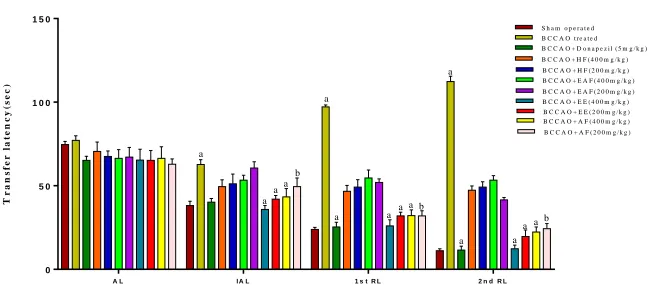
Figure 1, 2 & 3 shows, the rats in the disease control group exhibited severe memory impairment and loss of memory. All the group rats learning abilities were compared before and after induction of cerebral ischemia against BCCAO treated group rats on the same day. Furthermore, with the passage of time, the rats in the disease control group exhibited severe memory impairment and loss of memory. Especially, the EE and AF treated rats exhibited significant (p<0.01 and p< 0.05 respectively) increase locomotor activity and decrease latency period times in a concentration-dependent manner compared with the disease control group. But there were no significant differences were observed in HF, EAF treated animals of behavioral parameters as compared with BCCAO treated group. This result representing that EE and AF significantly repaired the spatial cognitive and memory deficits induced by ischemia. Specifically the EE treated rats showed similar results when compared with donapezil treated animals.
A L I A L 1 s t R L 2 n d R L
0 5 0 1 0 0 1 5 0
M o r r i s w a t e r m a z e t e s t
T r a n s f e r l a t e n c y ( s e c )
S h a m o p e r a t e d B C C A O t r e a t e d B C C A O + D o n a p e z i l ( 5 m g / k g ) B C C A O + H F ( 4 0 0 m g / k g ) B C C A O + H F ( 2 0 0 m g / k g ) B C C A O + E A F ( 4 0 0 m g / k g ) B C C A O + E A F ( 2 0 0 m g / k g ) B C C A O + E E ( 4 0 0 m g / k g ) B C C A O + E E ( 2 0 0 m g / k g ) B C C A O + A F ( 4 0 0 m g / k g ) B C C A O + A F ( 2 0 0 m g / k g )
a a a b a a a a a
a ab a
a aa
b
Values are expressed as Mean ± SD (n = 6); a p<0.05, b p<0.01 as compared with corresponding values of BCCAO group. (One way ANOVA
followed by dunnett’s test)
Fig. 1: Effect of Donepezil,cucumberof HF, EAF, EE and AF extracts on spatial navigation task compared to the BCCAO group
T L I T L 1 s t R T L 2 n d R T L
0 5 0 1 0 0 1 5 0 2 0 0
R e c t a n g u la r m a z e t e s t
T r a n s f e r l a t e n c y ( s e c )
S h a m o p e r a t e d B C C A O t r e a t e d B C C A O + D o n a p e z i l ( 5 m g / k g ) B C C A O + H F ( 4 0 0 m g / k g ) B C C A O + H F ( 2 0 0 m g / k g ) B C C A O + E A F ( 4 0 0 m g / k g ) B C C A O + E A F ( 2 0 0 m g / k g ) B C C A O + E E ( 4 0 0 m g / k g ) B C C A O + E E ( 2 0 0 m g / k g ) B C C A O + A F ( 4 0 0 m g / k g ) B C C A O + A F ( 2 0 0 m g / k g )
b a a a b a a
abb a b b a a a a a
Values are expressed as Mean ± SD (n = 6); b p<0.05, a p<0.01 as compared with corresponding values of BCCAO group. (One way ANOVA
followed by dunnett’s test)
Fig. 2: Effect of Donepezil,cucumberof HF, EAF, EE and AF extracts on memory performance in rectangular maze compared
D a y 1 D a y 7 D a y 1 2 D a y 2 1
0 5 0 1 0 0 1 5 0 2 0 0
L o c o m o t o r a c t iv it y
N
u
m
b
e
r
o
f
c
r
o
s
s
in
g
s
i
n
5
m
in
s
S h a m o p e r a t e d B C C A O t r e a t e d B C C A O + D o n a p e z i l ( 5 m g / k g ) B C C A O + H F ( 4 0 0 m g / k g ) B C C A O + H F ( 2 0 0 m g / k g ) B C C A O + E A F ( 4 0 0 m g / k g ) B C C A O + E A F ( 2 0 0 m g / k g ) B C C A O + E E ( 4 0 0 m g / k g ) B C C A O + E E ( 2 0 0 m g / k g ) B C C A O + A F ( 4 0 0 m g / k g ) B C C A O + A F ( 2 0 0 m g / k g )
a
aa a b
a
a a a b
a
aa ab a
a a
Values are expressed as Mean ± SD (n = 6); b p<0.05, a p<0.01 as compared with corresponding values of BCCAO group. (One way ANOVA
followed by dunnett’s test)
Fig. 3: Effect of Donepezil,cucumberof HF, EAF, EE and HEF successive extracts on locomotor activity compared to the
BCCAO group
Biochemical assays:
After cerebral ischemia followed by reperfusion, there was a significant increase in oxidative stress in BCCAO treated group, which is indicated (table 3) by increase in MDA, AchE and decreased in CAT, DPPH assay in the brain as compared to a normal control group. The animals of EE and AF treated had
showed significant (p< 0.01 and p< 0.05 respectively) decrease in MDA, AchE levels and increase in CAT, DPPH activities when compared with BCCAO treated animals. However, there were no significant differences were observed in HF, EAF treated animals of oxidative stress parameters like MDA, AchE, CAT and DPPH activities as compared with BCCAO treated group.
Table No. 3: Effect of donapezil, cucumber HF, EAF, EE and AF extracts on BCCAO induced alteration in animals brain MDA, CAT, DPPH and AchE levels
Treatment (mg/kg) MDA (nmol/g
tissue) CAT(µmol of Hmin/mg tissue)2O2 DPPH (Free radical scavenging % (µmol SH/g/min)AchE activity
Sham operated 16.76±1.30 10.8±0.6 65.31±1.20 7.72±1.32
BCCAO treated 29.35±1.56 3.65±1.30 25.13±1.20 18.62±2.44
Donapezil- 5 19.65±2.60*** 8.01±1.6*** 48.9±2.8*** 8.24±4.30***
HF – 400 27.5±6.6 ns 5.2±0.6ns 25.21±3.8 ns 17.56±2.10 ns
HF – 200 29.1±3.2 ns 4.1±2.1 ns 20.28±3.30ns 16.89±1.30ns
EAF – 400 28.1±3.2 ns 4.0±2.0 ns 22.15±3.10 ns 17.26±1.20 ns
EAF – 200 29.1±53 ns 3.7±1.3 ns 21.11±4.10 ns 15.9±2.5 ns
EE – 400 19.20±2.10*** 8.5±2.3*** 47.31±2.50*** 7.6±2.4***
EE – 200 20.1±1.4*** 7.9±0.9** 43.18±4.10*** 8.2±1.3***
AF – 400 20.5±3.6** 7.8±1.3** 40.01±2.61** 12.62±2.30**
AF – 200 21.4±2.1** 6.8±2.5* 37.20±2.41* 13.56±1.30*
Values are expressed as Mean ± SD (n=6) * p<0.05, ** p<0.01, *** p<0.001 as compared with corresponding values of disease control group (one
way ANOVA followed by dunnett’s test).
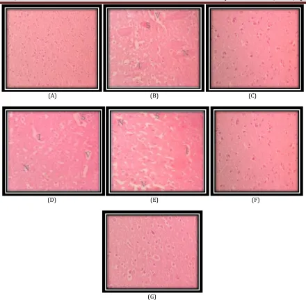
Histopathological studies:
From the figure 4 histopathological studies, it was observed that 30 minutes of BCCAO followed by 7 days reperfusion produced shrinkage, atrophy and necrosis of neurons along with the vacuolization and inflammatory infiltration in the hippocampal regions of disease control
(A) (B) (C)
(D) (E) (F)
(G)
Fig. 4: Histopathological representative photographs (H & E stain) of hippocampal brain sections
(A) sham operated (B) BCCAO treated (C) Donapezil (D, E, F, G) cucumber extracts of HF, EAF, EE and AF 400mg/kg treated ischemic groups respectively, which were observed under 40X magnification. [Inflammatory infiltration (I), Necrosis (N), shrinkage (S) vacuolization (V)]
DISCUSSION
T
he present study revealed neuroprotective potential of cucumber 70% EE than AF in dose dependent manner. We estimated LPO, CAT, DPPH and AchE levels in the brain tissue as an index to assess the severity of oxidative damage.The antioxidant and neuroprotective potential of cucumber was studied against BCCAO-induced oxidative stress in rats. Experimental models of stroke have been developed in animals in an attempt to mimic the events of human cerebral ischemia. It is well documented that transient global cerebral ischemia results in neurological abnormality. Therefore, global cerebral ischemia of short duration followed by reperfusion has been employed in the present study [21].
It was reported that the total phenolic content, antoxidant activity and flavonoid content of lemon and cucumber peels are good enough to extract which revealed that these materials thrown away everyday from households as waste can be used for extraction of valuable phenolic
compounds and can also be used as a good source of antioxidant activity which may play an important role in protecting cells and any organ from oxidative degeneration[22]. Another study found
that thephenolic compound content of hydroethanolic extract was higher than in the water extract of guava leaves [23].
According to another research study, the phenolic compound content was highest in 40% hydroethanolic extract [24].
Antioxidant activity is strongly correlated with reducing power, which increased depending on the concentration and reaction time of the extracts [25]. Our results
Ischemic-reperfusion injury is known to produce necrosis of brain, which can be directly visualized by histological study; biopsy of the rat brain subjected to ischemic-reperfusion injury showed significant necrosis [26]. The severe
neuronal loss, observed as shrinkage of neurons, vacuolization, inflammatory infiltration and necrosis, was observed in histopathological sections of ischemic reperfused brains. Interestingly the effect was attenuated by administrating 400 mg/kg of cucumber 70% EE > AF treated animals.
In the present study, BCCAO followed by reperfusion caused a significant increase in the acetyl cholinesterase activity thereby leading to learning and memory deficits. Cucumber 70% EE and AF was able to ameliorate the BCCAO induced decrease in AChE activity and there was no significant decrease in AchE activity in cucumber HF, EAF treated animals. In summary, the present study suggests that chronic administration of Cucumber 70% EE prevents maximally the BCCAO induced cognitive impairment and associated oxidative stress than AF treated animals. Thus, the use of Cucumber 70% EE is promising for the treatment of AD and other neurodegenerative disorders.
CONCLUSION
F
rom the above discussion it can be concluded that cucumber has neuroprotective property, which is responsible for beneficial effects in cerebral ischemia-reperfusion injury. It also reduced acetyl cholinesterase and free radical mediated neuronal damage. This study intended to find the best extraction solvent for high neuroprotective efficacy of cucumber peel using various solvents, in which neuroprotective activity of 70% EE was significantly higher than that of the AF extracts due to its valuable polyphenolic compounds.ACKNOWLEDGMENT
T
he authors would like to thank the Principal and Management of Vaagdevi College of pharmacy, Warangal for providing the necessary facilities to carry out this research work.REFERENCES:
1. Nyirenda KK, Saka JDK, Naidoo D, Maharaj VJ, Muller CJF. Antidiabetic, anti-oxidant and antimicrobial activities of Fadogia ancylantha extracts from Malawi. J Ethnopharmacol 2012;143:372–376.
2. Zini A, Tomas R, Grimaldi, Vannimi L, Agnati F. Detection of free radicals during brain ischemia and reperfution by spin trapping. Neurosci Lett 1992;138: 279.
3. Rice evans CA. Flavanoids and antioxidants. Curr med chem 2001;8:797.
4. Haung SS, Tsai MC, Chin CI,Hung LM, Tsai SK. Resverotol reduction of infract sizein Long evans rats subjected to focal cerebral ischemia. Life sci 2001;69: 1057.
5. Yodium K, Spencer JPE, Schroeter H, Rice-Evans C. Dietary flavonoids as potential neuroprotectants. Biol chem 2002;383:503.
6. Hisahiro K, Masaki B, Toru O. Inhibitory Effect of Cucumis sativus on melanin production in melanoma B16 Cells by down regulation of tyrosinase expression. Planta Med 2008;74:1785-1788.
7. Cao G, Sofic E, Prior RL. Antioxidant capacity of tea and common vegetables. J Agri & Food Chem 1996;44(11): 3426-3431.
8. Acute oral toxicity. Acute oral toxic class method guideline 423 adopted 17.12.2001 OECD, guidelines for the testing of chemicals organisation for economical co-operation and development, Paris, June,
2000.
9. Kalyani P, Ajith B, Sravanthi A, Goverdhan P. Neuroprotective Effect of Pyritinol and Fluvastatin in cerebral ischemic reperfusion injury and memory dysfunction: Adv in Biol Res 2014;8(2):68-78. 10. Prakash T, Kotresh D, Rama RN. Neuroprotective
Activity of Wedelia Calendulacea on cerebral ischemia/reperfusion induced oxidative stress in rats: Ind J. Pharmacol 2011;43:1-8.
11. Lin Y, Chen F, Zhang J, Wang T, Wei X, Wu J, et al. Neuroprotective effect of resveratrol on ischemia/reperfusion injury in rats through TRPC6/CREB pathways: J Mol Neurosci 2013 ;50:504-13.
12. Frautschy SA, Hu W, Kim P. Phenolererdhanic anti-inflammatory antioxidant reversal of Aβ-induced cognitive deficits and neuropathology: Neurobiol Aging 2001;22(6):993–1005.
13. Reddy DS, Kulkarni SK. Possible role of nitric oxide in the nootropic and antiamnesic effects of neurosteroids on aging- and dizocilpine-induced learning impairment: Brain Res 1998;799(2):215–29.
14. Goverdhan P, Sravanthi A, Mamatha T.
Neuroprotective Effects of Meloxicam and Selegiline in Scopolamine-Induced Cognitive Impairment and Oxidative Stress: Int J Alzheimer’s Dis 2012;1-8. 15. Agarwal A, Malini S, Bairy KL, Rao MS. Effect of
Tinospora cordifolia on learning and memory in normal and memory deficit rats: Ind J pharmacol 2002;34:339-349.
16. Priyadarshini A, Yeswanth reddy M, Goverdhan P. Neuroprotective Effect of Aegle marmelos Leaf Extract in Scopolamine Induced Cognitive Impairment and Oxidative Stress in Mice: Global J. Pharmacol 2016; 10(2):45-53.
17. Wills ED. Mechanisms of lipid peroxide formation in animal tissues: Biochem J 1966;99(3):667– 76. 18. Luck H. Catalase. In: Bergmeyer HU Editor. Methods of
Enzymatic Analysis. New York: Academic Press; 1971;
pp. 885–93.
19. Kaur IP, Geetha T. Screening Methods for Antioxidants: A Review Mini Rev Med Chem 2006;48:305-12. 20. Ellman GL, Courtney KD, Andres Jr V, Featherstone RM.
A new and rapid colorimetric determination of acetylcholinesterase activity: Biochem Pharmacol 1961;7(2):88–90.
21. Raghavendra M, Maiti R, Kumar S, Trigunayat A, Mitra S, Acharya SB. Role of Centella asiatica on cerebral
post-ischemic reperfusion and long-term
hypoperfusion in rats. Int J Green Pharm 2009 ;3:88-96.
22. Madhu A, Arvind kumar, Ragini gupta, Sushant U. Extraction of Polyphenol, Flavonoid from Emblica officinalis, Citrus limon, Cucumis sativus and Evaluation of their Antioxidant Activity.Orien J Chem
2012;28(2):993-998.
24. Ito T, Kakino M, Tazawa S, Watarai T, Oyama M, Maruyama H, et al. Quantification of polyphenols and pharmacological analysis of water and ethanol-based extracts of cultivated agarwood leaves. J Nutr Sci Vitaminol 2012;58:136–142.
25. Kwon TH, Kim TW, Kim CG, Park NH. Antioxidant activity of various solvent fractions from edible brown
alga, Eisenia bicyclis and its active compounds. J Food Sci 2013;78:679–684.
26. Facchinetti F, Dawson VL, Dawson TM. Free radicals as mediators of neuronal injury. Cell Mol Neurobiol 1998;18:667-682.
How to cite this article:
Goverdhan P, et al. STUDY TO FIND THE BEST EXTRACTION SOLVENT FOR USE WITH CUCUMBER PEEL (CUCUMIS SATIVUS) FOR HIGH NEUROPROTECTIVE ACTIVITY IN COGNITIVE IMPAIRED RATS. J Sci Res Pharm 2019;8(4):34-41.
DOI:https://doi.org/10.5281/zenodo.2652390