C A S E R E P O R T
Open Access
Primary hepatic mucosa-associated
lymphoid tissue lymphoma: a case report
and literature review
Shigeyuki Nagata
*, Norifumi Harimoto and Kiyoshi Kajiyama
Abstract
Primary hepatic mucosa-associated lymphoid tissue (MALT) lymphoma is an extremely rare disease. We herein
describe the findings in a 74-year-old man with elevated liver enzyme levels. Dynamic computed tomography
showed focal biliary dilation and atrophy in the posterior segment, while dynamic magnetic resonance images
revealed a small, highly enhanced small mass located at the root of posterior branch of the biliary ducts. As the
mass was not detected on abdominal ultrasonography, a biopsy could not be performed. Cholangiocellular
carcinoma was suspected, and surgery was performed. However, the surgically resected hepatic tumor was a
nodule of aggregated lymphocytes that formed a lymphoepithelial lesion. Immunohistochemical analysis
revealed that the lymphoma cells were positive for CD20 and CD79a, but negative for CD3. No other lymphoid
lesions were found during additional postoperative examinations. Therefore, the patient was diagnosed with
primary hepatic MALT lymphoma. He was also diagnosed with
Helicobacter pylori
infection, and thus, pylorus
eradication was performed. At the time of this report, the patient was free of disease for 2 years without any
additional treatment. The present case contributed to the diagnosis and management of this rare disease, as
previously published case reports described varying imaging features; it also suggested that preoperative
diagnosis was often difficult without biopsy.
Keywords:
Primary hepatic lymphoma; Mucosa-associated lymphoid tissue lymphoma; Hepatectomy;
Helicobacter pylori
Background
Mucosa-associated lymphoid tissue (MALT) lymphoma
is a low-grade malignant lymphoma that was first described
by Isaacson and Wright in 1983 [1]. The stomach is one of
the most common sites of MALT lymphoma development,
and gastric MALT lymphoma is commonly associated
with
Helicobacter pylori
(HP) infection. However, primary
hepatic lymphoma (PHL) is very rare, accounting for
ap-proximately only 0.016 % of all cases of all non-Hodgkin’s
lymphoma cases [2]. Furthermore, primary hepatic MALT
lymphoma is extremely rare among the diagnosed PHL
cases. In addition, the standard diagnostic method and
treatment strategy of this disease have yet to be
estab-lished. Herein, we describe a case of surgically resected
primary hepatic MALT lymphoma, which was initially
suspected to be a cholangiocellular carcinoma, and review
the relevant literature.
Case presentation
A 74-year-old man was referred to our department for
mild elevation of liver enzyme levels. He had no significant
medical history except for hypertension that was medically
managed. His family history was unremarkable. Physical
examination at presentation did not indicate any
abnormal-ities. The laboratory tests conducted at our hospital showed
the following findings: hemoglobin level of 17.4 g/dl, a
platelet count of 204,000/
μ
l, albumin level of 4.5 g/dl,
total bilirubin level of 0.6 mg/dl, aspartate
aminotrans-ferase level of 22 IU/L, alanine aminotransaminotrans-ferase level
of 34 IU/L, lactate dehydrogenase level of 160 IU/L,
γ
-glutamyltranspeptidase level of 36 IU/L, alkaline
phos-phatase level of 338 IU/L, C-reactive protein level of
0.43 mg/dl, IgG level of 2199 mg/dl, and IgM level of
268.7 mg/dl. Hepatitis B surface antigen and anti-hepatitis
* Correspondence:punchnagata@live.jp
Department of Surgery, Iizuka Hosipital, Yoshiomachi 3-83, Iizuka, Fukuoka 820-8505, Japan
C virus antibody in the serum were negative. Anti-nuclear
antibody and anti-mitochondrial antibody were also
nega-tive. Tumor marker levels including carcinoembryonic
antigen, carbohydrate antigen 19-9,
α
-fetoprotein, and
des-
γ
-carboxy prothrombin were within the normal ranges.
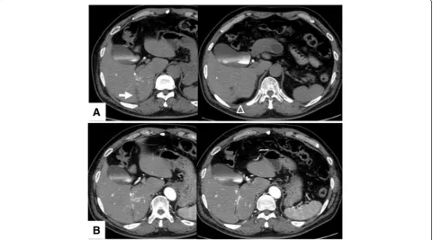
Dynamic computed tomography (CT) with drip infusion
cholangiography revealed focal dilatation of the biliary
ducts and atrophy in the posterior segments of the liver
without any observable mass (Fig. 1a, b). The magnetic
resonance imaging (MRI) scans, T1- and T2-weighted
im-ages, did not show any mass. However, when gadolinium
was used as a contrast agent, a 1.5-cm mass located in the
area adjacent at the main posterior biliary duct was highly
enhanced on T1-weighted images during the arterial phase
but demonstrated rapid withdrawal in the portal venous
and delayed phases (Fig. 2). Gastroscopic and colonoscopic
examinations showed no ulcerative or tumorous lesion. As
the mass was not detected on abdominal ultrasonography
(US) and it could possibly be a malignant tumor such
as cholangiocellular carcinoma, the patient consented
to
undergo
a right hepatectomy with lymph node
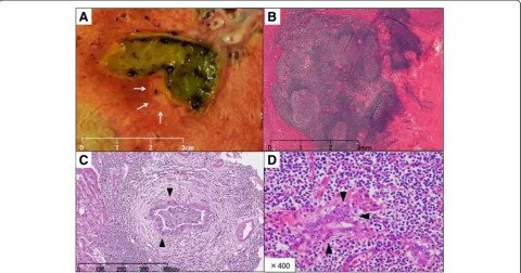
dissec-tion in the hepatic portal region. Grossly, a 7-mm white
mass detected along with the posterior biliary duct was
soft and non-encapsulated like a lymph follicle (Fig. 3a).
Histologically, dense lymphocyte infiltration with some
lymphoid follicles was observed in the portal area (Fig. 3b).
Small- to middle-sized lymphocytes showed no apparent
atypia but formed lymphoepithelial lesions on some bile
capillaries (Fig. 3c, d). Immunohistochemical studies
in-dicated that the lymphocytes were positive for CD20 and
CD79a (Fig. 4), but negative for CD3. The patient was
di-agnosed with low-grade hepatic MALT lymphoma based
on the abovementioned pathological findings.
Subsequently, the patient’
s level of interleukin-2 receptor
was found to be elevated at 1133 U/ml (normal range,
122–496 U/ml). He was also infected with HP and medical
Fig. 1Computed tomography findings.aDynamic computed tomography with drip infusion cholangiography revealed focal dilatation of the biliary ducts (arrow) and atrophy (arrowheads) in the posterior segments of the liver.bNo tumor was detected via enhanced computed tomography
treatment for pylorus eradication was provided. Biopsy
of the bone marrow revealed a normoplastic marrow.
Positron emission tomography demonstrated diffuse
accumulation in both the thyroid glands, with a maximum
standardized uptake value of 4.0. Biopsy of the thyroid
glands showed chronic thyroiditis without malignancy, and
the patient’s thyroid function was within normal limits. The
present case of MALT lymphoma was diagnosed a stage I
tumor, according to the Ann Arbor classification, and
care-ful follow-up without additional treatment was selected. At
the time of this report, the patient remained alive and free
of disease 2 years after surgery.
Discussion
MALT lymphoma often develops at several anatomic sites,
including the gastrointestinal tract, lungs, head and neck,
skin, thyroid glands, breasts, and liver. Gastric MALT
lymphoma is thought to be triggered by chronic
inflam-mation, which can occur in different diseases including
chronic gastritis associated with HP infection, Sjogren
syndrome, and Hashimoto thyroiditis [3]. The etiology
of primary hepatic MALT lymphoma is unclear, but it
has been reported that primary biliary cirrhosis [4–8],
hepatitis C viral infection [8–13], hepatitis B viral infection
[14–16], ascariasis [17, 18], and HP infection [19] are
possibly related with the pathogenesis of hepatic MALT
lymphoma.
At presentation, our patient was not infected with
hepatitis viruses, and his thyroid function and bone marrow
were normal. He was also negative for anti-nuclear and
anti-mitochondrial antibodies. However, his serum IgG and
IgM levels were elevated, and he showed HP infection.
Fig. 3Tumor characteristics.aGrossly, the 7-mm white mass along the posterior biliary duct was soft and non-encapsulated.bHistological findings on hematoxylin and eosin staining. The lesion consisted of dense lymphocyte infiltration with some lymph follicles.canddSmall to mid-sized lymphocytes formed lymphoepithelial lesions on some bile capillariesTable 1
Reported cases of hepatic MALT lymphoma
Case Sex/age HBV HCV Concomitant disease Tumor no. Treatment Outcome
1 M/66 ND ND Ureteral cancer 1 Resection 12 M/alive
2 F/73 ND ND (−) 1 Resection Lost to follow-up
3 M/85 ND ND Prostatic cancer 2 (−) Death after other surgery
4 F/60 ND ND Liver cirrhosis Multiple Transplantation 12 M/dead
5 F/57 (−) ND Ascariasis 1 Resection 55 M/alive
6 M/48 (+) ND Hepatitis 1 Resection + Chemotherapy 38 M/alive
7 F/47 (−) (−) Multiple biliary unilocular cysts 1 Resection + Radiation 30 M/alive
8 M/64 (−) (−) Colon cancer 1 Resection Lost to follow-up
9 F/62 (−) (−) Primary biliary cirrhosis 1 Resection 6 M/alive
10 F/64 (−) (+) Liver cirrhosis 1 Chemotherapy 24 M/alive
11 F/65 (−) (+) Hepatitis 1 Chemotherapy 48 M/alive
12 F/69 (−) (−) (−) 1 Resection Short time/alive
13 F/41 (−) (−) Primary biliary cirrhosis 1 (−) 12 M/alive
14 F/64 (−) (−) (−) 1 Resection 72 M/alive
15 F/57 (−) (−) Primary biliary cirrhosis 1 Transplantation 9 M/alive
16 F/64 (−) (−) Ascariasis 1 Resection Pulmonary recurrence after 96 M
17 F/59 ND ND ND Multiple Resection + Chemotherapy ND
18 M/61 (−) (−) Gastric cancer 1 Resection 18 M/alive
19 M/73 (−) (+) Liver cirrhosis 1 Resection 34 M/alive
20 M/59 (−) (+) Hepatitis 1 Resection 30 M/alive
21 F/50 (−) (−) (−) 1 Resection + Chemotherapy 30 M/alive
22 F/72 ND ND Colon cancer 1 (−) 1 M/dead
23 F/61 ND ND Rheumatoid arthritis 1 (−) Dead
24 F/58 ND ND (−) Multiple Resection + Chemotherapy 37 M/alive
25 F/62 ND ND Breast cancer 1 Resection 9 M/alive
26 F/65 (+) (−) Hepatocellular carcinoma 1 Resection 10 M/alive
27 F/60 (−) (−) Gastric MALT lymphoma 1 (−) 30 M/alive
28 M/59 (+) (−) Liver cirrhosis 2 Transplantation 6 M/Alive
29 M/36 (+) (−) Hepatitis 1 Resection Hepatic recurrence after 40 M
30 M/53 (−) (+) Liver cirrhosis Multiple Transplantation + Chemo ND
31 M/67 (−) (−) Hepatitis (drug) 1 Radiation Pulmonary recurrence after 72 M
32 M/69 (−) (−) (−) 2 RFA + Chemo 24 M/alive
33 F/74 (−) (−) (−) 1 Resection + Chemotherapy 6 M/alive
34 F/67 NA NA Gastric MALT lymphoma Multiple (−) 1 M/dead
35 F/72 (−) (−) Colon cancer 1 Resection 24 M/alive
36 M/64 (−) (−) Gastric cancer Multiple (−) 24 M/alive
37 M/71 (−) (−) (−) 1 Resection 15 M/alive
38 M/71 (−) (−) (−) 1 Resection + Chemotherapy 45 M/alive
39 F/56 (−) (−) (−) 1 Resection Pulmonary recurrence after 84 M
40 M/59 (−) (−) (−) 1 Resection 5 M/alive
41 M/86 (+) (−) Hepatitis 1 (−) 15 M/alive
42 M/58 (−) (+) Hepatitis 1 Resection + Chemotherapy 6 M/alive
43 F/43 (−) (−) Gastric cancer 1 Resection 24 M/alive
Such clinical findings suggested that the hepatic MALT
lymphoma might be strongly associated with chronic
in-flammation caused by HP infection. Subsequent treatment
for HP infection after surgery was successful.
For literature review, we searched PubMed and Ichushi
Web by Japan Medical Abstracts Society independently.
Key terms used included
“MALT lymphoma,” “liver,”
“hepatic MALT lymphoma,”
and
“primary hepatic
lymph-oma. To our knowledge, there are 37 reports including 51
patients with primary hepatic MALT lymphoma [4–41]
(Table 1). The mean age of these 22 men and 29 women
was 64.0 years. In most cases, the hepatic tumors were
incidentally detected during surgical resection or on
follow-up imaging examination for liver diseases or other
conditions. In 24 patients (47 %), liver diseases
concomi-tantly existed (ascariasis, 2; primary biliary cirrhosis, 5;
hepatitis B, 4; hepatitis C, 6; drug induced hepatitis, 1;
cirrhosis without hepatitis viral infection, 5; and
mul-tiple biliary cysts, 1). Thus, hepatic MALT lymphoma
development might be related to chronic liver
inflamma-tion, similar to gastric MALT lymphoma. Thirty-eight
patients (74 %) had solitary mass, and the tumor size
was
≤
3 cm in 22 of the 41 reported cases (53 %). Regarding
radiological characteristics, in 15 cases, the tumors were
de-scribed as detectable hypo-echoic masses via abdominal
US. In 21 cases, they were detected as low-density masses
via CT, including 6 cases with enhancement and 9 without.
In 16 cases with detailed MRI description, all tumors
showed high density on T1-weighted images and low
density on T2-weighted images. Two cases that described
contrast-enhanced MRI showed sickly enhancement in
the early phase. Both cases had solitary mass, and their
tumor sizes were 3 and 6.5 cm, respectively [27, 41]. Our
case showed highly enhanced mass in the early phase, but
not detected in abdominal US and CT. It is suggested that
these findings would be specific to small hepatic MALT
lymphoma. With regard to treatment, 31 patients (60.8 %)
underwent surgical resection with or without
chemother-apy or radiation therchemother-apy. Of these, 28 patients had a single
tumor, including 4 whose tumors were accidentally
discov-ered in the isolated liver from transplantation patients. In
addition, one patient underwent radiofrequency ablation,
five received chemotherapy only, and two received radiation
only. Eight patients did not receive any treatment, five of
whom died during the follow-up period. Recurrence was
reported in two patients.
According to the abovementioned case reports, primary
hepatic MALT lymphoma tends to be solitary and small.
Furthermore, it is often difficult to make a definite
diagno-sis of primary hepatic MALT lymphoma solely based
on the imaging findings as the disease seem to exhibit
variable imaging features. Therefore, it is necessary to
accumulate more cases and establish a therapeutic strategy
for primary hepatic MALT lymphoma.
Conclusions
In the present report, we described a case of primary
hepatic MALT lymphoma. Our experience in this case and
review of relevant literature indicated that preoperative
diagnosis of hepatic MALT lymphoma might be
chal-lenging because of the disease’s varying imaging
fea-tures. Thus, further study of this extremely rare disease
is necessary.
Consent
Written informed consent was obtained from the patient
for publication of this case report and accompanying
images.
Abbreviations
CT:computed tomography; HP:Helicobacter pylori; MALT: mucosa-associated lymphoid tissue; MRI: magnetic resonance imaging; PHL: primary hepatic lymphoma; US: ultrasonography.
Competing interests
The authors declare that they have no competing interests.
Authors’contributions
SN, NH, and KK drafted the manuscript. All authors read and approved the final manuscript.
Acknowledgements
We would like to thank Editage (www.editage.jp) for English language editing.
Received: 27 February 2015 Accepted: 17 September 2015
References
1. Isaacson P, Wright DH. Malignant lymphoma of mucosa-associated lymphoid tissue. Cancer. 1983;52:1410–6.
Table 1
Reported cases of hepatic MALT lymphoma
(Continued)
45 M/76 (−) (+) Hepatitis 1 Radiation 60 M/alive
46 F/74 (−) (−) Colon cancer 2 Resection 24 M/alive
47 F/74 (−) (−) Primary biliary cirrhosis Multiple Chemotherapy 36 M/alive, no relapse
48 M/73 (−) (+) Hepatitis Multiple Chemotherapy 24 M/alive, relapse
49 F/56 (−) (−) (−) 1 Resection 13 M/alive
50 M/77 (−) (+) Hepatitis 1 Resection 8 M/alive
Our case M/74 (−) (−) (−) 1 Resection 30 M/alive
2. Yang XW, Tan WF, Yu WL, Shi S, Wang Y, Zhang YL, et al. Diagnosis and surgical treatment of primary hepatic lymphomas. World J Gastroenterol. 2010;16:6016–9.
3. Thieblemont C, Bertoni F, Cople-Bergman C, Ferreri AJ, Ponzoni M. Chronic inflammation and extra-nodal marginal-zone lymphoma of MALT type. Semin Cancer Biol. 2014;24:33–42.
4. Prabhu RM, Medeiros LJ, Kumar D, Drachenberg CI, Papadimitriou JC, Appleman HD, et al. Primary hepatic low grade B-cell lymphoma of mucosa-associated lymphoid tissue (MALT) associated with primary biliary cirrhosis. Mod Pathol. 1998;11:404–10.
5. Sato S, Masuda T, Oikawa H, Satoh T, Suzuki Y, Takikawa Y, et al. Primary hepatic lymphoma associated with primary biliary cirrhosis. Am J Gastroenterol. 1999;94:1669–73.
6. Ye MQ, Suriawinata A, Black C, Min AD, Strauchen J, Thung SN. Primary hepatic marginal zone B-cell lymphoma of mucosa-associated lymphoid tissue type in a patient with primary biliary cirrhosis. Arch Pathol Lab Med. 2000;124:604–8.
7. Nakayama S, Yokote T, Kobayashi K, Hirata Y, Akioka T, Miyoshi T, et al. Primary hepatic MALT lymphoma associated with primary biliary cirrhosis. Leuk Res. 2010;34:e17–20.
8. Tanaka M, Fukushima N, Yamasaki F, Ohshima K. Primary hepatic extranodal marginal zone lymphoma of mucosa-associated lymphoid tissue type is associated with chronic inflammatory process. Open J Hematol. 2010. www.rossscience.org/ojhmt/articles/2075-907X-1-5.pdf. Accessed 14 Feb 2015. 9. Ascoli V, Lo Coco F, Artini M, Lerero M, Martelli M, Negro F. Extranodal
lymphomas associated with hepatitis C virus infection. Am J Clin Pathol. 1998;109:600–9.
10. Yago K, Shimada H, Itoh M, Ooba N, Itoh K, Suzuki M, et al. Primary low-grade B-cell lymphoma of mucosa-associated lymphoid tissue (MALT)-type of the liver in a patient with hepatitis C virus infection. Leuk Lymphoma. 2002;43:1497–500.
11. Mizuno S, Isaji S, Tabata M, Uemoto S, Imai H, Shiraki K. Hepatic mucosa-associated lymphoid tissue (MALT) lymphoma mucosa-associated with hepatitis C. J Hepatol. 2002;37:872–3.
12. Orrego M, Guo L, Reeder C, De Petris G, Balan V, Douglas DD, et al. Hepatic B-cell non-hodgkin’s lymphoma of MALT type in the liver explant of a patient with chronic hepatitis C infection. Liver Transpl. 2006;12:560–5. 13. Doi H, Horiike N, Hiraoka A, Koizumi Y, Yamamoto Y, Hasebe A, et al.
Primary hepatic marginal zone B cell lymphoma of mucosa-associated lymphoid tissue type: case report and review of the literature. Int J Hematol. 2008;88:418–23.
14. Takeshima F, Kunisaki M, Aritomi T, Osabe M, Akama F, Nakasone T, et al. Hepatic mucosa-associated lymphoid tissue and hepatocellular carcinoma in a patient with hepatitis B virus infection. J Clin Gastroenterol. 2004;38:823–6. 15. Nart D, Ertan Y, Yilmaz F, Yüce G, Zeytunlu M, Kilic M. Primary hepatic marginal
zone B-cell lymphoma of mucosa-associated lymphoid tissue type in a liver transplant patient with hepatitis B cirrhosis. Transplant Proc. 2005;37:4408–12. 16. Gockel HR, Heidemann J, Lugering A, Mesters RM, Parwaresch R, Domschke
W, et al. Stable remission after administration of rituximab in a patient with primary hepatic marginal zone B-cell lymphoma. Eur J Haematol. 2005;74:445–7. 17. Yamabe H, Haga H, Kashu I, Watanabe C, Kobashi Y. Malignant lymphoma
of mucosa-associated lymphoid tissue (MALT) type associated with ascariasis in the liver. Med Kagoshima Univ. 1995. http://ir.kagoshima-u.ac.jp/bitstream/ 10232/18332/1/AN00040104_v47s2_p137-139.pdf. Accessed 14 Feb 2015. 18. Chen F, Ike O, Wada H, Hitomi S. Pulmonary mucosa-associated lymphoid
tissue lymphoma 8 years after resection of the same type of lymphoma of the liver. Jpn J Thorac Cardiovasc Surg. 2000;48:233–5.
19. Iida T, Iwahashi M, Nakamura M, Nakamori M, Yokoyama S, Tani M, et al. Primary hepatic low-grade B-cell lymphoma of MALT-type associated with helicobacter pylori infection. Hepatogastroenterology. 2007;54:1898–901. 20. Isaacson PG, Banks PM, Best PV, McLure SP, Muller-Hermelink HK, Wyatt JI.
Primary low-grade hepatic B-cell lymphoma of mucosa-associated lymphoid tissue(MALT)-type. Am J Surg Pathol. 1995;19:571–5.
21. Ueda G, Oka K, Matsumoto T, Yatabe Y, Yamanaka K, Suyama M, et al. Primary hepatic marginal zone B-cell lymphoma with mantle cell lymphoma phenotype. Virchows Arch. 1996;428:311–4.
22. Maes M, Depardieu C, Dargent JL, Hermans M, Verhaeghe JL, Delabie J, et al. Primary low-grade B-cell lymphoma of MALT-type occurring in the liver: a study of two cases. J Hepatol. 1997;27:922–7.
23. Kirk CM, Lewin D, Lazarchick J. Primary hepatic B-cell lymphoma of mucosa-associated lymphoid tissue. Arch Pathol Lab Med. 1999;123:716–9.
24. Bouron D, Léger-Ravet MB, Gaulard P, Franco D, Capron F. Unusual hepatic tumor. Ann Pathol. 1999;19:547–8.
25. Raderer M, Traub T, Formanek M, Virgolini I, Osterreicher C, Fiebiger W, et al. Somatostatin-receptor scintigraphy for staging and follow-up of patients with extraintestinal marginal zone B-cell lymphoma of the mucosa associated lymphoid tissue (MALT)-type. Br J Cancer. 2001;85:1462–6. 26. Murakami J, Fukushima N, Ueno H, Saito T, Watanabe T, Tanosaki R, et al.
Primary hepatic low-grade B-cell lymphoma of the mucosa-associated lymphoid tissue type: a case report and review of the literature. Int J Hematol. 2002;75:85–90.
27. Arai O, Wani Y, Kaneyoshi T, Ikeda H, Kono Y, Tsukayama C. A case of primary hepatic low-grade B-cell lymphoma of mucosa-associated lymphoid tissue (MALT). Liver Cancer. 2003;9:144–9.
28. Streubel B, Lamprecht A, Dierlamm J, Cerroni L, Stolte M, Ott G, et al. T(14;18)(q32;q21) involving IGH and MALT1 is a frequent chromosomal aberration in MALT lymphoma. Blood. 2003;101:2335–9.
29. Shin SY, Kim JS, Lim JK, Hahn JS, Yang WI, Suh CO. Long-lasting remission of primary hepatic mucosa-associated lymphoid tissue (MALT) lymphoma achieved by radiotherapy alone. Korean J Intern Med. 2006;21:127–31. 30. Hamada M, Tanaka Y, Kobayashi Y, Takeshita E, Joko K. A case of MALT
lymphoma of the liver treated by RFA and Rituximab. Nippon Shokakibyo Gakkai Zasshi. 2006;103:655–60.
31. Yasui T, Okino H, Onitsuka K, Shono M, Watanabe J, Takeda S. A case of primary hepatic lymphoma. Nippon Rinsho Geka Gakkai Zasshi. 2006. www.ci.nii.ac.jp/naid/130003605234. Accessed 14 Feb 2015.
32. Chung YW, Sohn JH, Paik CH, Jeong JY, Han DS, Jeon YC, et al. High-grade hepatic mucosa-associated lymphoid tissue (MALT) lymphoma probably transformed from the low-grade gastric MALT lymphoma. Korean J Intern Med. 2006;21:194–8.
33. Chatelain D, Maes C, Yzet T, Brevet M, Bounicaud D, Plachot JP, et al. Primary hepatic lymphoma of MALT-type: a tumor that can simulate a liver metastasis. Ann Chir. 2006;131:121–4.
34. Ito T, Hiramatsu K, Machiki Y, Akagawa T, Miyata T, Hirata A, Hara T, Yoshida K, Kato K. A case of resected mucosa-associated lymphoid tissue lymphoma of the liver. Jpn J Gastroenterol Surg. 2008. www.journal.jsgs.or.jp/pdf/ 041091686.pdf. Accessed 14 Feb 2015.
35. Shito M, Kakefuda T, Omori T, Ishii S, Sugiura H. Primary non-Hodgkin’s lymphoma of the main hepatic duct junction. J Hepatobiliary Pancreat Surg. 2008;15:440–3.
36. Koubaa Mahjoub W, Chaumette-Planckaert MT, Murga Penas EM, Dierlamm J, Leroy K, Delfau MH, et al. Primary hepatic lymphoma of mucosa-associated lymphoid tissue type: a case report with cytogenetic study. Int J Surg Pathol. 2008;16:301–7.
37. Murata T, Uetsuka H, Uda M, Kawamata O, Nakai H, Ohta T. A case of mucosa-associated lymphoid tissue lymphoma of the liver mimicking a metastatic liver tumor of gastric cancer. Nippon Rinsho Geka Gakkai Zasshi. 2009. https://www.jstage.jst.go.jp/article/jjsa/70/6/70_6_1799/_pdf. Accessed 14 Feb 2015.
38. Yoshida M, Sekikawa S, Takanashi S, Kashiyama M, Ishigooka M, Kawashima H, et al. A resected primary hepatic mucosa-associated lymphoid tissue lymphoma with colon cancer. Nippon Rinsho Geka Gakkai Zasshi. 2010. www.ci.nii.ac.jp/naid/10026341055. Accessed 14 Feb 2015.
39. Hayashi M, Yonetani N, Hirokawa F, Asakuma M, Miyaji K, Takeshita A, et al. An operative case of hepatic pseudolymphoma difficult to differentiate from primary hepatic marginal zone B-cell lymphoma of mucosa-associated lymphoid tissue. World J Surg Oncol. 2011;9:3.
40. Miwa T, Yamamura Y, Fukuoka T, Mashita N, Inaoka K, Sawaki K, et al. A case of primary hepatic MALT lymphoma, in which hepatocellular carcinoma was diagnosed in preoperative images. Kanzo. 2011. www.ci.nii.ac.jp/naid/ 10029526600. Accessed 14 Feb 2015.


