O R I G I N A L A R T I C L E
Effect of alpha-tocopherol on bone formation during distraction
osteogenesis: a rabbit model
Mustafa Kurklu•Cemil Yildiz•Ozkan Kose•
Yuksel Yurttas• Ozgur Karacalioglu•
Muhittin Serdar•Salih Deveci
Received: 21 March 2011 / Accepted: 17 June 2011 / Published online: 15 July 2011 ÓThe Author(s) 2011. This article is published with open access at Springerlink.com
Abstract
Purpose The purpose of this study was to evaluate the effects of alpha-tocopherol on distraction osteogenesis. Materials and methods Right tibias of 30 New Zealand white rabbits were distracted at a rate of 0.5 mm/day for 20 days with a circular external fixator. Experimental group rabbits (n=15) were administered i.m. 20 mg/kg/ day alpha-tocopherol for 30 days. Radiographic examina-tions were performed at the 20th, 30th and 40th days. Bone scintigraphy was performed at the 5th and 20th days. Serum total antioxidant capacity (TAC) was measured at the 5th and 30th days. All animals were sacrificed and the right tibias of all animals were harvested for histopatho-logic examination at the 40th day.
Results Radiologic scores were statistically similar at the 20th day. However, the experimental group demonstrated higher radiologic scores at the 30th and 40th days. A scin-tigraphic baseline study at the 5th day of the study showed statistically similar osteoblastic activities in both groups. However, at the 20th day, osteoblastic activity was signif-icantly higher in the experimental group. Serum TAC val-ues were also significantly higher in the experimental group at the 30th day. At necropsy, histopathologic examination revealed statistically significantly higher scores in the experimental group.
Conclusion The results of this study show that alpha-tocopherol has beneficial effects on new bone formation during distraction osteogenesis.
Keywords Distraction osteogenesisAlpha-tocopherol Vitamin E Free radicalsAntioxidant therapyFracture healing—drug effects
Introduction
The technique of ‘‘distraction osteogenesis’’ is frequently used in the treatment of bony loss, pseudoarthrosis, chronic osteomyelitis, limb length discrepancy, biologic recon-struction after wide tumoral resection, and deformity [1–6]. One major problem with this method, however, is the prolonged time required for the newly formed bone in the distraction gap to consolidate and become strong enough for weight-bearing [7]. Various clinical and experimental investigations have been focused on the acceleration of bone formation and consolidation, and have thereby aimed to shorten the framing time [8–11].
Distraction osteogenesis is recognized as being ‘‘intra-membranous ossification,’’ which can be assumed to be a M. KurkluC. YildizY. Yurttas
Department of Orthopaedics and Traumatology, Gulhane Military Medical Academy, Ankara, Turkey
O. Kose (&)
Antalya Education and Research Hospital,
Orthopaedics and Traumatology Clinic, Pinarbas¸i mh. 758.sk. Nazlibahce Evleri A Blok D8, Konyaalti, Antalya, Turkey
e-mail: [email protected]
O. Karacalioglu
Department of Nuclear Medicine,
Gulhane Military Medical Academy, Ankara, Turkey
M. Serdar
Deparment of Clinical Biochemistry,
Gulhane Military Medical Academy, Ankara, Turkey
S. Deveci
Department of Pathology, Gulhane Military Medical Academy, Ankara, Turkey
special form of fracture healing [12]. Fracture healing after injury involves inflammation, repair and remodeling [13]. At the inflammatory stage, polymorphonuclear leukocytes (PMNLs), macrophages and mast cells migrate into the fracture site, and osteoclasts begin to remove necrotic bone [14,15]. Activation of PMNLs produces oxygen free rad-icals, which cause lipid peroxidation and are known to impair fracture healing [16, 17]. Antioxidant administra-tion has been shown to be beneficial in suppressing the damaging effects of oxygen free radicals in cells during fracture healing [18–21].
Consequently, we hypothesized that alpha-tocopherol, which is a potent antioxidant, may also have favorable effects on the quality of new bone formation during dis-traction osteogenesis and shorten the time required for consolidation. Thus, in this study, the effect of alpha-tocopherol on bone formation during distraction osteo-genesis was investigated.
Materials and methods
Animals
In this study, 30 adult New Zealand white rabbits (mean weight 1,800 g; range 1,500–2,000 g) were used. The animals were fed a standard laboratory diet and water and had a 12 h day/night cycle. The rabbits were housed separately in standard cages in a temperature-controlled room (20–22°C). Before initiating the study, approval from the local ethics committee was obtained. The study was carried out in the Center for Experimental Animals at the same institution. The rabbits were randomized into experimental and control groups each consisting of 15 animals.
Surgical procedures (day 1)
Rabbits were anesthetized with 0.2 mg/kg xylazine and 20 mg/kg ketamine hydrochloride. Infection prophylaxis was provided with 20 mg/kg/day cefazoline sodium preop-eratively and 2 days postoppreop-eratively. When the appropriate depth of anaesthesia had been achieved, a preconstructed four-ring circular external fixator was applied to the right tibia of each rabbit. 5/8 rings were used at the distal and proximal levels. Each ring was connected with three rods. Two Kirchner wires (1 mm in diameter) crossing at angles of 45–60°with respect to each other were applied to every level. The skin subdermis and periosteum were exposed with an anteromedial longitudinal incision, and a transverse osteot-omy was performed on the middle third of the right tibia. Subsequently, the periosteum and skin were closed properly (Fig.1). After then waiting for 5 days, distraction was
applied to each tibia on a schedule of 0.125 mm per 6 h a day (0.5 mm/day). Distraction continued for 20 days.
Experimental intervention (day 1–30)
The experimental group of rabbits received 20 mg/kg alpha-tocopherol intramuscularly starting on the first day of study, and a daily injection of alpha-tocopherol was given for 30 days thereafter. The control group did not receive any corresponding treatment.
Radiological follow-up and evaluation (days 20, 30 and 40)
Radiographs were taken at the 20th, 30th and 40th days of the study and evaluated by one of the investigators (who was blinded to the assignment of study groups) using the five-point grading scale described by Lane and Sandhu (Table1) [22].
Scintigraphic method (days 5 and 20)
the gamma camera (Millenium, General Electric, Milwau-kee, WI, USA) equipped with a low energy, high-resolution collimator, and planar acquisition was initiated for 10 min using a 15% window centered over the 140 keV photopeak. Rectangular regions of interest (ROIs) were drawn on both tibias (the region of distraction osteogenesis and the con-tralateral healthy leg) at approximately similar locations. Counts were derived from both ROIs in order to calculate the osteoblastic activity ratio (count for the lesion/count for the contralateral side). Scintigraphic assessments were performed on the 5th and 20th days of the study, before and after the distraction period.
Total antioxidant capacity (TAC) measurement (days 5 and 30)
Plasma TAC was measured using a Randox total antioxi-dant status kit (Total Antioxiantioxi-dant Status, Randox, Crumlin, UK) in which ABTS (2,2-azino-di-[3-ethylbenzthiazolin-6-sulfanate]) is incubated with a peroxidase and H2O2 to
produce the radical cation ABTS?. This has a stable blue-green color, which is measured at 600 nm. Antioxidants present in the sample suppress this color production to an extent that is proportional to their concentration. The suppression of the absorbance of the ABTS?radical cation by serum antioxidants was compared with the suppression caused by Trolox (6-hydroxy-2,5,7-tetramethylchroman-2-carboxylic acid), which is included as part of the TAC kit. The results are expressed as mmol/l of the Trolox equivalent [23].
Histopathologic evaluation (day 40)
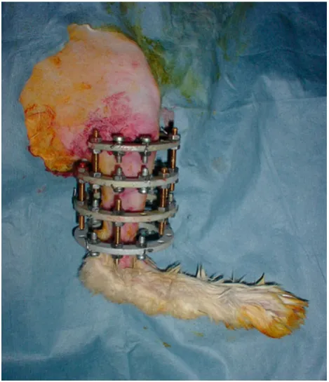
All rabbits in each group were sacrificed with a high-dose intraperitoneal sodium pentobarbital injection on the 40th day of the experiment. After sacrifice, all tibias were har-vested, stripped from their soft tissue, and sent to a pathologist, who was unaware of the allocation of rabbits into groups. For histological evaluation, extremities dis-sected from their soft tissues were fixed in 10% neutral buffered formaldehyde solution for ten days, and then treated with a rapid decalcification solution (formic acid) for five days. After the decalcification procedure, tibias were sampled longitudinally from distracted segments. For each tibial segment, four cuts were taken from the dis-traction area and routine histological preparation was car-ried out. Samples were embedded in paraffin, and sections of 4lm width were taken from the paraffin blocks and stained with hematoxylin–eosin. Sections were examined under a light microscope. Histopathological evaluation was done according to the grading system described by Huddlestone et al. (Table2) [24].
Statistical analysis
The Wilcoxon signed-rank test was used for repeated measurements of the same group and the Mann–WhitneyU test was employed to compare groups used. Statistical significance (P\0.05) was determined based on the 95% confidence interval.
Results
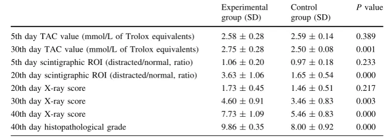
All rabbits in both groups completed the experiment without any complications, and the pertinent results are summarized in Table 3.
Table 1 Radiologic evaluation system [22]
Bone formation Score
Lack of bone formation 0
Bone formation filling 25% of the defect 1
Bone formation filling 50% of the defect 2
Bone formation filling 75% of the defect 3
Bone formation filling 100% of the defect 4
Union distal
No union 0
Onset of union 1
Complete radiologic union 2
Union proximal
No union 0
Onset of union 1
Complete radiologic union 2
Remodeling
None 0
Formation of intramedullary channel 1
Formation of cortex 2
Total score (maximum) 10
Table 2 Histopathologic evaluation system [24]
Tissue Score
Fibrous tissue 1
Predominantly fibrous tissue with some cartilage 2
Equal amounts of fibrous and cartilage tissue 3
All cartilage 4
Predominantly cartilage tissue with some woven bone 5
Equal amounts of cartilage and woven bone 6
Predominantly woven bone with some cartilage 7
Entirely woven bone 8
Woven bone and some mature bone 9
Radiologic results
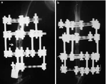
On the 20th day, radiologic scores were statistically similar in both groups (P=0.217). However, on the 30th and 40th days, the experimental group displayed statistically signif-icantly higher radiologic scores and visible callus forma-tion, maturation and remodeling (P=0.003 andP=0.000 respectively; Fig.2).
Scintigraphic results
Scintigraphic baseline study on the 5th day of study showed statistically similar osteoblastic activities in both groups (P=0.233). However, on the 20th day, the osteoblastic activity was significantly higher in the exper-imental group (0.000).
TAC measurement results
Serum TAC values were statistically similar in both groups on the 5th day of the study (P=0.389). However, on the 30th day, TAC values were significantly higher in the experimental group (P=0.001).
Histopathologic results
At necropsy, rabbits in the experimental group had statis-tically significantly higher scores in the histopathologic examination, and showed the formation of mature bone (P=0.000; Fig.3).
Discussion
In this study, possible favorable effects of alpha-tocopherol on the quality of new bone formation during distraction osteogenesis were investigated. We have shown that the administration of alpha-tocopherol provided better results as far as the radiologic, scintigraphic and histopathologic evaluations were concerned.
Oxygen-derived free radicals are highly toxic molecules that produce cellular damage by causing both structural and functional impairment in almost all components of the cell, but mainly the cell membrane. They initiate a chain reac-tion leading to cell membrane damage via lipid peroxida-tion, thereby causing cell lysis [25]. Alpha-tocopherol is a natural macromolecule that acts as a biological antioxidant in the cell membranes, inhibiting lipid peroxidation by scavenging peroxy and alkoxy radicals and thus breaking chain reactions [26,27].
Distraction osteogenesis is widely used for the treatment of various challenging musculoskeletal disorders. Pro-longed time spent with external fixation is one of the dis-advantages that can cause complications such as pin tract infection, loosening, muscle weakness and contractures [28]. Furthermore, prolonged framing time decreases the compliance of patients and causes psychological and behavioral problems [29].
Distraction osteogenesis is considered intramembranous ossification, which can be assumed to be a special form of fracture healing [12]. During the initial ischemic stage, considerable amounts of oxygen-derived free radicals are produced due to the activation of inflammatory cells [30, 31]. Likewise, Prasad et al. measured the predictors of oxidative stress in fracture patients and found that oxida-tive stress was directly proportional to the number of fractures, and that it peaked at the 3th week after the fracture and continued until the 4th week [32].
On the other hand, various experimental studies have been carried out to accelerate and shorten fracture healing with the administration of antioxidants. Go¨ktu¨rk et al. demonstrated that the administration of zymosan—which induces oxygen-free radicals through the stimulation of NADPH oxidase in polymorphonuclear leukocytes— impaired fracture healing in a rat model [17]. Yilmaz et al. have demonstrated the positive effects of ascorbic acid, a well-known antioxidant, on fracture healing [21]. Moreover, there are also studies that have shown the beneficial effects of alpha-tocopherol on fracture healing, whereas its effects on distraction osteogenesis have not been investigated Table 3 Summary of results
(mean radiologic and histopathologic scores, TAC values, and scintigraphic counts for the groups, as well as statistical significance)
SDstandard deviation
Experimental group (SD)
Control group (SD)
Pvalue
5th day TAC value (mmol/L of Trolox equivalents) 2.58±0.28 2.59±0.14 0.389
30th day TAC value (mmol/L of Trolox equivalents) 2.75±0.28 2.50±0.08 0.001
5th day scintigraphic ROI (distracted/normal, ratio) 1.06±0.20 0.97±0.18 0.233
20th day scintigraphic ROI (distracted/normal, ratio) 3.63±1.06 1.65±0.54 0.000
20th day X-ray score 1.73±0.45 1.46±0.51 0.217
30th day X-ray score 4.60±0.91 3.46±0.83 0.003
40th day X-ray score 7.73±1.09 5.46±0.83 0.000
[18–20]. Therefore, to our best knowledge, our study is the first report in this regard.
The fact that serum TAC values were not significantly different from those of the control group on the 5th day of our study, but they were significantly greater on the 30th day, suggests that alpha-tocopherol exerts a favorable effect during the ischemic stage but not during the inflammatory period. Radiologic and histologic evidence of callus for-mation and maturation were also found to be directly pro-portional to serum TAC values in the experimental group. It
is also worth noting that decreasing the ischemic stage using antioxidants would not only induce osteoblastic activity but it would also impede the osteoclastic resorption of newly formed bone due to oxygen-derived free radicals.
Overall, based on our study, we may conclude that the administration of supplemental alpha-tocopherol in patients treated with distraction osteogenesis may shorthen the framing time and increase the quality of the regenerated bone. Further clinical studies are necessary to check its effects on humans and also to ascertain whether it should Fig. 2 X-ray of a rabbit in the experimental group (a) and in the control group (b) on the 40th day of the study. Note the enhanced callus formation and consolidation in the experimental group (arrow)
Fig. 3 Histopathologic examination of a rabbit in the experimental group (a) and in the control group (b). Note the mature lamellar bone in the experimental group and the predominantly woven bone with
be used prophylactically or continuously until the end of the consolidation period. However, when compared with normal fracture healing, alpha-tocopherol may be much more effective at decreasing the repetitive ischemic cycles that are produced during distraction osteogenesis.
Acknowledgments This study was funded by the Animal Research Center at our institution. We, the authors and our affiliations, declare that we have no relevant financial involvement with any commercial organisation with direct financial interest in the subject or materials discussed in this manuscript.
Conflict of interest None.
Open Access This article is distributed under the terms of the Creative Commons Attribution License which permits any use, dis-tribution and reproduction in any medium, provided the original author(s) and source are credited.
References
1. D’Hooghe P, Defoort K, Lammens J, Stuyck J (2006) Manage-ment of a large post-traumatic skin and bone defect using an Ilizarov frame. Acta Orthop Belg 72:214–218
2. Paley D, Catagni MA, Argnani F, Villa A, Benedetti GB, Cattaneo R (1989) Ilizarov treatment of tibial nonunions with bone loss. Clin Orthop Relat Res 241:146–165
3. Kucukkaya M, Kabukcuoglu Y, Tezer M, Kuzgun U (2002) Management of childhood chronic tibial osteomyelitis with the Ilizarov method. J Pediatr Orthop 22:632–637
4. Aldegheri R (1999) Distraction osteogenesis for lengthening of the tibia in patients who have limb-length discrepancy or short stature. J Bone Joint Surg Am 81:624–634
5. Tsuchiya H, Abdel-Wanis ME, Sakurakichi K, Yamashiro T, Tomita K (2002) Osteosarcoma around the knee. Intraepiphyseal excision and biological reconstruction with distraction osteo-genesis. J Bone Joint Surg Br 84:1162–1166
6. Nakase T, Yasui N, Kawabata H et al (2007) Correction of deformity and shortening due to post traumatic epiphyseal arrest by distraction osteogenesis. Arch Orthop Trauma Surg 127: 659–663
7. Prokuski LJ, Marsh JL (1994) Segmental bone deficiency after acute trauma. The role of bone transport. Orthop Clin North Am 25:753–763
8. Sen C, Gunes T, Erdem M, Koseoglu RD, Filiz NO (2006) Effects of calcitonin and alendronate on distraction osteogenesis. Int Orthop 30:272–277
9. Chan CW, Qin L, Lee KM, Zhang M, Cheng JC, Leung KS (2006) Low intensity pulsed ultrasound accelerated bone remodeling during consolidation stage of distraction osteogene-sis. J Orthop Res 24:263–270
10. Kawamoto K, Kim WC, Tsuchida Y et al (2005) Effects of alternating current electrical stimulation on lengthening callus. J Pediatr Orthop B 14:299–302
11. Yamane K, Okano T, Kishimoto H, Hagino H (1999) Effect of ED–71 on modeling of bone in distraction osteogenesis. Bone 24:187–193
12. Aronson J (1997) Current concepts review. Limb lengthening, skeletal reconstruction, bone transport with the Ilizarov method. J Bone Joint Surg Am 79:1243–1253
13. Buckwalter JA, Einhorn TA, Bolander ME, Cruess RL (1996) Healing of the musculoskeletal tissues. In: Rockwood CA, Green DP, Bucholz RW et al (eds) Fractures in adults, 4th edn. Lip-pincott, Philadelphia, pp 261–304
14. Cornell CN, Lane JM (1992) Newest factors in fracture healing. Clin Orthop 277:297–311
15. Frost HM (1989) The biology of fracture healing. An overview for clinicians. Part II. Clin Orthop 248:294–308
16. Reilly PM, Schiller HJ, Bulkley GB (1991) Pharmacologic approach to tissue injury mediated by free radicals and other reactive oxygen metabolites. Am J Surg 161:488–503
17. Gokturk E, Turgut A, Baycu C, Gu¨nal I, Seber S, Gu¨lbas Z (1995) Oxygen-free radicals impair fracture healing in rats. Acta Orthop Scand 66:473–475
18. Turk C, Halici M, Guney A, Akgun H, Sahin V, Muhtaroglu S (2004) Promotion of fracture healing by vitamin E in rats. J Int Med Res 32:507–512
19. Durak K, Sonmez G, Sarisozen B, Ozkan S, Kaya M, Ozturk C (2003) Histological assessment of the effect of alpha-tocopherol on fracture healing in rabbits. J Int Med Res 31:26–30 20. Durak K, Bilgen OF, Kaleli T, Tuncel P, Ozbek R, Turan K (1996)
Antioxidant effect of alpha-tocopherol on fracture haematoma in rabbits. J Int Med Res 24:419–424
21. Yilmaz C, Erdemli E, Selek H, Kinik H, Arikan M, Erdemli B (2001) The contribution of vitamin C to healing of experimental fractures. Arch Orthop Trauma Surg 121:426–428
22. Lane JM, Sandhu HS (1987) Current approaches to experimental bone grafting. Orthop Clin North Am 18:213–225
23. Miller NJ, Rice-Evans C, Davies MJ, Gopinathan V, Milner A (1993) A novel method for measuring antioxidant capacity and its application for monitoring the antioxidant status in premature neonates. Clin Sci 84:407–412
24. Huddlestone PM, Steckelberg JM, Hanssen AD, Rouse MS, Bolander ME, Patel R (2000) Ciprofloxacin inhibition of exper-imental fracture healing. J Bone Joint Surg Am 82:161–173 25. Sheweita SA, Khoshhal KI (2007) Calcium metabolism and
oxidative stress in bone fractures: role of antioxidants. Curr Drug Metab 8:519–525
26. Burton GW, Ingold KU (1989) Vitamin E as an in vitro and in vivo antioxidant. Ann N Y Acad Sci 570:7–22
27. van Acker SA, Koymans LM, Bast A (1993) Molecular phar-macology of vitamin E: structural aspects of antioxidant activity. Free Radic Biol Med 15:311–328
28. Green SA (1990) Complications of pin and wire external fixation. Instr Course Lect 39:219–228
29. Yildiz C, Uzun O, Sinici E, Ates¸alp AS, Ozs¸ahin A, Bas¸bozkurt M (2005) Psychiatric symptoms in patients treated with an Ilizarov external fixator. Acta Orthop Traumatol Turc 39:59–63 30. Petrovich YA, Podorozhnaya RP, Kichenko SM, Kozlova MV
(2004) Effects of selenium-containing compounds and their metabolism in intact rats and in animals with bone fractures. Bull Exp Biol Med 137:74–77
31. Turgut A, Gokturk E, Kose N et al (1999) Oxidant status increased during fracture healing in rats. Acta Orthop Scand 70:487–490
![Table 2 Histopathologic evaluation system [24]](https://thumb-us.123doks.com/thumbv2/123dok_us/948474.1593727/3.595.49.286.70.326/table-histopathologic-evaluation-system.webp)