R E S E A R C H
Open Access
Functional morphological study of the
choana in different bird species
Fatma A. Mahmoud
1*, Ali G. Gadel-Rab
2and Nahed A. Shawki
1Abstract
Background:The anatomical information about the structure of the choana is lacking in literature, and its role in the olfactory and feeding mechanism is still unknown
Results:The present study discusses the adaptation of choana to cranial kinesis during feeding process in different bird species: kestrel, common moorhen, and hoopoe. Kestrel possesses a kinetic skull while the hoopoe and common moorhen have kinetic one; however, the common moorhen skull seems highly kinetic more than that of the hoopoe that properly effect on the choanal epithelium. The choana of kestrel and hoopoe are lined by pseudostratified ciliated columnar epithelium, while choana of common moorhen have transitional epithelium beside pseudostratified ciliated columnar epithelium. The choana epithelium of each bird species provides with simple alveolar glands and numerous goblet cells. In kestrel and hoopoe, the secretion products of choanal glands contain neutral and sulfated mucin, while in the common moorhen, these glands secret neutral and carboxylate mucopolysaccharides.
Conclusion:The choana of the three studied bird species apparents adaptation to the olfaction process but also affects the movement of skeletal elements of the skull during the feeding process
Keywords:Choana, Cranial, Kinesis, Bird, Feeding process
Background
Birds have variation in the form and function of their feeding system which are amenable to comparative analysis because they represent modifications of the same basic structure of this apparatus among different species without a doubt that reflected the success of birds to survival and occupy a wide range of habitats. Many of the literature has studied the avian feeding system (Jackowiak & Godynicki, 2004; Mahmoud, Shawki, & Wahba, 2009; El-Bakary, 2011, 2012; Al-Zahaby & Elsheikh,2014; Erdogan & Iwasaki,2014; and Shah & Aziz, 2014; Gadel-Rab, Shawki, & Saber, 2017), but still the feeding mechanics and functional morpho-logical information about most bird species remain poorly understood.
The roof of buccal cavity is considered as a part of feeding system and was given little attention in literature. It is interesting to state that only Shawki, Abdeen, and Mahmoud (2016) gave a clear description of the roof of buccal cavity of the kestrel and discussed its
role in the feeding mechanism. Mahmoud, Gad-Rab, and Shawki (2017) pointed out the effect of different feeding behaviors on the lining epithelium of the roof of buccal cavity of two bird species.
The feeding and sensory systems are considered in-tegrating systems and reflect a general historical trend of increasing cephalization in vertebrate evolution (Schwenk, 2000). The avian olfactory system consists of the choana which appears in birds and reptiles re-lated to the incomplete secondary palate, through which the nasal and oral cavities communicate with each other (Kent & Carr, 2001). The anatomical infor-mation about the structure of the choana is lacking in literature, and its role in the olfactory and feeding mechanism is still unknown.
Thus, the present study gives attention to the functional morphological structure of the choana in three different bird species to exhibit the adaptation of the choana to the feeding process.
The selected bird species are kestrel, hoopoe, and common moorhen that belong to different avian’s orders: Falconiformes, Bucerotiformes, and Gruiformes, respectively, each of them have own feeding behavior.
* Correspondence:[email protected]
1Department of Zoology, Faculty of Science, Assiut University, Assiut, Egypt
Full list of author information is available at the end of the article
Methods
The specimens of three bird species in the present study were collected from Abou-Rawash area of Egypt and brought alive to the comparative anatomy of vertebrate lab. The specimens were anesthetized by ether then dissected under wild stereomicroscope to separate the head from the rest of the body. Some specimens were carefully investigated by wild stereomicroscope to clarify the anatomical features of roof of mouth; the skull the was prepared; and then photos were photographed by using a digital camera. Some specimens were fixed in 10% neutral formalin for 3 days and prepared to paraffin embedding. Tissues were sectioned at 7μm and stained with Hematoxylin and Eosin, Masson’s trichrome, Bromophenol blue, PAS and PAS-Alcian blue (Bancroft & Stevens,1996).
Results
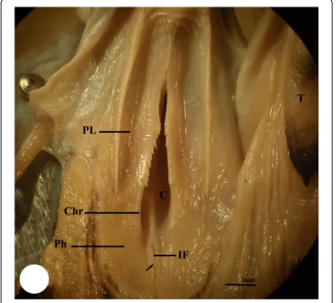
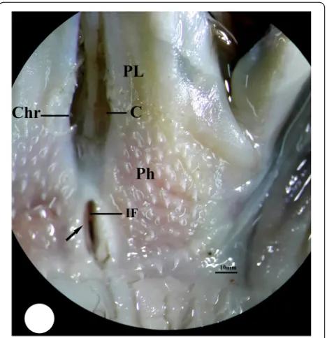
The buccal cavity of the three studied bird species is bounded externally by their beak while internally, it can be distinguished into the floor of mouth, which includes the tongue, and the roof of mouth which contains two regions: the palate and pharyngeal region. The palate re-gion (PL) occupies the choana that faces the glottis within the tongue. The choana (C) of each studied bird species exhibits different shape; in kestrel, it appears to be formed from two parts, a short narrow anterior part and long-wide posterior one separated by a constriction (Fig. 1). In common moorhen, the choana is formed from two equal parts, an anterior tapering part and a
posterior wide one (Fig. 2). Meanwhile, the choana of the hoopoe has pear-shape with posterior wide half and slightly anterior narrow one (Fig. 3). The pharyngeal region (Ph) of kestrel and common moorhen is situated posterior to the palatine wing which is not distin-guished in the hoopoe. The pharyngeal region in the three studied bird species incubates the oval-shaped fissure which is known as the infundibular fissure (IF). In the present study, the infundibular fissure of the kestrel and hoopoe is located posterior to the choana ridges (Chr), while in the common moorhen, it lies within the choana ridges. The margins of in-fundibular fissure of the kestrel carry a longitudinal row of posteriorly directed papillae, whereas these
Fig. 1Photomicrograph of the buccal cavity of kestrel shows palate region (PL), choana (C), pharyngeal region (Ph), infundibular fissure (IF), choana ridges (Chr), margins of infundibular fissure (arrow) with papillae, and tongue (T)
Fig. 2Photomacrograph of the buccal cavity of common moorhen shows upper jaw (UJ), palate region (PL), choana (C), pharyngeal region (Ph), choana ridges (Chr), and margins of infundibular fissure (arrow) without papillae
papillae disappear in the case of hoopoe and the common moorhen (Figs. 1, 2, and 3).
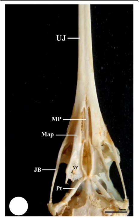
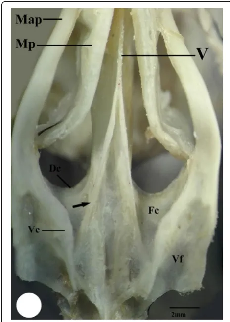
The roof of the mouth is supported by the palatine bone (Os palatinum) which exhibits anatomical variation in the three studied bird species. The palatine bone have paired anterior portion (the maxillary process of the pal-atine bone, Map) which appears thick and narrow in the kestrel and broader in the hoopoe (Figs. 4 and 5), while in the common moorhen, it is long, thin, and dorso-medially curved. Moreover, this portion is fused laterally with the posterior portion of the maxillae “upper jaw, UJ” through the maxillopalatine process (Fig. 6 and 7). Meanwhile, in the kestrel and hoopoe, this process is completely fused with the ventral surface of the upper jaw (UJ) (Figs.8, and9).
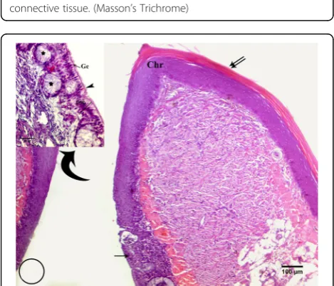
Dorsally, the vomer bone is located between the anter-ior portion of palatine and extends posteranter-iorly to fuse with the parasphenoid bone. In the kestrel, the vomer bone has club shape with rounded head which fuse an-teriorly with maxillopalatine process (MP), and extends posteriorly as rod-shape to fuse with the parasphenoid bone (Fig. 8). Meanwhile, in the common moorhen, the vomer bone initiates as narrow unpaired bone, then forks posteriorly towards the parasphenoid bone (Figs. 6 and 7), whereas in the hoopoe, the vomer bone is absent and replaced by a longitudinal ribbon of collagenous connective tissue which extends pos-teriorly cross the ventromedial surface of the para-sphenoid bone (Fig. 9).
Posteriorly, the palatine bone broads to form the pars lateralis which contains the ventral fossa. This fossa ap-pears deeper in the common moorhen than that of the kestrel, while in the hoopoe, it appears shallow (Figs. 7, 8, and 9). The pars lateralis possesses medial and ventral crest which enclose the choana fossa between them. The choana fossa is narrow and shallow in the kestrel, broad and slightly deep in the common moorhen, and narrow and deep in the hoopoe (Figs.7,8, and9).
Moreover, the kestrel and common moorhen have paired medial crest (Figs. 7 and 8) that is absent in the case of hoopoe and replaced by fibrous connective tissue (Fig. 9). The ventral crest that forms the lateral wall of the choana protrudes ventrally in the kestrel and the common moorhen more than in the hoopoe.
The histological investigation of the roof of the mouth of three studied bird species revealed that the roof is covered by stratified squamous epithelium with variation in keratinzation; in the kestrel and the common moor-hen, the lining epithelium of the roof is keratinized (Figs.10and11), while the roof of the hoopoe is covered by non-keratinized epithelium with appearance of keratinization on the palatine papillae (Fig.12). However, Fig. 3Photomicrograph of the buccal cavity of hoopoe shows
palate region (PL), choana (C), pharyngeal region (Ph), infundibular fissure (IF), choana ridges (Chr), and margins of infundibular fissure (arrow) with papillae
in the studied bird species, the epithelium layer of the roof lines the choana extends dorsally and changes grad-ually from stratified squamous epithelium into pseudos-tratified ciliated columnar epithelium (Figs. 10, 11, 12, and 13). Moreover, in the common moorhen, the dorsomedial surface of choana is covered by transitional epithelium (Fig.13b).
The simple alveolar gland and goblet cells are distributed within the lining epithelium of the choana of all species analyzed. In the common moorhen, compound-branched alveolar glands were observed embedding dorso-medially within the epithelium of the chaona (Fig.13a).
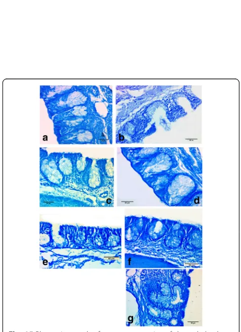
In all investigated species, the glandular cells of the choanal gland are strongly reacted with PAS indicating the presence of neutral mucopolysaccharides (Fig.14b, c) and give positive reaction with PAS-Alcian blue that indicate the presence of neutral and acid mucopolysaccharides (Fig.14a).
The glandular cells of the choanal glands of kestrel and the hoopoe exhibited a slight positive staining affin-ity with Alcian-blue PH 2(Fig.15a, c), while moderate in the common moorhen (Fig. 15b). This indicates the presence of carboxylate mucopolysaccharides. In kestrel and hoopoe, the glandular cells of the choanal glands give deep-bluish color with Alcian-blue PH1 (Fig.15d, f) which mean the presence of sulfated mucopolysaccha-rides. In the common moorhen, the glandular cells within the epithelium covering the dorso-medial surface of choana give deep blue color with Alcian-blue PH1 while that covering the lateral surface give faint blue color (Fig.15e, g).
Moreover, in kestrel and hoopoe, the choanal glands showed moderate reaction with mercuric bromophenol blue (Fig. 14d, f) indicating the presence of protein secretory materials, while in the common moorhen, the choanal glands gives slightly react with mercuric bromo-phenol blue (Fig.14e).
Fig. 6Photograph of ventral surface of skull of common moorhen shows upper jaw (UJ), the maxillary process of the palatine bone (Map), maxillopalatineprocess (MP), vomer bone (V), choana fossa (Fc), ventral crest (Vc), dorsal crest (Dc), ventral fossa (Vf), Pterygoid (Pt), and parasphenoid (Sph)
Fig. 5Photomacrograph of ventral surface of skull of hoopoe shows upper jaw (UJ), Jugal bar (JB), the maxillary process of the palatine bone (Map), maxillopalatine process (MP), ventral fossa (Vf), Pterygoid (Pt), and parasphenoid (Sph)
Discussion
The kestrel, common moorhen, and hoopoe are three different bird species with different feeding behaviors: kestrel is prey–predator bird which possesses hard curved beaks to cut their prey while the hoopoe and common moorhen are probing birds that catch their prey by different strategies. Shawki and Ismail (2006) and Mahmoud et al. (2009) mentioned the functional morphological structure of the lingual apparatus of the common moorhen and kestrel respectively and confirmed that these bird species mainly rely on their lingual apparatus in feeding process, while the hoopoe depends on the jaw apparatus in manipulating their prey than its tongue (Gadel-Rab, Shawki, & Saber, 2012) which agree with those mentioned by Rawal (1968) who concluded that the long arms of hoopoe’s bill confer greater advantage in thrusting the bill into soil.
The cranial kinesis is an important character of the feeding behavior of birds (Bout & Zweers, 2001). The kestrel, hoopoe, and common moorhen are prokinetic birds like most bird species, in which the bending
zone is situated at the nasal-frontal hinge that allows the movement of the upper jaw up/down relative to the skull.
Mahmoud, Shawki, and Abdeen (2017) clarified that there is no flexible zone appearing at the nasal-frontal hinge in the kestrel’s skull. In the hoopoe, this hinge is movable that confirmed practically during the skull preparation; also in the common moorhen, the upper jaw can depress downward relative to skull. The move-ment of the upper jaw (depression) occurs as resulting of the contraction of pterygoid muscle complex of each bird species. The pterygoid muscles are originated from the palatine bone“pars lateralis”and insert on the lower jaw“posterior portion of the mandible”. The contraction of this muscle pulls the palatine bone backward, thereby depressing the upper jaw, as well as, adducting the man-dible by elevating the anterior portion of manman-dible that leads to closing the beak. Physically, the naso-frontal hinge determines the ability of the gliding movement of the palatine bone beside its shape and position. The skull of kestrel have immovable naso-frontal hinge; this leads to the mechanical force which is produced during the contraction of the pterygoid muscle closing the man-dible quickly to produce a strong bit force; this result agree with those mentioned by Mahmoud et al. (2017). Fig. 7High magnification of ventral surface of skull of common
moorhen shows the maxillary process of the palatine bone (Map), maxillopalatine process (MP), vomer bone (V), choana fossa (Fc), medial crest (arrow), ventral crest (Vc), and ventral fossa (Vf)
Meanwhile, in hoopoe and the common moorhen, the movable naso-frontal hinge gives flexibility for depression of the upper jaw by pulling the palatine bone during the contraction of the pterygoid muscle. During depression of upper jaw in the common moorhen, the force transferred to curve the anterior portion of the palatine bone“ maxil-lary process of the palatine bone”. This force enables bird to widen the choana. Morphologically, widening of choana Fig. 9High magnification of ventral surface of skull of hoopoe
shows the maxillary process of the palatine bone (Map), maxillopalatine process (MP), vomer bone (arrow) is absent and replaced by a longitudinal ribbon of collagenous connective tissue, choana fossa (Fc), ventral crest (Vc), ventral fossa (Vf), Pterygoid (Pt), and parasphenoid (Sph). Upper right, photomicrograph of a transverse section of roof of hoopoe shows the collagenous connective tissue. (Masson’s Trichrome)
Fig. 10Photomicrograph of a transverse section of kestrel shows the epithelium roof is keratinized (double arrows) which covers the choanal ridges (Chr), then extends dorsally and changes gradually into pseudostratified ciliated columnar epithelium (arrow). Upper left shows pseudostratified columnar epithelium (Psc) with cilia (head arrow), goblet cell (Gc), and simple alveolar gland (star). (H& E)
Fig. 11Photomicrograph of a transverse section of common moorhen shows the epithelium roof is keratinized (arrow) then extends dorsally and changes gradually into pseudostratified ciliated columnar epithelium (double arrows) with simple alveolar gland (star). Upper left shows pseudostratified columnar epithelium (Psc) with cilia (head arrow), goblet cell (Gc) and simple alveolar gland (star). (H& E)
Fig. 12Photomicrograph of a transverse section of hoopoe shows the epithelium roof is non-keratinized (double arrows), appears only to cover the papillae (head arrow), then changes gradually into pseudostratified ciliated columnar epithelium (arrow) with simple alveolar gland (star). Upper right shows goblet cells (Gc) within pseudostratified ciliated columnar epithelium (head arrow) and simple alveolar gland (star). (H& E)
may lead to increase air-flow that drain to the buccal cav-ity during closing of the mouth after catching prey or may be related to the uptake of large food items. In the hoopoe, the anatomical structure of the palatine bone maxillary process of the palatine bone is none allowable to change the diameter of choana. In fact, the hoopoe catches small insects and worms in no need to change the diameter of choana like that of kestrel which feeds on small items cut-ting before by their beak.
However, the morphological analysis of choana of these bird species suggests that the ability of movement of upper jaw relative to skull (cranial kinesis) affects the type
Fig. 13Photomicrograph of a transverse section of common moorhen.aTransitional epithelium with cilia (arrow) and
compound-branched alveolar gland (star).bTransitional epithelium (Te) covers the vomer bone (V). (H& E)
Fig. 14Photomicrograph of a transverse section of choanal gland,a
in kestrel with PAS-Alcian blue,bin hoopoe with PAS,cin common moorhen with PAS,din kestrel with bromophenel blue,ein common moorhen with bromophenel blue, andfin hoopoe with bromophenel blue
Fig. 15Photomicrograph of a transverse section of choanal gland,a
of epithelium that lining the choana. The choana of kestrel and hoopoe is lined by pseudostratified ciliated columnar epithelium. This type of epithelium usually placed within the nasal passages of birds (Bang & Wenzel, 1995) and some part of respiratory system of other animals (Junqueira & Carneiro, 1980) formed the mucosa of the olfactory and respiratory tracts. The presence of pseudos-tratified ciliated columnar epithelium within the choana in all studied bird species may help in increasing the effi-ciency of smell in these bird species. In addition, in the common moorhen, there is another type of epithelium be-side the pseudostratified ciliated columnar epithelium, transitional epithelium type which lining the dorso-medial surface of choana. This surface of choana is more exposed to the mechanical forces during the movement of upper jaw explaining the presence of this type of epithelium in this region. The transitional epithelium has stretching ability during exposure to any pressure, if it is external (food items) or internal (mechanical performance of the feeding apparatus) pressure.
Moreover, the epithelium of choana in the three stud-ied bird species provides with simple alveolar glands and numerous goblet cells which lack in most bird species (Bang & Wenzel, 1995). In kestrel and hoopoe, the secretion products of glands contain neutral and sulfated mucin, while in the common moorhen, secretion is neu-tral and carboxylate mucopolysaccarides. The natural secretion of choanal gland of these bird species confirms that these glands have no role in the digestion of food items. Moreover, these birds feed on meat; thereby, the digestion process occurs by secretion of preventriculus portion of the alimentary tract of the bird (McLelland, 1979), so their secretions of choanal gland in these bird species may help in lubrication of ingested food for ease of swelling process and provide an effective barrier against enzymatic acidic elements in contact with the buccal mucosal surface. In addition, we suggested that the choanal glands may share in detection the odoriferous molecules through dissolving in it as an olfactory stimulus.
Conclusions
The choana of the three studied bird species apparents adaptation to the olfaction process but also affects the movement of skeletal elements of the skull during the feeding process. We suggest that the different epithelium which appears within the choana and the natural of their gland secretions may be specialized for different func-tions. Thus, it is clear that we still have much to know about the structure and function of the choana in birds.
Acknowledgements
I would like to express our thanks to the microscopical unit in Zoology Dept., Faculty of Science, Assiut University, for their help in specimen preparations.
Funding
No funding sources
Availability of data and materials
No dataset was analyzed in the current study.
Authors’contributions
FM, AG, and NS designed and performed this work. All authors read and approved the final manuscript.
Ethics approval and consent to participate
The authors declare that this work is carried out within an appropriate ethical framework.
Consent for publication
Not applicable.
Competing interests
The authors declare that they have no competing interests.
Publisher’s Note
Springer Nature remains neutral with regard to jurisdictional claims in published maps and institutional affiliations.
Author details
1Department of Zoology, Faculty of Science, Assiut University, Assiut, Egypt.
2Department of Zoology, Faculty of Science, AL-Azhar University, Assiut,
Egypt.
Received: 2 November 2017 Accepted: 15 January 2018
References
Al-Zahaby, S. A., & Elsheikh, E. H. (2014). Ultramorphological and histological studies on the tongue of the common kingfisher in relation to its feeding habit.The Journal of Basic and Applied Zoology,67, 91–99.
Bancroft, J. D., & Stevens, A. (1996).Theory and practice of histological techniques, (4th ed., ). Edinburgh: Churchill Livingstone.
Bang, B.G. and Wenzel, B.M. (1995). Nasal cavity and olfactory system. In: King, A. S. and McLelland, J.( eds), Form and function in birds, (Vol. 3, Ch. 4 ,p.195-225). Academic Press, San Diego.
Bout, R. G., & Zweers, G. A. (2001). The role of cranial kinesis in birds.Comparative Biochemistry and Physiology, Part A,131, 197–205.
El-Bakary, N. E. R. (2011). Surface morphology of the tongue of the hoopoe ( UpupaEpops).Journal of American Science,7(1), 394–399.
El-Bakary, N. E. R. (2012). Scanning electron microscope study of the dorsal lingual surface of Halcyon smyrnensis (white breasted kingfisher).Global Veterinaria,9(2), 192–195.
Erdogan, S., & Iwasaki, S. (2014). Function- related morphological characteristics and specialized structures of the avian tongue.Annals of Anatomy,196, 75–87.
Gadel-Rab, A. G., Shawki, N. A., & Saber, S. A. A. (2012).Functional anatomy of the lingual apparatus of two bird species performing different feeding habits,PH. D. Thesis(). Assuit: Zoology Department, Faculty of Science, AL-Azhar University. Gadel-Rab, A. G., Shawki, N. A., & Saber, S. A. A. (2017). Morpho-functional
adaptations of the lingual epithelium of two bird species which have different feeding habits.The Eygptian journal of Hospital Medicine,69(3), 2115–2127.
Jackowiak, H., & Godynicki, S. (2004). Light and scanning electron microscopic study of the tongue in the white tailed eagle (Haliaeetusalbicilla,Accipitridae, Aves).Annals of Anatomy,187, 197–322.
Junqueira, L. C., & Carneiro, J. (1980).Basic histology,Ch. 10(3rd ed., pp. 190–192). Lange Medical Publications.
Kent, G. C., & Carr, R. K. (2001).Comparative anatomy of the vertebrates, (9th ed., ). New York: McGraw-Hill.
Mahmoud, F. A., Gad-Rab, A. G., & Shawki, N. A. (2017). Effect of different feeding behaviors on the lining epithelium of the roof of buccal cavity of two bird species.Egypt. J. Zool.,67, 175–190.
Mahmoud, F. A., Shawki, N. A., & Abdeen, A. M. (2017). Biomechanics analysis of jaw musculature of the common kestrel (Falco tinnunculus) and the budgerigar (Melopsittacus undulates).Bio Bulletin,3(1), 39–55.
Mahmoud, F. A., Shawki, N. A., & Wahba, M. T. (2009).Functional morphology of the feeding system of Falco tinnunculus,Master thesis(). Assuit: Zoology Department, Faculty of Science, Assuit University.
McLelland, J. (1979). Digestive system. In A. S. King, & J. McLelland (Eds.),Form and function in birds, (vol. 3, pp. 69–181). London: Academic Press. Rawal, U. M. (1968). Anatomy of the feeding apparatus of hoopoe-Upupa
epops epopsLinnaeus.Proceedings of the Indian Academy of Sciences Section B,68(2), 79–90.
Schwenk, K. (2000).Feeding: Form, function and evolution in tetrapod vertebrates. San Diego: Academic Press.
Shah, S. W., & Aziz, N. A. (2014). Morphology of the lingual apparatus of the Swiftlet,Aerodramus fuciphagus(Aves, Apodiformes, Apodidae).Journal of Microscopy and Ultrastructure,2, 100–103.
Shawki, N. A., Abdeen, A. M., & Mahmoud, F. A. (2016). Functional-morphological study of the oropharyngeal cavity and the tongue of the common kestrel, (Falco tinnunuculus), part I: The roof of the oropharyngeal cavity.IJRSZ.,1(3), 16–26. Shawki, N. A., & Ismail, A. I. (2006). Functional morphology of the lingual