RESEARCH
Airway pressure release ventilation
during acute hypoxemic respiratory failure:
a systematic review and meta-analysis
of randomized controlled trials
Andrea Carsetti
1*, Elisa Damiani
2, Roberta Domizi
1, Claudia Scorcella
1, Simona Pantanetti
1, Stefano Falcetta
1,
Abele Donati
2and Erica Adrario
2Abstract
Background: Airway pressure release ventilation (APRV) has been considered a tempting mode of ventilation during acute respiratory failure within the concept of open lung ventilation. We performed a systematic review and meta-analysis to verify whether adult patients with hypoxemic respiratory failure have a higher number of ventilator-free days at day 28 when ventilated in APRV compared to conventional ventilation strategy. Secondary outcomes were difference in PaO2/FiO2 at day 3, ICU length of stay (LOS), ICU and hospital mortality, mean arterial pressure (MAP), risk of barotrauma and level of sedation. We searched MEDLINE, Scopus and Cochrane Central Register of Controlled Trials database until December 2018.
Results: We considered five RCTs for the analysis enrolling a total of 330 patients. For ventilatory-free day at day 28, the overall mean difference (MD) between APRV and conventional ventilation was 6.04 days (95%CI 2.12, 9.96, p = 0.003; I2= 65%, p = 0.02). Patients treated with APRV had a lower ICU LOS than patients treated with conven-tional ventilation (MD 3.94 days [95%CI 1.44, 6.45, p = 0.002; I2= 37%, p = 0.19]) and a lower hospital mortality (RD 0.16 [95%CI 0.02, 0.29, p = 0.03; I2= 0, p = 0.5]). PaO
2/FiO2 at day 3 was not different between the two groups (MD 40.48 mmHg [95%CI − 25.78, 106.73, p = 0.23; I2= 92%, p < 0.001]). MAP was significantly higher during APRV (MD 5 mmHg [95%CI 1.43, 8.58, p = 0.006; I2= 0%, p = 0.92]). Then, there was no difference regarding the onset of pneu-mothorax under the two ventilation strategies (RR 1.94 [95%CI 0.54, 6.94, p = 0.31; I2= 0%, p = 0.74]). ICU mortality and sedation level were not included into quantitative analysis.
Conclusion: This study showed a higher number of ventilator-free days at 28 day and a lower hospital mortality in acute hypoxemic patients treated with APRV than conventional ventilation, without any negative hemodynamic impact or higher risk of barotrauma. However, these results need to be interpreted with caution because of the low-quality evidence supporting them and the moderate heterogeneity found. Other well-designed RCTs need to be conducted to confirm our findings.
Keywords: Airway pressure release ventilation, Acute respiratory failure, Acute respiratory distress syndrome, Meta-analysis
© The Author(s) 2019. This article is distributed under the terms of the Creative Commons Attribution 4.0 International License (http://creat iveco mmons .org/licen ses/by/4.0/), which permits unrestricted use, distribution, and reproduction in any medium, provided you give appropriate credit to the original author(s) and the source, provide a link to the Creative Commons license, and indicate if changes were made.
Introduction Acute hypoxemic respiratory failure is a common
rea-son for patients to be admitted to the intensive care unit (ICU). An international study showed an incidence of acute respiratory distress syndrome (ARDS) of 10.4% in ICU critically ill patients with an hospital mortality
reaching 46.1% for most severe cases [1].
Open Access
*Correspondence: [email protected]
1 Anesthesia and Intensive Care Unit, Azienda Ospedaliero Universitaria
Ospedali Riuniti, Ancona, Italy
A protective ventilation strategy using low tidal
vol-ume (LTV) and a plateau pressure lower than 30 cmH2O
is widely accepted to limit ventilator-induced lung injury
[2], and it currently represents the intervention able to
reduce mortality supported by the strongest evidences
[3].
Airway pressure release ventilation (APRV) was
described for the first time by Stock and Downs [4] and
consists in a triggered, pressure-limited and time-cycled ventilation mode in which the pressure was
alter-nated from a high level (Phigh) applied for a prolonged
time (Thigh) to maintain adequate lung volume and
alveo-lar recruitment, to a low level (Plow) for a short period of
time (Tlow) where most of ventilation and CO2 removal
occurs. In contrast to pressure-controlled inverse-ratio ventilation, APRV uses a release valve that allows spon-taneous breathing during any phase of respiratory cycle. The rationale behind this approach is to maintain a pres-sure above the closing prespres-sure of recruitable alveoli for
a sustained time, limiting the release time to allow CO2
removal but avoiding de-recruitment. Another concep-tual advantage to APRV over controlled modes is the preservation of spontaneous breathing, which may pro-mote a redistribution of aeration to the dependent lung regions, less need for neuromuscular blockade and seda-tion, improved venous return and a better ventilation/
perfusion (V/Q) matching. For this reason, APRV has
been considered a tempting mode of ventilation during acute respiratory failure within the concept of open lung ventilation. However, the benefits of APRV over conven-tional ventilation need to be verified.
The aim of our systematic review and meta-analysis was to verify whether adult patients with hypoxemic res-piratory failure have a higher number of ventilator-free days when ventilated in APRV compared to conventional ventilation strategy.
Methods
The methods and reporting of the systematic review fol-lowed Preferred Reporting Items for Systematic Reviews
and Meta-analyses (PRISMA) guidelines [5].
Eligibility criteria
The population of interest included adults (age ≥ 18 years)
who were diagnosed with acute hypoxemic respiratory
failure (PaO2/FiO2 < 300 mmHg) and excluded those
with severe chronic lung diseases and asthma. The intervention included APRV compared with any type of conventional ventilation. The primary outcome was ven-tilator-free days at day 28. Secondary outcomes were
dif-ference in oxygenation (PaO2/FiO2 at day 3), ICU length
of stay (LOS), ICU and hospital mortality, hemodynamics
(mean arterial pressure), risk of barotrauma and level of sedation.
Eligible studies were randomized controlled trials (RCTs). We excluded observational studies, case series and case reports, studies published in abstracts, literature reviews, editorials and studies not conducted in humans. Language was restricted to English.
Search strategy and data extraction
We searched MEDLINE, Scopus and Cochrane Central Register of Controlled Trials database from their incep-tion to December 2018 for eligible studies. We combined the terms “airway pressure release ventilation,” “APRV,” “acute respiratory distress syndrome,” “ARDS,” “acute lung injury,” “ALI,” “acute respiratory failure.” Results were then filtered for adult human’s studies.
Study selection and data collection
Two investigators (AC and ED) independently performed the first screen (title and abstract), and the full-text screen of the studies retrieved by our search. The same investigators independently extracted the data. Discrep-ancies at any step of the process (first screening, full-text screening and data extraction) were resolved by consen-sus or by the opinion of a third investigator (EA).
Assessment of risk of bias and quality of evidence
Two trained reviewers (AC and ED) independently assessed the quality of the included studies. We used the Cochrane Collaboration’s tool for assessing risk of bias in
RCTs [6]. The included RCTs were assessed for
random-sequence generation, allocation random-sequence concealment, blinding of participants and personnel, blinding of out-come assessment, completeness of outout-come data, selec-tive reporting and other sources of bias. Each domain was assessed as low, unclear or high risk of bias. The highest risk of bias for any criteria was used to reflect the overall risk of bias for the study.
Data synthesis and statistical analysis
The statistical analyses were performed using RevMan, version 5.3 (Cochrane Collaboration). The random-effects model was used for all analyses. Dichotomous variables were analyzed using the Mantel–Haenszel method and were expressed as risk ratio (RR) or risk difference (RD). Continuous variables were analyzed using the inverse variance random-effects model and were expressed as mean differences (MD). For stud-ies that only reported medians, we estimated the mean and standard deviation (SD) using the methods
pro-posed by Wan et al. [7] A two-tailed p value of less
than 0.05 was set for statistical significance.
with I2 greater than 50% being considered substantial
[8]. The possibility of publication bias was assessed by
visual estimate of funnel plot and by the regression test
of Egger test when 10 or more trials were pooled [6, 9].
As the ventilatory strategy for ARDS patients has been significantly changed after the publication of
ARD-SNetwork trial [3], a sensitivity analysis for the
pri-mary outcome has been performed excluding studies not in line with low tidal volume ventilation.
Results
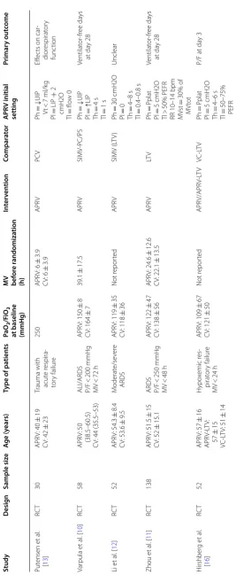
Study selection and study characteristics
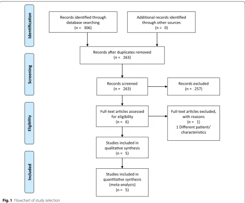
We identified 306 titles. After removal of duplicates, we screened the titles/abstracts of 263 records and assessed the full text of six articles. Finally, we
con-sidered five RCTs for the analysis (Fig. 1) enrolling a
total of 330 patients. Table 1 describes the main
char-acteristics of the selected studies. The studies were
published from 2001 to 2018. All of them are single-centered RCT. Three studies specifically enrolled only patients with ALI/ARDS including overall 248 patients
(75% of the total population considered) [10–12]. One
trial specifically included only traumatic patients with
acute respiratory failure [13]. Two studies defined ALI/
ARDS according to the American-European Consensus
Conference on ARDS of 1994 [14], two studies applied
the Berlin definition of ARDS [15], and one study did
not clearly declare the definition used [12]. One trial
compared three ventilation modalities in three groups of patients: APRV, APRV-LTV and volume-controlled
LTV (VC-LTV) [16]. For the purpose of our study, we
extrapolated data regarding APRV-LTV and VC-LTV. Three studies clearly enrolled patients within the early phase of respiratory failure, counting less than 24 h of mechanical ventilation before randomization
for the majority of patients [11, 13, 16]. Varpula et al.
Records idenfied through database searching
(n = 306)
Screenin
g
Included
Elig
ib
ilit
y
Idenficao
n
Addional records idenfied through other sources
(n = 0)
Records aer duplicates removed (n = 263)
Records screened
(n = 263) Records excluded(n = 257)
Full-text arcles assessed for eligibility
(n = 6)
Full-text arcles excluded, with reasons
(n = 1) 1 Different paents’
characteriscs
Studies included in qualitave synthesis
(n = 5)
Studies included in quantave synthesis
(meta-analysis) (n = 5)
Table
1
C
har
ac
teristics of the studies included in the meta-analy
sis ALI acut e r espir at or y injur y, APR V air w ay pr essur e r elease v en tila tion, ARDS acut e r espir at or y distr ess syndr ome , CV c on ven tional v en tila tion, LIP lo w er inflec tion poin t, LT V lo
w tidal v
olume v en tila tion, MV mechanical ven tila tion, MV st spon taneous minut e v en tila tion, MV tot total minut e v en tila tion, PEFR peak e xpir at or y flo w r at e, Ph high lev
el of pr
essur
e,
Pl
lo
w lev
el of pr
essur e, RCT randomiz ed c on tr olled tr ial , RR respir at or y r at e, Th time f
or high pr
essur e, Tl time f or lo w pr essur e, UIP upper inflec tion poin t, Vt tidal v olume Study Desig n Sample siz e A ge (y ears) Type of pa tien ts Pa O2 /F iO2
at baseline (mmHg) MV bef
or e r andomiza tion (h) In ter ven tion Compar at or APR V initial setting Primar y out come Put ensen et al . [ 13 ] RC T 30 APR V: 40 ± 19 C V: 42 ± 23
Trauma with acut
e r espira -tor y failur e 250 APR V: 6 ± 3.9 C V: 6 ± 3.9 APR V PC V Ph = ↓ UIP Vt < 7 ml/k g Pl = LIP + 2 cmH2O Tl = flo w 0 Eff ec
ts on car
-dior espirat or y func tion Var pula et al . [ 10 ] RC T 58 APR V: 50 (38.5–60.5) C
V: 44 (35.5–53)
ALI/ARDS P/F < 200 mmHg MV < 72 h APR V: 150 ± 8 C V: 164 ± 7 39.1 ± 17.5 APR V SIMV -PC/PS Ph = ↓ UIP Pl = ↑ LIP Th = 4 s Tl = 1 s Ventilat or -fr ee da ys at da y 28 Li et al . [ 12 ] RC T 52 APR V: 54.3 ± 8.4 C V: 53.6 ± 9.5 M oderat e/se ver e ARDS APR V: 119 ± 35 C V: 118 ± 36 Not r epor ted APR V SIMV (L TV ) Ph = 30 cmH2O Pl = 0 Th = 4–8 s Tl = 0.4–0.8 s Unclear Zhou et al . [ 11 ] RC T 138 APR V: 51.5 ± 15 C V: 52 ± 15.1 ARDS P/F < 250 mmHg MV < 48 h APR V: 122 ± 47 C V: 138 ± 56 APR V: 24.6 ± 12.6 C V: 22.1 ± 13.5 APR V LT V Ph = P plat Pl = 5 cmH2O Tl > 50% PEFR RR 10–14 bpm MV st = 30% of MV tot Ventilat or -fr ee da ys at da y 28 H irshber g et al . [ 16 ] RC T 52 APR V: 57 ± 16 APR V-L TV : 57 ± 15 VC-L TV : 51 ± 14 H ypo xemic r es -pirat or y failur e MV < 24 h APR V: 109 ± 67 C V: 121 ± 50 Not r epor ted APR V/APR V-L TV VC-L TV Ph = P plat Pl = 5 cmH2O Th = 4–6 s Tl = 50–75% PEFR
P/F at da
[10] allowed randomization until 72 h from starting of
ventilation. Finally, Li et al. [12] did not report the time
under ventilator before randomization.
About the initial APRV setting, three studies
meas-ured a static pressure–volume (P–V) curve to
iden-tify lower (LIP) and upper (UIP) inflection points and
used these data to set pressures [10, 12, 13]. Putensen
et al. [13] and Varpula et al. [10] set Phigh below UIP
and Plow above LIP allowing to reach zero flow during
the release phase. Li et al. set Plow to 0 but used P–V
curve to set Tlow to obtain an intrinsic end expiratory
pressure (PEEP) of 2 cmH2O above LIP. On the other
hand, the other two studies [11, 16] set Phigh according
to plateau pressure measured during conventional
ven-tilation, with a Plow of 5 cmH2O. Then, Tlow was set to
reach 50–75% of peak expiratory flow rate.
About outcomes, all studies reported length of mechanical ventilation. Two studies reported hospital
mortality [11, 16], and one studied reported ICU
mor-tality [11].
Risk of bias and quality of evidence
The results of the quality assessment of included
stud-ies are given in Table 2. Two studies have a low bias for
random-sequence generation using a computer-based
randomization [11, 16]. Three studies have a low risk of
bias for allocation concealment as they use sealed
enve-lopes [10, 11, 16]. Li et al. [12] did not clearly define
inclusion and exclusion criteria. Due to the nature of the intervention investigated, blindness was not possible for
any studies exposing to a high risk of performance bias. None of the studies included mentioned a blindness of outcome assessment.
Primary outcome
Five RCTs including 313 patients reported
ventilator-free day at day 28 [10–13, 16]. The overall MD between
APRV and conventional ventilation was 6.04 days (95%CI
2.12, 9.96, p = 0.003; I2= 65%, p = 0.02) (Fig. 2). The
sen-sitivity analysis performed excluding two studies [10, 13]
confirmed the result (MD 8.03 days [(95%CI 3.42, 12.65,
p < 0.001; I2= 52%, p = 0.12]). As the studies included
were less than ten, a publication bias analysis was not performed.
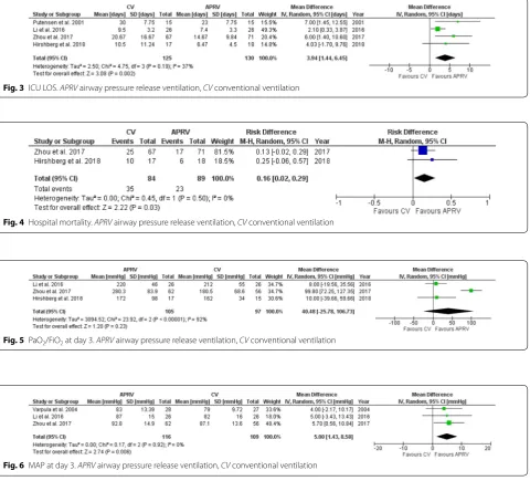
Secondary outcomes
Patients treated with APRV had a lower ICU LOS than patients treated with conventional ventilation (MD
3.94 days [95%CI 1.44, 6.45, p = 0.002; I2= 37%, p = 0.19],
Fig. 3) and a lower hospital mortality (RD 0.16 [95%CI
0.02, 0.29, p = 0.03; I2= 0, p = 0.5], Fig. 4). PaO
2/FiO2
at day 3 was not different between the two groups (MD
40.48 mmHg [95%CI − 25.78, 106.73, p = 0.23; I2= 92%,
p < 0.001] Fig. 5). MAP at day 3 was significantly higher
during APRV (MD 5 mmHg [95%CI 1.43, 8.58, p = 0.006;
I2= 0%, p = 0.92] Fig. 6). Four studies did not show any
differences in vasoactive drugs use and dosage between
groups [10–12, 16]. Only one study demonstrated a lower
dose of norepinephrine and dobutamine in APRV group
[13]. Then, there was no difference regarding the onset of
Table 2 Quality assessment of the included studies
+, low risk of bias; –, high risk of bias; ?, unclear risk
Random-sequence generation
Allocation
concealment Blinding of participants and personnel
Blinding of outcome assessment
Incomplete
outcome data Selective reporting Other bias
Putensen et al. [13] ? ? – ? + ? –
Varpula et al. [10] ? + – ? + ? ?
Li et al. [12] ? ? – ? + ? ?
Zhou et al. [11] + + – ? + + +
Hirshberg et al. [16] + + – ? + + +
pneumothorax under the two ventilation strategies (RR
1.94 [95%CI 0.54, 6.94, p = 0.31; I2= 0%, p = 0.74] Fig. 7).
All studies reported data about sedation. However, the heterogeneity for reporting this outcome was very high and a quantitative analysis was not possible. Two studies have shown no difference in the dosage of
analgo-seda-tive drugs [10, 16]. On the other hand, two studies
dem-onstrated a lower sedation depth for patients treated with APRV than conventional ventilation and a lower dose of
sedative drugs [11, 13]. Finally, Li et al. [12] have recorded
more days without sedative drugs in APRV group. Three studies reported data about spontaneous
breath-ing in APRV patients. Zhou et al. [11] reported that the
amount of spontaneous breathing was about 24% of total
ventilation at day 1, increasing progressively up to about
26% at day 7. Similarly, Putensen et al. [13] showed that
spontaneous breathing accounted for 10% of total ven-tilation at day 1 and raised up to 35% at day 10. Finally,
Hirshberg et al. [16] reported that patients on APRV were
able to develop a spontaneous tidal volume of 6.2 ml/kg over a total volume of 8.3 ml/kg at day 1.
ICU mortality was reported only by Zhou et al. [11]
with no statistical significance between the two groups
(APRV 23.9%, conventional ventilation 37.3%; p = 0.088).
Fig. 3 ICU LOS. APRV airway pressure release ventilation, CV conventional ventilation
Fig. 4 Hospital mortality. APRV airway pressure release ventilation, CV conventional ventilation
Fig. 5 PaO2/FiO2 at day 3. APRV airway pressure release ventilation, CV conventional ventilation
Discussion
This study was the first systematic review and meta-analysis investigating the clinical effects of APRV in adult patients affected by acute hypoxemic respiratory failure compared with conventional ventilation. We demon-strated that APRV was associated with higher ventilator-free days at 28 day, lower ICU LOS and lower hospital mortality, without any major impact on cardiovascular system and risk of barotrauma.
APRV may be considered a general approach of ventila-tion rather than a single unified ventilatory strategy. The conceptual aim of APRV is to maximize and maintain
alveolar recruitment applying the Phigh for the most time
of ventilatory cycle and allowing spontaneous breathing. The purpose is to stabilize and rest the open lung reduc-ing the repetitive alveolar collapse and expansion limitreduc-ing the ventilator-induced lung injury. The rational and the optimal application of APRV has been recently reviewed
by Nieman et al. [17] Some evidences suggest that this
strategy resulted in better oxygenation and respiratory compliance with less applied pressure than conventional
ventilation [11]. Spontaneous breathing allowed during
any phase of ventilatory cycle may have several
advan-tages [18]. A better V/Q match has been demonstrated
with a reduction in dead space and intrapulmonary shunt
[19] due to the increased aeration in dependent lung
regions and the increased lung perfusion [20]. Lower
value of pleural pressure during spontaneous breathing may also be responsible for better hemodynamics param-eters observed during APRV, with increased venous return, increased preload and consequently increased
cardiac output [13]. In our study, although patients
treated with APRV had a statistically significant higher value of MAP than patients on conventional ventila-tion, the difference was not probably clinically significant
(MD 5 mmHg; 95%CI 1.43, 8.58, p = 0.006) and we can
substantially state that APRV has no adverse effect on blood pressure. Although we have considered only MAP in our quantitative analysis, clinical evidences showed that APRV has no negative effects on cardiac index (CI).
Varpula et al. [10] showed no difference between groups,
while other two studies demonstrated higher values of CI
when APRV was used [12, 19]. Then, APRV needs less
sedation level than conventional ventilation, which may explain the lower ICU LOS of these patients than whom heavily sedated to allow total controlled ventilation. Finally, another beneficial effect of spontaneous breath-ing is maintenance of diaphragmatic function.
However, despite the numerous positive aspects of APRV, several concerns have been raised about sponta-neous breathing, especially during the most severe cases
of ARDS [18]. Spontaneous breathing effort is able to
increase trans-pulmonary pressure, and then, increasing
the lung stretch, it increases the risk of lung injury [21,
22]. A great variability in tidal volume during APRV has
been also demonstrated that may be responsible for the development of larger tidal volume exceeding the pro-tective ventilation. Furthermore, edema may be worse because of increased trans-vascular pressure promoting
by negative pleural pressure [23]. Thus, it has been
dem-onstrated that early short term neuromuscular block-ade was associated with lower mortality in patients with
severe ARDS [24].
Taking this concern in mind, APRV may probably be considered in case of mild/moderate ARDS, when the potential benefits may overcome the potential risks.
This meta-analysis has several limitations. We included a low number of studies, and the overall quality level of them is low. This is due to the lack of blindness and the lack in reporting the management of other potential bias
(Table 2). Only two studies have a low risk of bias [11, 16].
Moreover, the heterogeneity for the primary outcome (ventilator-free days at 28 day) was quite high, probably because of the methodological differences between trials.
Two studies [10, 19] enrolled patients before the
publica-tion of ARDSNetwork trial [3], showing the benefit of low
tidal volume ventilation. Thus, for these studies, a higher tidal volume than protective ventilation was permitted. Excluding them in the sensitivity analysis, the heteroge-neity decreased without any significant impact on overall effect. Patient’s selection may also explain some
hetero-geneity. Stratification based on PaO2/FiO2 was not
be considered all together. Moreover, the different time of ventilation before application of APRV may affect the outcome. Finally, another aspect to consider is the differ-ent initial setting of APRV. Despite it was described for the first time in 1987, a standard setting is not reached yet and some variability in its used is described in the literature. The lower hospital mortality of APRV group than patients on conventional ventilation shown by our study must be interpreted with caution. In fact, only two trials were included in this analysis with an overall popu-lation of only 173 patients.
Conclusion
This study showed a higher number of ventilator-free days at 28 day and a lower hospital mortality in acute hypoxemic patients treated with APRV than conventional ventilation, without any negative hemodynamic impact or higher risk of barotrauma. However, these results need to be interpreted with caution because of the low-quality evidence supporting them and the moderate heterogene-ity found. Thus, based on these evidences, it is difficult to draw a clinical message about APRV in this specific set-ting and other well-designed RCTs need to be conducted to confirm our findings.
Abbreviations
ALI: acute lung injury; APRV: airway pressure release ventilation; ARDS: acute respiratory distress syndrome; ICU: intensive care unit; LIP: lower inflection points; LOS: length of stay; LTV: low tidal volume; MD: mean differences; PEEP:
positive end expiratory pressure; Phigh: pressure high; Plow: pressure low; P–V:
pressure–volume curve; RCTs: randomized controlled trials; RD: risk difference;
RR: risk ratio; SD: standard deviation; Thigh: time high; Tlow: time low; UIP: upper
inflection points; V/Q: ventilation/perfusion matching.
Authors’ contributions
AC wrote the manuscript. AC, ED and EA performed the literature review. AC performed the statistical analysis. RD, CS, SP, SF and AD revised the text. All authors read and approved the final manuscript.
Author details
1 Anesthesia and Intensive Care Unit, Azienda Ospedaliero Universitaria
Ospedali Riuniti, Ancona, Italy. 2 Anesthesia and Intensive Care Unit, Università
Politecnica delle Marche, Ancona, Italy.
Acknowledgements Not applicable.
Competing interests
The authors declare that they have no competing interests.
Availability of data and materials
All data generated or analyzed during this study are included in this published article.
Consent for publication Not applicable.
Ethics approval and consent to participate Not applicable.
Funding None.
Publisher’s Note
Springer Nature remains neutral with regard to jurisdictional claims in pub-lished maps and institutional affiliations.
Received: 17 January 2019 Accepted: 30 March 2019
References
1. Bellani G, Laffey JG, Pham T, Fan E, Brochard L, Esteban A, et al. Epidemiol-ogy, patterns of care, and mortality for patients with acute respira-tory distress syndrome in intensive care units in 50 countries. JAMA. 2016;315:788–800.
2. Fan E, Del Sorbo L, Goligher EC, Hodgson CL, Munshi L, Walkey AJ, et al. An official American Thoracic Society/European Society of Intensive Care Medicine/Society of Critical Care Medicine clinical practice guideline: mechanical ventilation in adult patients with acute respiratory distress syndrome. Am J Respir Crit Care Med. 2017;195:1253–63.
3. Acute Respiratory Distress Syndrome Network, Brower RG, Matthay MA, Morris A, Schoenfeld D, Thompson BT, et al. Ventilation with lower tidal volumes as compared with traditional tidal volumes for acute lung injury and the acute respiratory distress syndrome. N Engl J Med. 2000;342:1301–8.
4. Stock MC, Downs JB, Frolicher DA. Airway pressure release ventilation. Crit Care Med. 1987;15:462–6.
5. Liberati A, Altman DG, Tetzlaff J, Mulrow C, Gøtzsche PC, Ioannidis JPA, et al. The PRISMA statement for reporting systematic reviews and meta-analyses of studies that evaluate health care interventions: explanation and elaboration. Ann Intern Med. 2009;151:W65–94.
6. Higgins J, Green S. Cochrane handbook for systematic reviews of inter-ventions. 5.1.0. 2011. www.handb ook.cochr ane.org. Accessed Dec 2018. 7. Wan X, Wang W, Liu J, Tong T. Estimating the sample mean and standard
deviation from the sample size, median, range and/or interquartile range. BMC Med Res Methodol. 2014;14:135.
8. Higgins JPT, Thompson SG. Quantifying heterogeneity in a meta-analysis. Stat Med. 2002;21:1539–58.
9. Egger M, Smith GD, Schneider M, Minder C. Bias in meta-analysis detected by a simple, graphical test. BMJ. 1997;315:629–34.
10. Varpula T, Valta P, Niemi R, Takkunen O, Hynynen M, Pettilä V. Airway pres-sure release ventilation as a primary ventilatory mode in acute respiratory distress syndrome. Acta Anaesthesiol Scand. 2004;48:722–31.
11. Zhou Y, Jin X, Lv Y, Wang P, Yang Y, Liang G, et al. Early application of air-way pressure release ventilation may reduce the duration of mechanical ventilation in acute respiratory distress syndrome. Intensive Care Med. 2017;43:1648–59.
12. Li J, Li N, Han G, Pan C, Zhang Y, Shi X, et al. Clinical research about airway pressure release ventilation for moderate to severe acute respiratory distress syndrome. Eur Rev Med Pharmacol Sci. 2016;20:2634–41. 13. Putensen C, Zech S, Wrigge H, Zinserling J, Stüber F, Von Spiegel T,
et al. Long-term effects of spontaneous breathing during ventilatory support in patients with acute lung injury. Am J Respir Crit Care Med. 2001;164:43–9.
14. Bernard GR, Artigas A, Brigham KL, Carlet J, Falke K, Hudson L, et al. The American-European Consensus Conference on ARDS. Definitions, mecha-nisms, relevant outcomes, and clinical trial coordination. Am J Respir Crit Care Med. 1994;149:818–24.
15. ARDS Definition Task Force. Acute respiratory distress syndrome: the Berlin definition. JAMA. 2012;307:2526–33.
16. Hirshberg EL, Lanspa MJ, Peterson J, Carpenter L, Wilson EL, Brown SM, et al. Randomized feasibility trial of a low tidal volume-airway pressure release ventilation protocol compared with traditional airway pressure release ventilation and volume control ventilation protocols. Crit Care Med. 2018;46:1943–52.
18. Yoshida T, Fujino Y, Amato MBP, Kavanagh BP. Fifty years of research in ARDS. Spontaneous breathing during mechanical ventilation. Risks, mechanisms, and management. Am J Respir Crit Care Med. 2017;195:985–92.
19. Putensen C, Mutz NJ, Putensen-Himmer G, Zinserling J. Spontaneous breathing during ventilatory support improves ventilation–perfusion distributions in patients with acute respiratory distress syndrome. Am J Respir Crit Care Med. 1999;159:1241–8.
20. Neumann P, Wrigge H, Zinserling J, Hinz J, Maripuu E, Andersson LG, et al. Spontaneous breathing affects the spatial ventilation and perfu-sion distribution during mechanical ventilatory support. Crit Care Med. 2005;33:1090–5.
21. Yoshida T, Uchiyama A, Matsuura N, Mashimo T, Fujino Y. Spontaneous breathing during lung-protective ventilation in an experimental acute
lung injury model: high transpulmonary pressure associated with strong spontaneous breathing effort may worsen lung injury. Crit Care Med. 2012;40:1578–85.
22. Yoshida T, Uchiyama A, Matsuura N, Mashimo T, Fujino Y. The comparison of spontaneous breathing and muscle paralysis in two different severities of experimental lung injury. Crit Care Med. 2013;41:536–45.
23. Kallet RH, Alonso JA, Luce JM, Matthay MA. Exacerbation of acute pul-monary edema during assisted mechanical ventilation using a low-tidal volume, lung-protective ventilator strategy. Chest. 1999;116:1826–32. 24. Papazian L, Forel J-M, Gacouin A, Penot-Ragon C, Perrin G, Loundou A,