LETTER
Patternable particle microarray utilizing
controllable particle delivery
Sanghyun Lee
1†, Hojin Kim
2†, Wonhyung Lee
1and Joonwon Kim
1*Abstract
In this study, we demonstrate an on-demand delivering and sequential arraying of single microparticles utilizing multiple pneumatic pressure-driven elastomer valves and a deterministic particle arraying mechanism. Two types of separate microfluidic devices are combined: (i) a particle transfer device and (ii) a particle arraying device, to construct a desired particle pattern. The elastomer valve integrated in the transfer device acts as a removable particle trap that enables the trapping and on-demand releasing of a particle depending on the application of the pneumatic pressure. The arraying device is composed of highly packed particle trapping sites to deterministically array incoming particles that are released and transferred from a transfer device. Repeating the “trapping-transfer-and-array” sequence can construct an array of different types of particles in a certain pattern. One-dimensional linear and two-dimensional planar microparticle-based patterns were demonstrated using bare and red-fluorescent microparticles.
Keywords: Microfluidics, Microparticle, Pneumatic microvalve, Controllable particle delivery, Patterning
© The Author(s) 2019. This article is distributed under the terms of the Creative Commons Attribution 4.0 International License (http://creat iveco mmons .org/licen ses/by/4.0/), which permits unrestricted use, distribution, and reproduction in any medium, provided you give appropriate credit to the original author(s) and the source, provide a link to the Creative Commons license, and indicate if changes were made.
Introduction
Recently, particle-incorporated microfluidic platforms have emerged as effective tools to conduct various anal-yses in biological and chemical research fields [1–5]. When compared with flat substrates or supports, micro-particles can serve as a mobile substrate and provide multiple functionalities including huge analytical surface and the capability of effective mixing, sorting, and trans-porting of molecules of interest [6]. For most cases in these platforms, particles are trapped in an array format within microchannel networks with embedded physical barrier structures (e.g., weirs or micropillars) or external active forces (e.g., electric forces). Depending on the tar-get applications, surfaces of particles can be functional-ized (e.g., DNA or antibody conjugation). To construct the particle array, the particles (i.e., array elements) are introduced from an off-chip environment and subse-quently arrayed within microfluidic devices [7–11].
Generally, different types of functionalized particles are arrayed randomly to perform analyses; thus, the encod-ing and decodencod-ing of individual particles must be per-formed for their identification and readout of the results after their reactions (i.e., mix-and-match). Although several strategies for particle encoding exist [12–14], a high-resolution imaging system is typically required to identify individual particles based on their encod-ing method. Meanwhile, a position-based signal readout method facilitates an easy readout of the results such as the wellplate-based enzyme-linked immunosorbent assay (ELISA). Thus, the advantages of particle-based analysis and position-based easy readout can be combined in an integrated system.
Several studies have been carried out to generate spe-cific particle patterns using microfluidic platforms. A 1-D microfluidic bead array was constructed by depos-iting particles one by one using vacuum tweezers [15, 16]. This approach can form a desired particle pattern in predefined channels, but is a cumbersome process. As an alternative approach, an additional microbead loading channel was integrated to deposit the func-tionalized microparticles into predetermined positions [17, 18]. This requires additional components and has limitation to enable a single particle level patterning.
Open Access
*Correspondence: [email protected]
†Sanghyun Lee and Hojin Kim contributed equally to this work 1 Department of Mechanical Engineering, Pohang University of Science and Technology (POSTECH), 77 Cheongam-Ro, Nam-Gu, Pohang, Gyeongbuk 37673, South Korea
Without using physical trap structures, dynamic parti-cle patterning was demonstrated using standing surface acoustic waves (SSAWs) [19, 20]. However, complicated device fabrication and system setup are required.
In this regard, we present a method to construct a particle array in a desired pattern (i.e., patternable particle array) instead of a random pattern, to enable a position-based easy readout using different types of particles with a single-particle-level resolution. Our strategy combines two types of separate microflu-idic devices that offer different functions: (i) a particle transfer device and (ii) a particle arraying device. Pneu-matic microvalve-based removable trap techniques and deterministic particle arraying techniques are used in the transfer and arraying devices, respectively. A trans-fer device can selectively transtrans-fer the particles of inter-est; these can be patterned in the arraying device in a desired pattern. Many studies have demonstrated parti-cle manipulation (e.g., trapping, releasing, and pairing) using pneumatic pressure-driven elastomer microv-alve structures [21–25]. Depending on the state of the valve (i.e., either “on” or “off”), particles can be either trapped or released. An arraying device can capture the introduced particles deterministically (i.e., from the transfer device) based on flow fractionation [10, 26]. As a proof-of-concept of our method, we demonstrated particle patterns of one-dimensional (1D) linear (e.g., “dot-dash” line) and two-dimensional (2D) planar (e.g.,
Braille numbers) using bare and red-fluorescent poly-styrene particles.
Materials and methods Device design and operation
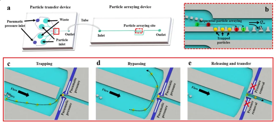
To demonstrate on-demand single particle delivery and the sequential arraying of particles, two types of micro-fluidic devices are integrated: (i) a particle transfer device and (ii) a particle arraying device (Fig. 1a). The outlet of the transfer device and the inlet of the arraying device are connected using a tube for particle transfer. The particle transfer device is composed of pneumatic pressure inlets, particle inlets, wastes, and an outlet. A pneumatic chan-nel is used to operate the elastomeric pneumatic microv-alve. The particle arraying device is composed of an inlet, a particle arraying site, and an outlet.
In the transfer device, particles are introduced through the particle inlet and abundant particles are washed out toward waste. The particle transfer function can be ena-bled by operating the elastomeric pneumatic microvalve that acts as a removable particle trap depending on the applied pneumatic pressure [25]. When a positive pneu-matic pressure is applied through the pneupneu-matic channel, a thin membrane (i.e., channel wall) is deflected and as a result, a narrowed branch pocket that can trap a single particle is formed (Fig. 1c). The pillar structure can guide the particles to migrate close to the channel wall; this facilitates particle trapping [27]. Once a single particle
is trapped, subsequent particles will migrate toward the waste without any additional trapping (Fig. 1d). The trapped particles can be released and transferred to the arraying device in an on-demand manner when the applied pneumatic pressure is removed (Fig. 1e).
The target particle can be transferred from the trans-fer device to the arraying device through the tube con-nection. Based on the flow fractionation at each vacant trap sites (i.e., flow is distributed into main flow Qm and trapping flow Qt), the transferred particle is trapped deterministically at each vacant trap site (sequentially from upstream to downstream) of the particle arraying device (Fig. 1b) [26]. By repeating the “trapping-transfer-and-array” sequence, we can construct the particle array in a controllable manner (i.e., patternable particle array). We can select specific particles to be transferred using different types of particle suspensions at different parti-cle inlets. The numbers of partiparti-cle inlets and pneumatic pressure inlets can be varied based on the types of parti-cles that are arrayed.
Device fabrication
Both microfluidic devices were fabricated using polydi-methylsiloxane (PDMS) (Sylgard 184, Dow Corning Inc.) by standard soft lithography [28]. Negative tone photore-sist (KMPR 1025, MicroChem, Inc.) was used to prepare the master molds. It was deposited onto two four-inch silicon wafers with the same thickness (32 µm) by spin coating and soft baking (100 °C for 15 min). Ultraviolet (UV) exposure through a photomask and post-exposure baking (100 °C for 3 min) and development (SU-8 devel-oper, MicroChem, Inc.) were proceeded to define the patterns.
Two different PDMS prepolymer mixtures (i.e., differ-ent PDMS monomer base to crosslinker ratio) were used to prepare PDMS replicas using the master molds: (i) 12:1 w/w ratio mixture for the particle transfer device and (ii) a 10:1 w/w ratio mixture for the particle arraying device. Regardless of the PDMS mixture ratio, PDMS replicas were prepared by the same procedure. The PDMS mix-ture was poured onto the master mold and degassed; this was then thermally cured at 100 °C for 20 min. The cured PDMS replica was peeled off from the master mold and holes were punched using a disposable biopsy punch. The holes were rinsed with isopropanol. The PDMS replica and glass substrate were irreversibly bonded by contact after air plasma treatment (CUTE-MP, FemtoScience). The prepared devices were stored at room temperature for 24 h for a reliable device operation.
Experimental setup
For system operation, separate custom-built pneumatic pressure supply systems are used: (i) positive pressure
supply system to operate the elastomeric valve and infuse particles and (ii) a negative pressure supply system to facilitate particle transfer and arraying. A positive pres-sure application system (connected with the pneumatic/ particle inlet of the transfer device) consists of a pressure pump (i.e., positive pressure generation), solenoid valves, a pressure regulator, and a pressure monitor. A negative pressure application system (connected with the outlet of the arraying device) consists of a pressure pump (i.e., negative pressure generation), pressure regulator, and pressure monitor. Microscopic images were acquired using an inverted microscope (IX 73, Olympus) with a charge-coupled device (CCD) camera (DP80, Olym-pus). For fluorescence detection, a SOLA light engine (SM 365, lumencor) and filter cube (U-FGWA, Olym-pus) were used as the light source and fluorescence filter, respectively.
Results and discussion 1D linear patterning of particles
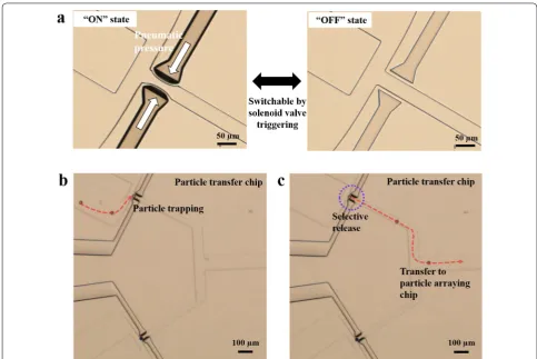
On-demand single particle release and delivery was dem-onstrated with elastomeric microvalve operation. The valve deflection state (i.e., ON or OFF) can be controlled using the applied pneumatic pressure and solenoid valve operation (Fig. 2a). When the valve state is “ON” (i.e., the wall of the valve is deflected under the application of pneumatic pressure), a single particle can be trapped at the narrow channel created by valve deflection (Fig. 2b). The average diameter of the particle used in this test was approximately 25 μm. Particle suspensions were intro-duced into the particle transfer chip under a pneumatic pressure of 5 kPa. To trap a single particle, we applied a pneumatic pressure of 200 kPa to deflect the wall of the microvalve. After particle trapping, subsequent particles passed the valve and migrated toward the waste chan-nel owing to a sudden increase in hydraulic resistance of the channel through the microvalve. When the applied pneumatic pressure was removed by triggering a solenoid valve (switching time of 100 ms) in an on-demand man-ner, the trapped single particle was selectively released and migrated to the outlet of the transfer device (Fig. 2c). This particle was subsequently transferred to the parti-cle arraying device under an applied vacuum pressure of − 15 kPa at the outlet of the arraying device.
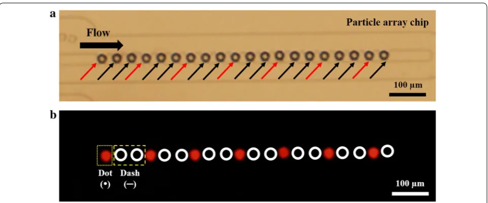
particle and two times bare particles) using individual solenoid valve triggering, we can construct a line pattern, as shown in Fig. 3a. The fluorescence image reveals that the particle line pattern is constructed as a “dot-dash” line (Fig. 3b).
2D planar patterning of particles
The multiple row patterning of 1D particle lines can yield 2D planar particle patterns. Selective and on-demand particle transfer and sequential arraying of particles were used similar to the 1D linear particle patterning method described above. Inspired by Braille patterns (Fig. 4a), which are used by the visually impaired, we constructed 2D particle patterns to display Braille numbers using con-trollable transfer and arraying of bare and red-fluorescent particles (Fig. 4b). Fluorescence detection revealed that several Braille number patterns (1, 2, 3, 4, and 5) were displayed by the proper combination of particles (Fig. 4c). Schematic of generating Braille-inspired particle patterns
in the particle arraying device are described in Additional file 1: S1.
Conclusion
In this study, we demonstrated a patternable particle microarray by trapping, releasing, transferring, and sequentially arraying two types of particles. A parti-cle transfer device and a partiparti-cle arraying device were combined to construct desired particle patterns. In the particle trapping device, the integration of a pneu-matic pressure-driven elastomer microvalve enabled single-particle trapping and selective, on-demand releasing of particles of interest. The released parti-cle was transferred to the arraying device and subse-quently trapped at vacant sites sequentially from the upstream direction. 1D linear (e.g., “dot-dash” line) and 2D planar (e.g., Braille numbers) particle patterns were constructed in a controllable manner by repeat-ing the “trapprepeat-ing-transfer-and-array” sequence. Even though we demonstrated particle patterns using two
types of particles (i.e., bare and red-fluorescent parti-cles), more complex particle patterns can be achieved with different types (e.g., > three types) of particles by operating additional particle inlets. The programmable operation of multiple pneumatic valves can facilitate the construction of desired particle patterns composed of different types of particles. We believe that the par-ticle patterning technique presented herein has poten-tial for various applications such as microarrays, drug screening, and cellular studies. Furthermore, proper integration of functionalized particles (e.g., DNA or
protein-conjugated particles) can facilitate a pattern-based easy-readout multiplex immunoassay platform.
Additional file
Additional file 1. Additional information is available about schematics of generating Braille-inspired patterns.
Acknowledgements Not applicable.
Fig. 3 1D linear pattern using two types of particles by repeating the “trapping-transfer-and-array” sequence. a Bright-field microscopic image of arrayed particles. Bare (indicated with black arrows) and red-fluorescent particles (indicated with red arrows) are deterministically trapped in an arraying device. b Fluorescent image revealing the linear pattern of the “dot-dash” line
Authors’ contributions
SL and HK performed the experiments, analyzed the data and wrote the manuscript. WL carried out device fabrication. JK supervised the research and reviewed the manuscript. All authors read and approved the final manuscript.
Funding
This research was supported by Basic Science Research Program through the National Research Foundation of Korea (NRF) funded by the Ministry of Sci-ence and ICT (No. NRF-2017R1A2B4003328).
Availability of data and materials
The datasets supporting the conclusions of this article are included within the article and its additional file.
Competing interests
The authors declare that they have no competing interests.
Author details
1 Department of Mechanical Engineering, Pohang University of Science and Technology (POSTECH), 77 Cheongam-Ro, Nam-Gu, Pohang, Gyeong-buk 37673, South Korea. 2 BioMEMS Lab, RadianQbio, 53 Gasan digital 2-ro, Geumcheon-gu, Seoul 08588, South Korea.
Received: 20 May 2019 Accepted: 18 July 2019
References
1. Birtwell S, Morgan H (2009) Microparticle encoding technologies for high-throughput multiplexed suspension assays. Integr Biol 1:345–362 2. Liu L, Wu S, Jing F, Zhou H, Jia C, Li G, Cong H, Jin Q, Zhao J (2016)
Bead-based microarray immunoassay for lung cancer biomarkers using quantum dots as labels. Biosens Bioelectron 80:300–306
3. Kim H, Lee S, Lee W, Kim J (2017) High-density microfluidic particle-clus-ter-array device for parallel and dynamic study of interaction between engineered particles. Adv Mater 29:1701351
4. Han SW, Jang E, Koh W-G (2015) Microfluidic-based multiplex immunoas-say system integrated with an array of QD-encoded microbeads. Sens Actuators B Chem 209:242–251
5. Hung L-Y, Huang T-B, Tsai Y-C, Yeh C-S, Lei H-Y, Lee G-B (2013) A micro-fluidic immunomagnetic bead-based system for the rapid detection of influenza infections: from purified virus particles to clinical specimens. Biomed Microdevice 15:539–551
6. Derveaux S, Stubbe B, Braeckmans K, Roelant C, Sato K, Demeester J, De Smedt S (2008) Synergism between particle-based multiplexing and microfluidics technologies may bring diagnostics closer to the patient. Anal Bioanal Chem 391:2453
7. Carlo DD, Wu LY, Lee LP (2006) Dynamic single cell culture array. Lab Chip 6:1445–1449
8. Jung Y, Hyun J-C, Choi J, Atajanov A, Yang S (2017) Manipulation of cells’ position across a microfluidic channel using a series of continuously vary-ing herrvary-ingbone structures. Micro Nano Syst Lett 5:6
9. Tan W-H, Takeuchi S (2007) A trap-and-release integrated microflu-idic system for dynamic microarray applications. Proc Natl Acad Sci 104:1146–1151
10. Chung K, Rivet CA, Kemp ML, Lu H (2011) Imaging single-cell signaling dynamics with a deterministic high-density single-cell trap array. Anal Chem 83:7044–7052
11. Teshima T, Ishihara H, Iwai K, Adachi A, Takeuchi S (2010) A dynamic microarray device for paired bead-based analysis. Lab Chip 10:2443–2448 12. Burger R, Reith P, Kijanka G, Akujobi V, Abgrall P, Ducrée J (2012)
Array-based capture, distribution, counting and multiplexed assaying of beads on a centrifugal microfluidic platform. Lab on a Chip 12:1289–1295 13. Lee H, Kim J, Kim H, Kim J, Kwon S (2010) Colour-barcoded magnetic
microparticles for multiplexed bioassays. Nat Mater 9:745
14. Appleyard DC, Chapin SC, Srinivas RL, Doyle PS (2011) Bar-coded hydro-gel microparticles for protein detection: synthesis, assay and scanning. Nat Protoc 6:1761
15. Zhang H, Yang X, Wang K, Tan W, Li H, Zuo X, Wen J (2008) On-chip oligonucleotide ligation assay using one-dimensional microfluidic beads array for the detection of low-abundant DNA point mutations. Biosens Bioelectron 23:945–951
16. Zhou L et al (2006) Quantitative intracellular molecular profiling using a one-dimensional flow system. Anal Chem 78:6246–6251
17. Zhang H, Fu X, Liu L, Zhu Z, Yang K (2012) Microfluidic bead-based enzymatic primer extension for single-nucleotide discrimination using quantum dots as labels. Anal Biochem 426:30–39
18. Zhang H, Liu L, Fu X, Zhu Z (2013) Microfluidic beads-based immunosen-sor for sensitive detection of cancer biomarker proteins using multi-enzyme-nanoparticle amplification and quantum dotslabels. Biosens Bioelectron 42:23–30
19. Shi J, Ahmed D, Mao X, Lin S-CS, Lawit A, Huang TJ (2009) Acoustic twee-zers: patterning cells and microparticles using standing surface acoustic waves (SSAW). Lab on a Chip 9:2890–2895
20. Ding X, Shi J, Lin S-CS, Yazdi S, Kiraly B, Huang TJ (2012) Tunable pattern-ing of microparticles and cells uspattern-ing standpattern-ing surface acoustic waves. Lab Chip 12:2491–2497
21. Tonooka T, Teshima T, Takeuchi S (2013) Clustering triple microbeads in a dynamic microarray for timing-controllable bead-based reactions. Microfluidics Nanofluidics 14:1039–1048
22. Ma C et al (2011) A clinical microchip for evaluation of single immune cells reveals high functional heterogeneity in phenotypically similar T cells. Nat Med 17:738
23. Kim HS, Devarenne TP, Han A (2015) A high-throughput microfluidic single-cell screening platform capable of selective cell extraction. Lab Chip 15:2467–2475
24. Kim H, Kim J (2014) A microfluidic-based dynamic microarray system with single-layer pneumatic valves for immobilization and selective retrieval of single microbeads. Microfluidics Nanofluidics 16:623–633
25. Kim H, Lee S, Kim J (2012) Hydrodynamic trap-and-release of single particles using dual-function elastomeric valves: design, fabrication, and characterization. Microfluidics Nanofluidics 13:835–844
26. Sochol RD, Dueck ME, Li S, Lee LP, Lin L (2012) Hydrodynamic reset-tability for a microfluidic particulate-based arraying system. Lab Chip 12:5051–5056
27. Iwai K, Tan W-H, Ishihara H, Takeuchi S (2011) A resettable dynamic micro-array device. Biomed Microdev 13:1089–1094
28. Xia Y, Whitesides GM (1998) Soft lithography. Angewandte Chem Int Edn 37:550–575
Publisher’s Note