Open Access
Review
Brachydactyly
Samia A Temtamy* and Mona S Aglan
Address: Department of Clinical Genetics, Human Genetics and Genome Research Division, National Research Centre (NRC), El-Buhouth St., Dokki, 12311, Cairo, Egypt
Email: Samia A Temtamy* - [email protected]; Mona S Aglan - [email protected] * Corresponding author
Abstract
Brachydactyly ("short digits") is a general term that refers to disproportionately short fingers and toes, and forms part of the group of limb malformations characterized by bone dysostosis. The various types of isolated brachydactyly are rare, except for types A3 and D. Brachydactyly can occur either as an isolated malformation or as a part of a complex malformation syndrome. To date, many different forms of brachydactyly have been identified. Some forms also result in short stature. In isolated brachydactyly, subtle changes elsewhere may be present. Brachydactyly may also be accompanied by other hand malformations, such as syndactyly, polydactyly, reduction defects, or symphalangism.
For the majority of isolated brachydactylies and some syndromic forms of brachydactyly, the causative gene defect has been identified. In isolated brachydactyly, the inheritance is mostly autosomal dominant with variable expressivity and penetrtance.
Diagnosis is clinical, anthropometric and radiological. Prenatal diagnosis is usually not indicated for isolated forms of brachydactyly, but may be appropriate in syndromic forms. Molecular studies of chorionic villus samples at 11 weeks of gestation and by amniocentesis after the 14th week of
gestation can provide antenatal diagnosis if the causative mutation in the family is known. The nature of genetic counseling depends both on the pattern of inheritance of the type of brachydactyly present in the family and on the presence or absence of accompanying symptoms.
There is no specific management or treatment that is applicable to all forms of brachydactyly. Plastic surgery is only indicated if the brachydactyly affects hand function or for cosmetic reasons, but is typically not needed. Physical therapy and ergotherapy may ameliorate hand function. Prognosis for the brachydactylies is strongly dependent on the nature of the brachydactyly, and may vary from excellent to severely influencing hand function. If brachydactyly forms part of a syndromic entity, prognosis often depends on the nature of the associated anomalies.
Background
DefinitionThe term brachydactyly is derived from the ancient Greek (brachy-: short; dactylos: digit). It indicates shortening of digits due to abnormal development of phalanges,
meta-carpals, or both. Brachydactyly is one of the ten categories of hand malformations classified by Temtamy & McKu-sick [1] in their original work on the genetics of hand mal-formations. In the latest International nosology and classification of genetic skeletal dysplasias, brachydactyly
Published: 13 June 2008
Orphanet Journal of Rare Diseases 2008, 3:15 doi:10.1186/1750-1172-3-15
Received: 4 April 2008 Accepted: 13 June 2008
This article is available from: http://www.ojrd.com/content/3/1/15
© 2008 Temtamy and Aglan; licensee BioMed Central Ltd.
was included as one of the dysostosis groups affecting the limbs [2]. Dysostoses refer to abnormalities of individual bones, either in isolation or in combination with various abnormally formed bones. They are usually static and arise during blastogenesis (1st 8 weeks of embryonic life)
[3], thus they differ from osteochondrodysplasias that usually present at a later stage of development, typically affect the skeleton in general, and may continue to evolve as a result of continuous gene functioning throughout life [3].
Epidemiological data
The various types of isolated brachydactyly are rare, except for types A3 and D, which are common, prevalence being around 2% [1].
A search of Online Mendelian Inheritance in Man [4] using brachydactyly as key term provided 232 entries (iso-lated forms; syndromes with brachydactyly; skeletal dys-plasias). A similar search using brachydactyly as search term in the London Medical Databases (LMD) [5] yielded 386 entities. The incidence and prevalence of these entries varied considerably.
Classification
As in all congenital anomalies, brachydactyly can occur either as an isolated malformation or as a part of a com-plex malformation syndrome. In isolated brachydactyly (Table 1), subtle changes elsewhere may still be present. Brachydactyly may also go along with other hand malfor-mations, such as syndactyly, polydactyly, reduction defects, or symphalangism.
One of the most commonly used classifications of brach-ydactyly based on anatomic grounds was provided by Bell [6] and further elaborated by Temtamy & McKusick [1]. This system of classification has the advantage of being malleable to accommodate addition of any other recently
discovered forms of brachydactyly mentioned in the liter-ature. No other classifications of brachydactyly, apart from a few appearing in the Russian and Romanian liter-ature, are known to us. Therefore, we follow here the def-initions of the various types of brachydactyly as described by Temtamy and McKusick [1]. We will focus on isolated brachydactyly, although selected syndromic forms will also be discussed.
General considerations of brachydactylies • Antenatal diagnosis and genetic counseling
Prenatal diagnosis is usually not indicated for isolated forms of brachydactyly, but may be appropriate in syndro-mic forms. Short phalanges may not be clearly evident by fetal ultrasonography early in development, and will only be visible later on, if expressed. Molecular studies of cho-rionic villus sampling at 11 weeks of gestation and by amniocentesis after the 14th week of gestation can provide
antenatal diagnosis if the causative mutation in the family is known.
• Genetic counseling
The nature of genetic counseling depends both on the pat-tern of inheritance of the type of brachydactyly present in the family and on the presence or absence of accompany-ing symptoms.
If the brachydactyly follows an autosomal dominant pat-tern of inheritance, the chance of recurrence in offspring of affected individuals is 50% regardless of sex. Variability in severity and incomplete penetrance are common in sev-eral types of brachydactyly.
If the brachydactyly follows an autosomal recessive pat-tern of inheritance, the risk of recurrence in the sib of an affected case is 25%. Although variable symptoms may still be present in affected sibs, variability is usually smaller in autosomal recessive forms.
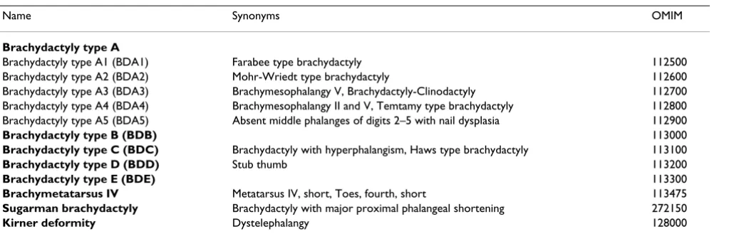
Table 1: Types of isolated brachydactyly
Name Synonyms OMIM
Brachydactyly type A
Brachydactyly type A1 (BDA1) Farabee type brachydactyly 112500 Brachydactyly type A2 (BDA2) Mohr-Wriedt type brachydactyly 112600 Brachydactyly type A3 (BDA3) Brachymesophalangy V, Brachydactyly-Clinodactyly 112700 Brachydactyly type A4 (BDA4) Brachymesophalangy II and V, Temtamy type brachydactyly 112800 Brachydactyly type A5 (BDA5) Absent middle phalanges of digits 2–5 with nail dysplasia 112900
Brachydactyly type B (BDB) 113000
Brachydactyly type C (BDC) Brachydactyly with hyperphalangism, Haws type brachydactyly 113100
Brachydactyly type D (BDD) Stub thumb 113200
Brachydactyly type E (BDE) 113300
Brachymetatarsus IV Metatarsus IV, short, Toes, fourth, short 113475
Sugarman brachydactyly Brachydactyly with major proximal phalangeal shortening 272150
• Management including treatment
There is no general, specific management or treatment that is applicable to all forms of brachydactyly. Plastic sur-gery is only indicated if the brachydactyly affects hand function or for cosmetic reasons, but is typically not needed. Physical therapy and ergotherapy may ameliorate hand function.
• Prognosis
Prognosis for the brachydactylies is strongly dependent on the nature of the brachydactyly, and may vary from excellent to severely influencing hand function. If brachy-dactyly forms part of a syndromic entity, prognosis often depends on the nature of the associated anomalies.
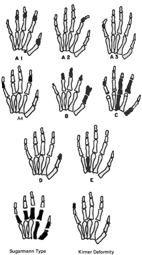
Types of isolated brachydactyly
Figure 1 illustrates the changes in the main types of lated brachydactyly. Table 2 shows the genomics of iso-lated brachydactyly.
1. Brachydactyly type A (BDA)
shortening is confined to middle phalanges. Depending on the affected digits, BDA is subdivided into:
• Brachydactyly type A1 (BDA1)
(OMIM: 112500) (syn: FARABEE TYPE
BRACHYDAC-TYLY)
Clinical description
Early in 1900's, Farabee and Drinkwater described a number of families with BDA1. In BDA1, middle phalanges of all digits are variably short or rudimentary and are occasionally fused with terminal phalanges. The proximal phalanges of the thumbs and big toes are short.
According to the degree of shortening, Drinkwater [7-9] identified two varieties, severe and minor brachydactyly. Many reports described families in which both varieties occurred in various affected members. In the severe vari-ety, the fingers are about half of the normal length, the middle phalanges of all digits are either absent or very
A diagram showing main types of isolated brachydactyly
Figure 1
A diagram showing main types of isolated brachydac-tyly. Modified from Temtamy & McKusick [1].
Table 2: Genomics of isolated brachydactyly
Type OMIM Gene Name Gene Locus Reference
BDA1 112500 IHH BDA1B 2q35-q35 5p13.3-p13.2 Gao et al. [12] Kirkpatrick et al. [14] BDA2 112600 BMPR1B GDF5 4q21-q20 20q11.2 Lehmann et al. [17]
Kjaer et al. [18] BDA4 112800 HOXD13 2q31-q32 Zhao et al. [26] BDB 113000 ROR2 NOG 9q22 4q23-q24 Schwabe et al. [30]
hypoplastic and might be fused with terminal phalanges (terminal symphalangism). In the mild variety of brachy-dactyly, the hypoplasia of the middle phalanges is less severe with more affection of the index and little fingers. Distal symphalangism, when present, is confined to the little finger. In both varieties of brachydactyly, the affected individuals are significantly shorter than their normal sibs. Armour et al. [10] reported a family with mild brach-ydactyly type A1 that, except for short stature, was not associated with additional clinical features. The authors illustrated the usefulness of the metacarpophalangeal pro-files.
Epidemiology
No epidemiological studies have been reported. It is a rare hand malformation with only few pedigrees reported in the literature [4].
Diagnosis and diagnostic methods
Type A1 brachydactyly can be diagnosed by clinical, anthropometric and radiologic evaluation of both hands. X-ray of hands on postero-anterior (PA) view show the selective distribution of the hypoplasia and aplasia of the middle phalanges. Molecular studies are helpful (see below).
Inheritance
This type of hand deformity was the first human trait interpreted in terms of Mendelian dominant inheritance [11]. Figure 2 illustrates the autosomal dominant inherit-ance in an Egyptian family with type A1 brachydactyly.
Molecular genetics
Type A1 brachydactyly can be caused by mutations in the Indian hedgehog gene (IHH) located on chromosome 2q35-36 [12]. This supports the hypothesis that IHH plays a pivotal role in normal human skeletogenesis [13]. Another locus for this phenotype, designated BDA1B has been identified on chromosome 5p13.3-p13.2 [14].
• Brachydactyly type A2 (BDA2)
(OMIM: 112600) (syn: MOHR-WRIEDT TYPE
BRACHY-DACTYLY)
Clinical description
BDA2 is characterized by hypoplasia/aplasia of the 2nd
middle phalanx of the index finger and sometimes little finger. It was first described by Mohr and Wriedt [15]. Characteristically, affected individuals have a triangular-shaped middle phalanx in the index fingers and second toes. In severely affected cases, the index finger is curved
Pedigree of an Egyptian family with BDA1 denoting autosomal dominant inheritance
Figure 2
radially. Deformity of the 2nd toe is a more consistent
find-ing than deformity of the index ffind-inger.
The big toes show malformation of the proximal phalanx resulting in fibular deviation of the distal phalanx, while all other toes have rudimentary middle phalanges causing tibial deflection of their distal phalanges.
Jones [16] suggested the designation "delta phalanx" for the triangular middle phalanx. On surgical exploration, he found that the epiphysis of the triangular phalanx was continuous, running from the proximal to the distal end of the phalanx along the shortened side. Thus, growth can occur only outward leading to persistent angulations and little gain in length. Temtamy and McKusick [1] reported a family, the fourth in the literature, with 16 affected members in four generations. One member of the family had severe bilateral digital anomalies and may have repre-sented homozygosity for the mutant gene because his father, who had the usual phenotype of A2 brachydactyly, was married to a normal relative.
Epidemiology
A very rare digital malformation. No epidemiologic stud-ies available. Reported pedigrees are summarized in OMIM [4].
Diagnosis and diagnostic methods
Clinical evaluation of the hands and curved index finger, together with the second toe, also seen in X-rays. This is seen as triangular shaped middle phalanx of the index fin-gers and second toes.
Inheritance
Studied families demonstrate autosomal dominant inher-itance.
Molecular genetics
Type A2 brachydactyly can be caused by mutation in the human bone morphogenetic protein receptor 1B gene (BMPR1B) on chromosome 4q, that affect cartilage for-mation in a dominant-negative manner [17]. Kjaer et al. [18] found that sparing of the 4th finger distinguishes the
Mohr-Wriedt type BDA2 from BDA2 caused by mutations in BMPR1B and concluded that the growth and differenti-ation factor 5 gene (GDF5) on chromosome 20q11 is a novel BDA2 causing gene. Mutations in GDF5 alter the receptor binding affinities and can also cause sym-phalangism [19].
• Brachydactyly type A3 (BDA3)
(OMIM: 112700) (syn: BRACHYMESOPHALANGY V,
BRACHYDACTYLY-CLINODACTYLY)
Clinical description
BDA3 is characterized by shortening of the middle pha-lanx of the little finger. Slanting of the distal articular sur-face of the middle phalanx leads to radial deflection of the distal phalanx. It is not always associated with clinodac-tyly. A single flexion crease of the little finger indicates a short or absent middle phalanx. This type should be dif-ferentiated from other types of crooked little fingers, namely Kirner deformity and camptodactyly, in the former there is radial bowing of the terminal phalanx due to curving of its shaft. Camptodactyly is a flexure contrac-ture deformity of the interphalangeal joints.
Epidemiology
Type A3 brachydactyly is very common. Sugiura et al. [20] found a frequency of 21% among Japanese school chil-dren. The frequency varied in different populations between 3.4% and 21% [1].
Diagnosis and diagnostic methods
Hertzog [21] defined it as middle phalanx V less than half the length of middle phalanx IV. Garn et al. [22] provided percentiles for the length of the phalanges and metacar-pals for various age groups making the diagnosis of brach-ydactyly possible in doubtful cases. Clinodactyly is not always associated with a short middle phalanx and vice-versa. A single flexion crease of the little finger indicates a short or absent middle phalanx. Other associated anoma-lies should be sought to diagnose syndromic associations [1].
Inheritance
This type is autosomal dominant with reduced pene-trance.
Molecular genetics
no gene or locus for BDA3 has yet been identified.
• Brachydactyly type A4 (BDA4)
(OMIM: 112800) (syn: BRACHYMESOPHALANGY II
AND V, TEMTAMY TYPE BRACHYDACTYLY)
Clinical description
Temtamy and McKusick [1] studied a pedigree with an unusual type of brachydactyly in four generations. The main features were brachymesophalangy affecting mainly the 2nd and 5th digits. When the 4th digit was affected, it
fin-Hands and X-ray hands of an Egyptian patient with BDE
Figure 3
gers due to brachymesophalangy, and one affected mem-ber had club foot. Stiles and Schalck [24] described a family in which many members of four generations had ulnar curvature of the second finger. Usually the 5th finger,
and sometimes also the 4th, showed at least mild radial
curvature. Ohzeki et al. [25] reported this form of brachy-dactyly in a Japanese mother and daughter who were also short of stature.
Epidemiology
This type is apparently rare. Only few pedigrees were reported [4].
Diagnosis and diagnostic methods
Clinical evaluation of digits. Radiological examination and pattern profile analysis for the length of the phalanges.
Molecular genetics
Mutations in the homeobox containing gene (HOXD13) can give rise to limb malformations with variable expres-sivity and a wide spectrum of clinical manifestations including synpolydactyly and brachydactyies types D and E. Zhao et al. [26] found a link between HOXD13 and two additional limb phenotypes – syndactyly type V and brachydactyly type A4, and suggested the term "HOXD13 limb morphopathies" for the spectrum of limb disorders caused by HOXD13 mutations.
• Brachydactyly type A5 (BDA5)
(OMIM: 112900) (syn: ABSENT MIDDLE PHALANGES
OF DIGITS 2–5 WITH NAIL DYSPLASIA)
Absence of the middle phalanges and nail dysplasia with duplicated terminal phalanx of the thumb was reported by Bass [27] and Cuevas-Sosa and Garcia-Segur [28]. We think that this type can be included in type B brachydac-tyly.
2. Brachydactyly type B (BDB) (OMIM: 113000)
Clinical description
There is absence or hypoplasia of the terminal parts of the index to little fingers with complete absence of fingernails. The thumbs are always intact but frequently show flatten-ing, splitting or duplication of the distal phalanges. Digits are less severely affected on the radial side of the hand compared to those on the ulnar side. The feet are similarly but less severely affected. The deformity is symmetric. There is soft tissue syndactyly, symphalangism, carpal and/or tarsal fusions and shortening of metacarpals and/ or metatarsals.
Epidemiology
This is a rare hand malformation with only a few pub-lished pedigrees in the world literature [4].
Diagnosis and diagnostic methods
Clinical examination of the thumb anomalies and distal phalangeal hypoplasia, and absence of nails sometimes with associated distal symphalangism. Radiological exam-ination of the hands and pattern profile analysis.
Inheritance
MacKinder [29] described this hand deformity in six gen-erations of a family. All studied cases in the literature demonstrated an autosomal dominant pattern of inherit-ance with variable severity. The range of variability of the trait is less extreme among various affected members of the same pedigree. Temtamy and McKusick [1] reported a pedigree with 11 affected individuals in 6 generations, also with autosomal dominant inheritance and variable expressivity.
Molecular genetics
In the majority of cases, BDB is caused by heterozygous truncating mutations in the receptor kinase-like orphan receptor 2 gene (ROR2) on 9q22. Patients affected with the distal mutations have a more severe phenotype than those with the proximal mutations [30]. In a subset of
ROR2-negative patients with BDB and additional occur-rence of proximal symphalangism and carpal synostosis, Lehmann et al. [31] identified different mutations in the bone morphogenetic protein (BMP) antagonist NOGGIN (NOG). The authors argued for a functional connection between BMP and ROR2 signaling and support the find-ings of a modulating effect of ROR2 on the BMP-receptor pathway through the formation of a heteromeric complex of the receptors at the cell surface. Autosomal recessive Robinow syndrome is also caused by mutations in the
ROR2 gene. The BDB phenotype, as well as the location and nature of the BDB mutations, suggest a specific muta-tional effect that can not be explained by simple haploin-sufficiency and that is distinct from that in Robinow syndrome.
3. Brachydactyly type C (BDC)
(OMIM: 113100) (syn: BRACHYDACTYLY WITH
HYPER-PHALANGISM, HAWS TYPE)
Clinical description
The hand deformity is characterized by brachymes-ophalangy of the index, middle and little fingers with hyperphalangy of the index and middle finger and short-ening of the 1st metacarpal. The ring finger is usually the
occasion-ally present. The feet are either normal or show ordinary brachydactyly. Temtamy and McKusick [1] reported one family with 10 affected members from 3 generations rep-resenting autosomal dominant inheritance and variable expressivity. The proband and her identical twin were sim-ilarly affected, with mild variability. Considerable intra-and interfamilial variation has been observed in type C brachydactyly [32].
Radiographic findings suggest a disturbance in chondrifi-cation and ossifichondrifi-cation with maximal effect on the devel-opment of the epiphyses and shafts of certain bones of the digits. Schwabe et al. [33] found mild shortening in hand radiographs of heterozygous mutation carriers. Castriota-Scanderbeg et al. [34] described a woman and her daugh-ter with BDC. The child had angle-shaped appearance of the proximal phalanges of index and middle fingers in one hand and more typical triangular epiphyses with elongation of their radial side at the opposite hand sug-gesting that this peculiar phalangeal configuration occurs as a transitory event in early or mid childhood in phalanges that are marked by severe ossification delay.
Epidemiology
This type of brachydactyly is rare with only few reported pedigrees [4].
Diagnosis and diagnostic methods
Clinical evaluation of hands. X-rays of hands and pattern profile analysis. The ring finger is always the longest, longer than the index (hyperphalangism detected on radi-ological examination).
Inheritance
Many studies support an autosomal dominant trait. Baraitser and Burn [35] described an affected brother and sister whose Iraqi first-cousin parents were unaffected, raising the possibility of autosomal recessive inheritance of this phenotype. Schwabe et al. [33] identified a muta-tion in the GDF5 gene in a consanguineous Turkish kin-dred with BDC. Homozygous offspring exhibited brachymesophalangy and hyperphalangy of the second, third, and fifth fingers with some phenotypic variability while all heterozygous mutation carriers showed mild shortening of metacarpals 4 and 5, suggesting a
semidom-Egyptian sibs with autosomal recessive Robinow syndrome
Figure 4
inant pattern of inheritance. Intra-familial variability and the observation of skipped generations in some families indicate that BDC may not be a true autosomal dominant condition due to mutation of a single gene [36].
Molecular genetics
Heterozygous mutations of cartilage derived morphoge-netic protein 1 (CDMP1), also known as growth/differen-tiation factor-5 gene (GDF5), resulting in loss of function have been reported in BDC [37]. Galjaard et al. [32] sug-gested that factors other than locus heterogeneity, such as genetic modifiers and/or environmental factors must play a role in phenotypic variability. Everman et al. [38] described heterozygous mutations in additional families with BDC and revealed non-penetrance in a mutation car-rier. Schwabe et al. [33] described a novel missense muta-tion in the prodomain of CDMP1 indicating an important function of the prodomain for the folding, secretion and availability of biologically active CDMP1.
There is a considerable degree of phenotypic overlap between BDA2, BDB and BDC. Mutations in the BMPR1B gene cause BDA2, mutations in the ROR2 gene cause BDB and mutations in the GDF5 gene cause BDC. Sammar et al. [39] demon-strated that all three components are involved in GDF5-induced regulation of chondrogenesis. They found that the functional interaction of ROR2 with GDF5 and BMPR1B is genetically confirmed by the presence of epistatic effects in crosses of ROR2, BMPR1B and GDF5 deficient mice. Leh-mann et al. [40]demonstrated that disturbances of the NOG-GDF5-BMPR1B signaling cascade can result in either a BDC/ symphalangism like phenotype or BDA2 depending on the quantitative effect and mode of action of the specific mutations within the same functional pathway.
4. Brachydactyly type D (BDD)
(OMIM: 113200) (syn: STUB THUMB)
Clinical description
Characteristically, the distal phalanx of the thumb alone is shortened. There are various degrees of thumb shorten-ing, either unilaterally or bilaterally. It has been noted that the base of the distal phalanx is broader than the surface of the proximal phalanx to which it articulates, and that
the distal end of the bone often shows some hyperplasia. Other investigations confirmed that the short distal pha-lanx of the thumb results from early closure of its epiphy-ses.
Epidemiology
This type of brachydactyly is common. The prevalence varies in different populations, ranging from 0.41% to 4.0%. Particularly high prevalence was reported among Israeli Arabs and in the Japanese population [1].
Diagnosis and diagnostic methods
Clinical evaluation and radiographs show a broad and short distal phalanx of thumbs, unilateral or bilateral. The distal phalanges of other digits may also be broad and short. Great toes may be similarly affected.
Inheritance
Breitenbecher [41] described the trait as autosomal dom-inant. Various other studies supported an autosomal dominant pattern with reduced penetrance.
Molecular genetics
no gene or locus for BDD has yet been identified.
5. Brachydactyly type E (BDE) (OMIM: 113300)
Clinical description
Variable shortening of the metacarpals with more or less normal length of phalanges (Fig. 3). Occasionally, the metatarsals are also short. This results from hypoplastic and partially fused metacarpal epiphyses, visible on radi-ographs. The terminal phalanges are often short. Hyperex-tensibility of the hand joints is frequently a striking feature. The axial triradius is often relatively high. Affected individuals are slightly short of stature. Temtamy and McKusick [1] reported a family with 17 affected members in three generations confirming autosomal dominant inheritance and variable expressivity.
Short metacarpals and metatarsals are frequent findings in other types of brachydactyly and are features of several genetic syndromes. Some individuals with BDE are mod-Table 3: Genomics of some syndromes with brachydactyly
Type OMIM Gene Name Gene Locus Reference
Robinow syndrome (autosomal recessive) 268310 ROR2 9q22 Stickler et al. [57] Rubinstein-Taybi syndrome 180849 CREBBP EP300 16p13.3 22q13 Roelfsema & Peters [58] Albright hereditary osteodystrophy 103580 GNAS1 (imprinting) 20q13.2-20q13.3 Davies & Hughes [59] Brachydactyly type E with hypertension 112410 PDE3A 12p12.2-p11.2 Gong et al. [60]
erately short of stature and have round facies but do not have ectopic calcification, mental retardation or cataract as in pseudopseudohypoparathyroidism (PPHP) which is otherwise a clinically similar entity. Poznanski et al. [42] concluded that 'brachydactyly E is indistinguishable radi-ologically from the PHP-PPHP syndrome'. Short stature, round facies, secundum type of atrial septal defect and BDE (in which shortening of the metacarpals was most pronounced but not limited to the 4th metacarpal), were described in a kindred by Czeizel and Göblyös [43].
Hertzog [44] suggested that there are at least three sub-types of BDE: E1, in which shortening is limited to the fourth metacarpals and/or metatarsals [45]; E2, in which variable combinations of metacarpals are involved, with shortening also of the first and third distal and the second and fifth middle phalanges [46] and E3, a dubious cate-gory which may have a variable combination of short metacarpals without phalangeal involvement.
Pitt and Williams [47] described a 'new' type of brachy-dactyly in 12 members of four generations combining fea-tures of types B and E with hypoplasia of the distal phalanges of the ulnar side of the hand and shortening of one or more metacarpals. The subjects were, however, not short of stature as in type E. Male-to-male transmission was noted in several instances. The authors called this phenotype brachydactyly type Ballard after the name of the family. Jensen and Hoo [48] described a similar family but pointed out that the manifestations in both families strongly resembled type E brachydactyly and were likely to represent a variant form of this phenotype. In particu-lar, they considered the condition in both families to be compatible with the E2 subtype, as suggested by Hertzog [44].
Epidemiology
Rare as an isolated anomaly [1].
Autosomal recessive Robinow syndrome
Figure 5
Diagnosis and diagnostic methods
Clinical and radiological evaluation of hand bones. Short 4th metacarpal can be diagnosed by a positive metacarpal
sign. On clinical examination (using a pencil touching the heads of the 3rd and 5th metacarpals of the back of the
closed fist), the 4th metacarpal is receding. The same sign
can be identified on X-rays by drawing a line to touch tips of the heads of the 3rd and 5th metacarpals (Fig. 3).
Inheritance
As an isolated anomaly, type E brachydactyly is inherited as an autosomal dominant trait with variable expressivity and variable involvement of the metacarpals and/or met-atarsals, with frequent bilateral asymmetry. This pheno-type is a useful example of genetic heterogeneity, because in addition to the autosomal dominant isolated type and Albright hereditary osteodystrophy, it also occurs with Turner syndrome.
Brachydactyly type E occurs with short stature and severe hypertension as an autosomal dominant syndrome (OMIM: 112410). Mollica et al. [49] reported a kindred in which six individuals had anetodermia (macular atrophy of the skin), eight had multiple exostoses, and two had type E brachydactyly seemingly unrelated to exostoses. In all, 10 members had one or more of these features. The authors suggested that this is an autosomal dominant syn-drome.
Molecular genetics
Brachydactyly type E as an isolated abnormality has not been mapped with confidence. Its occurrence was one fea-ture of what appeared to be a contiguous gene syndrome due to a cytogenetically visible de novo deletion of 2q37. Johnson et al. [50] described missense mutations in the homeodomain of HOXD13 in two cases with distinctive limb phenotypes exhibiting overlap with BDD and BDE.
An Egyptian patient with Rubinstein-Taybi syndrome
Figure 6
6. Brachymetatarsus IV
(OMIM: 113475) (syn: METATARSUS IV, SHORT TOE, FOURTH, SHORT)
Clinical description
Ray and Haldane [51] described short fourth metatarsi resulting in unilateral or bilateral short fourth toes in 206 persons in Northeastern India with no instance of short metacarpals, distinguishing this form from BDE. The authors concluded that the trait is autosomal dominant with approximately 27% penetrance.
Epidemiology
Not known, probably not rare.
Diagnosis and diagnostic methods
Clinical and radiological evaluation of feet will show short 4th metatarsals.
7. Sugarman brachydactyly
(OMIM: 272150) (syn: BRACHYDACTYLY WITH MAJOR
PROXIMAL PHALANGEAL SHORTENING)
Clinical description
Sugarman et al. [52] described a new form of brachydac-tyly of which a conspicuous feature was a non-articulating great toe which was set dorsal and proximal to the usual position. The great toes were amputated. The fingers were very short and had no motion at the proximal inter-phalangeal joints. The consanguinity in the family and the presence of seven other affected members among the patient's relatives made autosomal recessive inheritance likely.
Radiographs of the case described by Sugarman et al. [52] were provided by Fujimoto et al. [53]. The hands had bilateral double first metacarpals. The fifth fingers had
An Egyptian patient with du Pan syndrome
Figure 7
only two phalanges; the proximal and distal phalanges did not show bony fusion. The mother's hands were nor-mal. Fujimoto et al. [53] described an infant girl, sister of Sugarman's proband (with the same mother but a differ-ent father) who showed brachydactyly with major short-ening in the proximal phalanges. The first toes were proximally placed and medially curved. A paternal aunt of the mother had three children with the same anomaly. Consanguinity of the paternal aunt and her husband was claimed by Sugarman et al. [52], but could not be con-firmed by Fujimoto et al. [53] who proposed autosomal dominant inheritance with reduced penetrance.
Epidemiology
Extremely rare. Only one affected family reported twice by both Sugarman et al. [52] and by Fujimoto et al. [53].
Diagnosis and diagnostic methods By clinical and radiological changes.
8. Kirner deformity
(OMIM: 128000) (syn: DYSTELEPHALANGY)
Clinical description
This malformation of the little finger was first described by Kirner in 1972 [54]. It consists of radial bowing of the terminal phalanx. The tip of the little finger points towards the thenar eminence. This malformation is usu-ally bilateral.
Epidemiology
In an isolated Japanese village, Saito [55] found a fre-quency of 0.46% in males and 0.63% in females. David and Burwood [56] estimated an incidence of 1 in 410, with a higher prevalence in females than in males.
Diagnosis and diagnostic methods
Clinical examination shows radial bowing of the terminal phalanx of the little finger, which is short. Radiologically,
An Egyptian patient with du Pan syndrome
Figure 8
the following manifestations were listed by David and Burwood [56]:
• A well-defined radiolucent nidus of between 1 and 2 mm in diameter present in the terminal tuft.
• The diaphysis of the distal phalanx is short relative to the middle phalanx in a ventroradial angulation.
• The diaphysis is sclerotic with sparing of both the epiph-ysis and the tuft.
• The dorsopalmar diaphysis is reduced.
• Apparent overgrowth of the epiphysis of the distal pha-lanx is demonstrated at the ventral margin. They noted an anterior spur, which in the AP projection may mimic a cone shaped epiphysis. They considered the deformity of the little finger as congenital with lack of the radiological
signs before epiphyseal ossification at about the age of 2 years.
Inheritance
Autosomal dominant with incomplete penetrance.
Molecular genetics
no gene or locus for Kirner deformity has yet been identi-fied.
Brachydactyly as part of a syndrome
The number of syndromes with brachydactyly is exten-sive. We have selected some of these syndromes in which we have special expertise and those that are most com-monly included in their differential diagnosis. Additional file 1 shows different manifestations in the hands and in other parts of the body in the selected syndromes with brachydactyly (Figures 4, 5, 6, 7, 8 and 9). Table 3 shows the genomics of some syndromes with brachydactyly.
An Egyptian patient with Temtamy preaxial brachydactyly syndrome
Figure 9
Conclusion
The hand phenotype in isolated forms of brachydactyly allows identification of 11 types, with minimal degrees of phenotypic overlap. Typically, they show an autosomal dominant pattern of inheritance with variable expression and penetrance. The number of syndromic forms of brachydactyly is extensive, and, dictated by our expertise, only 21 entities were included in this review.
Molecular dysmorphology studies the abnormal function of molecules leading to disturbed development. Molecu-lar analysis of isolated forms of brachydactyly has shed some light on the role of certain genes in normal human skeletogenesis and limb formation. Examples include
BMPR1B gene mutations resulting in cartilage malforma-tion and transmitted in a dominant negative manner. The
IHH gene plays a pivotal role in human skeletogenesis.
HOXD13 mutations result in a spectrum of limb disorders including brachydactyly. A modulating effect of the ROR2
gene on the BMP receptor pathway has been suggested.
CDMP1 gene mutations with loss of function were found in certain types of isolated brachydactyly. It can be con-cluded that these genes affect the size and shape of indi-vidual bones or group of hand and foot bones. The mechanisms involve signaling molecules (IHH); convey-ing polarity information to cells (ROR2); and encoding the cartilage-derived morphogenetic protein (DMPCI). Further molecular studies are needed to unravel other underlying mechanisms of fine tuning of distal skeletal structures.
It is of utmost importance in cases of brachydactyly to proceed with clinical evaluation of both hands and feet including radiological evaluation, clinical evaluation of the whole body of the patient, family history (taking non-penetrance into account), and, if possible, clinical evalua-tion of at least first degrees relatives. If symptoms in other parts of the body are present, specific studies may be needed depending on the accompanying symptoms. Molecular analysis in isolated forms or syndromic cases of brachydactyly should be carried out if results could have consequences for patient care and/or for genetic coun-seling, or if indicated on research grounds.
Competing interests
The authors declare that they have no competing interests.
Authors' contributions
The two authors equally contributed to this review article.
Consent
Written consent for publication of the clinical pictures was obtained from the parents of the patients.
Additional material
Acknowledgements
We thank all patients seen at the Limb Malformations and Skeletal Dysplasia Clinic at the Medical Services Unit, National Research Centre in Cairo for their cooperation.
References
1. Temtamy SA, McKusick VA: The Genetics of Hand Malformations New York: Alan R Liss, INC; 1978.
2. Superti-Furga A, Unger S: Nosology and Classification of Genetic Skeletal Disorders: 2006 revision. Am J Med Genet A
2007, 143A(1):1-18.
3. Hall CM: International nosology and classification of constitu-tional disorders of bone. Am J Med Genet 2002, 113:65-77. 4. OMIM, Online Mendelian Inheritance in Man: [http://
www.ncbi.nlm.nih.gov/Omim/searchomim.html].
5. Winter RM, Baraitser M: The London Medical Database Oxford, Oxford University Press; 2006.
6. Bell J: On brachydactyly and symphalangism. In Treasury of Human InheritanceVolume 5. Edited by: Pensore LS. London: Cam-bridge University Press; 1951:1-31.
7. Drinkwater H: An account of a brachydactylous family. Proc Royal Soc Edin 1908, 28:35-57.
8. Drinkwater H: Account of a family showing minor-brachydac-tyly. J Genet 1912, 2:21-40.
9. Drinkwater H: A second brachydactylous family. J Genet 1914,
4:323-339.
10. Armour CM, Bulman DE, Hunter AG: Clinical and radiological assessment of a family with mild brachydactyly type A1: the usefulness of metacarpophalangeal profiles. J Med Genet 2000,
37:292-296.
11. Farabee WC: Hereditary and sexual influence in meristic var-iation: a study of digital malformations in man. In PhD thesis
Harvard University; 1903.
12. Gao B, Guo J, She C, Shu A, Yang Z, Guo S, Feng G, He L: Mutations in IHH, encoding Indian hedgehog, cause brachydactyly type A-1. Nat Genet 2001, 28:386-388.
13. McCready ME, Sweeney E, Fryer AE, Donnai D, Baig A, Racacho L, Warman ML, Hunter AG, Bulman DE: A novel mutation in the IHH gene causes brachydactyly type A1: a 95-year-old mys-tery resolved. Hum Genet 2002, 111:42-44.
14. Kirkpatrick TJ, Au KS, Mastrobattista JM, McCready ME, Bulman DE, Northrup H: Identification of a mutation in the Indian hedge-hog (IHH) gene causing brachydactyly type A1 and evidence for a third locus [letter]. J Med Genet 2003, 40:42-44.
15. Mohr OL, Wriedt C: A New Type of Hereditary Brachyphalangy in Man
Washington: Carnegie Inst; 1919:5-64.
16. Jones GB: Delta phalanx. J Bone Joint Surg 1964, 45A:1704. 17. Lehmann K, Seemann P, Stickler S, Sammar M, Meyer B, Suring K,
Majewski F, Tinschert S, Grzescik KH, Muller D, Knaus P, Nurnberg P, Mundlos S: Mutations in bone morphogenetic protein receptor 1B cause brachydactyly type A2. Proc Natl Acad Sci USA 2003, 100:12277-12282.
18. Kjaer KW, Eiberg H, Hansen L, Hagen CB van der, Rosendahl K, Tommerup N, Mundlos S: A mutation in the receptor binding site of GDF5 causes Mohr-Wriedt brachydactyly type A2. J Med Genet 2006, 43:225-231.
19. Seemann P, Schwappacher R, Kjaer KW, Krakow D, Lehmann K, Dawson K, Stickler S, Pohl J, Ploger F, Staub E, Nickel J, Sebald W, Knaus P, Mundlos S: Activating and deactivating mutations in
Additional file 1
Manifestations in some selected syndromes with associated brachydactyly. The data represent the digital phenotype, associated anomalies and mode of inheritance in some selected syndromes with brachydactyly.
Click here for file
the receptor interaction site of GDF5 cause symphalangism or brachydactyly type A2. J Clin Invest 2005, 115:2373-2381. 20. Sugiura Y: Abnormalities of músculo-skeletal system
observed in Shizuoka school children. J Hum Genet 1962, 7:10. 21. Hertzog K: Shortened fifth medial phalanges. Am J Phys Anthropol
1967, 27:113.
22. Garn SM, Poznanski AK, Nagy JM, McCann MB: Independence of brachymesophalangia-5 from brachymesophalangia-5 with cone mid-5. Am J Phys Anthropol 1972, 36:295.
23. Jeanselme (NI), Blamoutier (NI), Joannon (NI): Brachydactylie symetrique familiale: etude des lesions anatomique et de la transmission hereditaire. Rev Anthrop 1923, 33:1-23.
24. Stiles KA, Schalck J: A pedigree of curved forefingers. J Hered
1945, 36:211-216.
25. Ohzeki T, Hanaki K, Motozumi H, Ohtahara H, Shiraki K, Yoshioka K: Brachydactyly type A-4 (Temtamy type) with short stat-ure in a Japanese girl and her mother. Am J Med Genet 1993,
46:260-262.
26. Zhao X, Sun M, Zhao J, Leyva JA, Zhu H, Yang W, Zeng X, Ao Y, Liu Q, Liu G, Lo WH, Jabs EW, Amzel LM, Shan X, Zhang X: Mutations in HOXD13 underlie syndactyly type V and a novel brachy-dactyly-syndactyly syndrome. Am J Hum Genet 2007, 80:361-371. 27. Bass HN: Familial absence of middle phalanges with nail
dys-plasia: a new syndrome. Pediatrics 1968, 42:318-323.
28. Cuevas-Sosa A, Garcia-Segur F: Brachydactyly with absence of middle phalanges and hypoplastic nails: a new hereditary syndrome. J Bone Joint Surg Br 1971, 53B(1):101-105.
29. MacKinder D: Deficiency of fingers transmitted through six generations. Brit Med J 1857, 1:845-846.
30. Schwabe GC, Tinschert S, Buschow C, Meinecke P, Wolff G, Gil-lessen-Kaesbach G, Oldridge M, Wilkie AO, Komec R, Mundlos S:
Distinct mutations in the receptor tyrosine kinase gene ROR2 cause brachydactyly type B. Am J Hum Genet 2000,
67:822-831.
31. Lehmann K, Seemann P, Silan F, Goecke TO, Irgang S, Kjaer KW, Kjaergaard S, Mahonev MJ, Morlot S, Reissner C, Kerr B, Wilkie AO, Mundlos S: A new subtype of brachydactyly type B caused by point mutations in the bone morphogenetic protein antago-nist NOGGIN. Am J Hum Genet 2007, 81:388-396.
32. Galjaard RJ, Ham LI van der, Posch NA, Dijkstra PF, Oostra BA, Hov-ius SE, Timmenga EJ, Sonneveld GJ, Hoogeboom AJ, Heutink P: Dif-ferences in complexity of isolated brachydactyly type C cannot be attributed to locus heterogeneity alone. Am J Med Genet 2001, 98:256-262.
33. Schwabe GC, Turkmen S, Leschik G, Palanduz S, Stover B, Goecke TO, Mundlos S: Brachydactyly type C caused by a homozygous missense mutation in the prodomain of CDMP1. Am J Med Genet 2004, 124(4):356-363.
34. Castriota-Scanderbeg A, Garaci FG, Beluffi G: Angel-shaped phalanges in brachydactyly C: a case report, and speculation on pathogenesis. Pediatr radiol 2005, 35:535-538.
35. Baraitser M, Burn J: Recessively inherited brachydactyly type C. J Med Genet 1983, 20:128-129.
36. Debeer P, De Smet L, Fryns JP: Intrafamilial clinical variability in type C brachydactyly. Genet Couns 2001, 12:353-358.
37. Polinkovsky A, Robin NH, Thomas JT, Irons M, Lynn A, Goodman FR, Reardon W, Kant SG, Brunner HG, Burgt I van der, Chitayat D, McGaughran J, Donnai D, Luyten FP, Warman ML: Mutations in CDMP1 cause autosomal dominant brachydactyly type C [Letter]. Nature Genet 1997, 17:18-19.
38. Everman DB, Bartels CF, Yang Y, Yanamandra N, Goodman FR, Men-doza-Londono JR, Savarirayan R, White SM, Graham JM Jr, Gale RP, Svarch E, Newman WG, Kleckers AR, Francomano CA, Govindaiah V, Singh L, Morrison S, Thomas JT, Warman ML: The mutational spectrum of brachydactyly type C. Am J Med Genet 2002,
112:291-296.
39. Sammar M, Stickler S, Schwabe GC, Sieber C, Hartung A, Hanke M, Oishi I, Pohl J, Minami Y, Sebald W, Mundlos S, Knaus P: Modulation of GDF5/BRI-b signaling through interaction with the tyro-sine kinase receptor Ror2. Genes Cells 2004, 9:1227-1238. 40. Lehmann K, Seemann P, Boergermann J, Morin G, Reif S, Knaus P,
Mundlos S: A novel R486Q mutation in BMPR1B resulting in either a brachydactyly type C/symphalangism-like pheno-type or brachydactyly pheno-type A2. Eur J Hum Genet 2006,
14:1248-1254.
41. Breitenbecher JK: Hereditary shortness of thumbs. J Hered 1923,
14:15-21.
42. Poznanski AK, Werder EA, Giedion A: The pattern of shortening of the bones of the hand in PHP and PPHP – a comparison with brachydactyly E, Turner syndrome, and acrodysostosis. Radiology 1977, 123:707-718.
43. Czeizel A, Göblyös P: Familial combination of brachydactyly, type E and atrial septal defect, type II. Eur J Pediatr 1989,
149:117-119.
44. Hertzog KP: Brachydactyly and pseudo-pseudohypoparathy-roidism. Acta Genet Med Gemellol 1968, 8:7.
45. Hortling H, Puupponen E, Koski K: Short metacarpal or metatar-sal bones: pseudo-pseudohypoparathyroidism. J Clin Endocr
1960, 20:466-472.
46. McKusick VA, Milch RA: The clinical behavior of genetic dis-ease: selected aspects. Clin Orthop 1964, 33:22-39.
47. Pitt P, Williams I: A new brachydactyly syndrome with similar-ities to Julia Bell types B and E. J Med Genet 1985, 22:202-204. 48. Jensen K, Hoo JJ: Is brachydactyly type Ballard a variant of
brachydactyly type E? [Letter]. Am J Med Genet 2004,
129A:95-97.
49. Mollica F, Li Volti S, Guarneri B: New syndrome: exostoses, ane-todermia, brachydactyly. Am J Med Genet 1984, 19:665-667. 50. Johnson D, Kan SH, Oldridge M, Trembath RC, Roche P, Esnouf RM,
Giele H, Wilkie AO: Missense mutations in the homeodomain of HOXD13 are associated with brachydactyly types D and E. Am J Hum Genet 2003, 72:984-997.
51. Ray AK, Haldane JBS: The genetics of a common Indian digital abnormality. Proc Nat Acad Sci 1965, 53:1050-1053.
52. Sugarman GI, Hager D, Kulik WJ: A new syndrome of brachydac-tyly of the hands and feet with duplication of the first toes. Birth Defects Orig Art Ser 1974, X(5):1-8.
53. Fujimoto A, Smolensky LS, Wilson MG: Brachydactyly with major involvement of proximal phalanges. Clin Genet 1982,
21:107-111.
54. Kirner J: Doppelseitige Verkrummungen des Kleinfingerend-gliedes als selbstandiges Krankheitsbild. Fortschr Geb Roentgen-str Nuklearmed 1927, 36:804.
55. Saito TA: Genetic study on the abnormal shortenings of the finger. Jap J Hum Genet 1963, 8:177.
56. David TJ, Burwood RL: The nature and inheritance of Kirner's deformity. J Med Genet 1972, 9:430.
57. Stickler S, van Wijk Verhev, Witte F, Brieske N, Seidel K, Mundlos S:
Cloning and expression pattern of chicken Ror2 and func-tional characterization of truncating mutations in Brachy-dactyly type B and Robinow syndrome. Dev Dyn 2006,
235:3456-3465.
58. Roelfsema JH, Peters DJ: Rubinstein-Taybi syndrome: clinical and molecular overview. Expert Rev Mol Med 2007, 20(23):1-16. 59. Davies SJ, Hughes HE: Imprinting in Albright's hereditary
osteodystrophy. J Med Genet 1993, 30:101-103.
60. Gong M, Zhang H, Schulz H, Lee A-A, Sun K, Bahring S, Luft FC, Nurnberg P, Reis A, Rohde K, Ganten D, Hui R, Hubner N:
Genome-wide linkage reveals a locus for human essential (primary) hypertension on chromosome 12p. Hum Molec Genet 2003, 12:1273-1277.