International Journal of Health Sciences and Research
www.ijhsr.org ISSN: 2249-9571
Case Report
BCG Osteitis of the Distal Tibia: A Case Report
Svemir Custovic1,2, Sahmir Sadic1,2, Mahir Jasarevic1,2, Amela Pasic3, Ferid Krupic4,5
1
Clinic for Orthopaedics and Traumatology, University Clinical Centre Tuzla,
2
Department of Orthopedics, School of Medicine, University of Tuzla,
3
Department of Pediatrics, University Clinical Centre Tuzla,Tuzla, Bosnia and Herzegovina;
4
Department of Orthopaedics Institute of Clinical Sciences, The Sahlgrenska Academy, University of Gothenburg, Gothenburg,
5
Department of Orthopaedics, Sahlgrenska University Hospital Molndal; Sweden
Corresponding Author: Svemir Custovic
ABSTRACT
The authors report the case of a 19-month-old girl with an osteolytic lesion in the distal metaphysis of the left tibia. Based on clinical, radiological and histological findings, the authors suspected osteitis following Bacillus Calmette-Guérin (BCG) vaccination. After surgical management and antituberculosis treatment for 12 months, radiological and clinical findings suggest an improvement in the diagnosis. This uncommon case in paediatric practice may be an area of interest to orthopaedic surgeons as well, since the long bones are most frequently affected.
Key words: osteitis of tibia, BCG, case report
INTRODUCTION
The BCG vaccine contains a live attenuated strain of Mycobacterium bovis, which effectively protects against severe infant tuberculosis, including tuberculosis meningitis. (1) This vaccine is recognized as one of the most reliable vaccines against tuberculosis available today. (2,3) Although considered a safe procedure, local and systemic complications may occur, including osteitis of long bones. (4-7) Mycobacterium reaches a bone through lymphatic and haematogenous dissemination, while clinical manifestations occur in 1-6 % of cases. (8) Complications occur as a consequence of the side effects of the BCG vaccine, or due to the presence of both the BCG vaccine and a lack of immunobiological resistance, especially cellular immunity. (9) Osteitis, following BCG vaccination, is extremely rare with an incidence of 0.39/1,000,000, depending of the bacillus used. (10) The lesions are
localized to the metaphysis or epiphysis of long bones. (11) In countries such as Finland, Sweden, the Czech Republic and Slovakia, the incidence of osteitis following BCG vaccination varies, depending on the strain used for vaccination. (12) The lesion site on the bone is not necessarily associated with the injection site. (13) Osteitis after BCG vaccination is a poorly understood disease, with slow progression and mild symptoms, making diagnosis difficult. Although clinical manifestations usually occur 18 months after vaccination, this interval may range from several months to five years. The initial symptoms are sensitivity, pain and limited movement in the affected region, when the present fever is low and does not affect the patient’s general status.
(10,14)
CASE REPORT
limping on the left leg for four days. At the time of the examination, the girl had daily episodes of fever of 38°C and inflammation of the middle ear while a blood test revealed the following: leukocyte 12.2, SE 42 mm/h. An orthopaedic surgeon observed a normal general status of the patient on examination. The girl walked with slight limping on her left leg, with the passive range of motion in her lower extremities rated as normal and without pain on palpation. The girl was hospitalized at the orthopaedic clinic to be further examined for suspected coxitis transitoria of the left hip. The RTG and ultrasound examinations of her hips were normal. A further blood test (ESR 30 mm/hour, CRP 3.6 mg/L, leukocyte 12.7, antistreptolysin O antibodies (ASTO)<55.3, rheumatoid factor (RF)<8.0) and a throat swab culture showed the presence of the klebsiella pneumoniae bacteria, which were treated with antibiotics. A paediatrician and an ENT specialist also examined the patient. Five days later, the girl was referred to the clinic for children’s diseases. A re-evaluation of the tests was done within the following five days. An increase in ESR and leukocyte was observed (ESR 60 mm/h, leukocyte 21.15, CRP 5.30 mg/L), while a chest X-ray examination showed an initial infiltration on the right side. Neither fever nor limping was reported; and, after consulting the ENT specialist, the girl was discharged from hospital on the seventh day for home care treatment and regular follow-ups with a paediatrician. Four weeks after the initial onset of symptoms, the child’s
mother noticed a smaller oedema without redness above the inside-left ankle area. Daily episodes of fever were not reported, while the last throat swab culture was negative and a chest X-ray was without signs of infiltration. The girl was again referred to an orthopaedic surgeon, who performed an X-ray examination of the left lower extremity. The X-ray showed an osteolytic lesion in the distal tibia metaphysis with oedema of the soft tissue (Figure 1).
Figure 1: AP and lateral X-ray, showing an osteolytic lesion in the distal tibia metaphysis with oedema of the soft tissue
The repeated laboratory tests showed no significant changes (ESR62mm/h, CRP3.1mg/L). A surgery procedure was performed by removing granulation tissue and a smaller sequestrum of the bone from the distal tibia metaphysic lesion. Seven days after the surgical treatment, a radiological examination showed a decrease in oedema of the soft tissue (Figure 2).
Pathohistologic analysis showed chronic caseous inflammation of the bone, indicating BCG osteomyelitis. Anamnesis data revealed that the child had received a BCG vaccine in the neonatal period and that there had been no contact with people with chronic cough or pulmonary tuberculosis. The parents’ chest X-rays were normal. The child was again referred to the clinic for children’s diseases, where antituberculosis therapy was administered (rifampicin, isoniazid, pyrazinamide). Routine blood tests and a functional evaluation of the liver and kidneys were also performed. The chest X-ray showed regression of the previously described infiltration. The Quanti FERON test was negative (<0.35 IU/ml). Scintigraphy of the bones confirmed a pathologic finding on the distal tibia. Immunoglobulin levels were within the
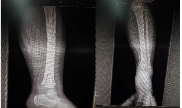
reference values, while the flow cytometry-cellular immunity test was normal. The HIV and brucellosis tests were negative. MRI of the endocranium was normal. Six gastric lavage analyses were performed, all of which were TB negative. The initial antituberculosis treatment included a combination of three drugs (isoniazid, rifampicin and pyrazinamide) for three months, followed by a further nine months of rifampicin and isoniazid. This antituberculosis treatment lasted 12 months in total. At the last examination, the girl had no complications, such as limping, deformities or shortening of the extremity. The last X-ray of the left lower leg showed the replacement of the osteolytic lesion on the tibia with the restoration of the cortical contours (Figure 3).
Figure 3: AP and lateral X-ray, 12 months after the treatment, showing the replacement of the osteolytic lesion with the restoration of the cortical contours
DISCUSSION
The 19-month-old girl with BCG osteitis in the distal tibia metaphysis responded well to the surgical treatment and antituberculostatic therapy, administered over 12 months. To the best of our knowledge, this is the first case report on osteitis of tibiae following BCG vaccination from Bosnia and Herzegovina. According to the literature, the age at which the onset of osteitis occurs significantly varies. For instance, a study of 222 children from Finland suggested that the age varies from 0.25 to 5.7 years. (10) In our case study, the
development in most patients is favourable.
(5,6,10)
The prognosis of this disease is good and sequelae of the bone or growth deficit are described in only 3% of cases. (10)
CONCLUSION
BCG osteitis is a very rare complication of the BCG vaccination. It is difficult to make a diagnosis and, with initial symptoms, rarely recognized. Therefore, we should be extremely cautious during the examination of small children with atypical symptoms in extremities, especially if they do not respond to initial treatment. To avoid possible further complications, early BCG osteitis diagnosis, treatment with antituberculostatics and surgical intervention are of great importance.
Declarations
ACKNOWLEDGEMENTS
The authors would like to thank the family of the child mentioned in this report for their consent to report this case and use the images.
Funding
There is no financial support for this case report.
Availability of data and materials
Data and materials supporting this report are available in the records of the hospital where this patient was treated and can be requested directly from the corresponding author to some extent, without revealing a patient’s identity.
Authors’ contributions
SC, MJ, AP and FK interpreted the patient data regarding the disease, designed the case report, and contributed in writing the manuscript. SS reported the X-ray images and contributed in writing the manuscript. All authors read and approved the final manuscript.
Ethics approval and consent to participate Not applicable.
Consent for publication
The author obtained from the child’s parents the written permission to report this case and images.
Competing interests
The authors declare that have no competing interest
Abbreviations
BCG: Bacillus Calmette-Guerin CRP: C-reactive protein
CXR: Chest X-ray L: leukocytes
ESR: Erythrocyte sedimentation rate RF: reuma factor
ASTO: antistreptolysin test ENT: ear, nose, and throat
REFERENCES
1. Hotokezaka H, Kitamura A, Matsumoto S, Hanazawa S, Amono S, Yamada T. Internalization of Mycobacterium bovis Bacillus Calmette-Guérin into osteoblast-like MC3T3-E1 cells and bone resorptive responses of the cells against the infection. Scand J Immunol. 1998; 47:453–8.
2. Murphy D, Corner LAL, Gormley E: Adverse reactions to Mycobacterium bovis bacilli Calmette-Guerin (BCG) vaccination against tuberculosis in humans, veterinary animals and wildlife species. Tuberculosis. 2008; 88 (4):344-57.
3. Goraya JS, Virdi VS: Bacille Calmette-Guerin lymphadenitis. Postgrad Med J. 2002; 78(920):327-9.
4. Bricks LF. Vacina BCG: via percutâneaouintradérmica? J. Pediatr (Rio J). 2004; 80(2):93-8.
5. KheirAEM, Ibrahim SA, Abdelsatir A, Bahar ME. Osteitis of the radius following Bacillus Calmette-Guerin vaccination at birth: a case report. J Med Case Rep. 2017; 11(1):283.
6. Yamada AF, Pellegrini JB, Cunha LM, Fernandes Ada R. Osteitis after BCG vaccination. J Bras Pneumol. 2009; 35(3):285-9.
7. Al Namshan M, Oda O, Almaarry J, Al Jadaan S, Crankson S, Al Banyan E, Al Shaalan M, Zamakhshary M. Bacilluss Calmette-Guerin - related cold thigh abscess as an unusual cause of thigh swelling in infants following BCG vaccine administration: a case series. J Med Case Rep 2011; 22(5):472.
8. Farhat CK, Carvalho ES, Carvalho LH, Succi RC, editors. Infectologia Pediatrica. Sao Paulo: Atheneu. 1999; p343-51.
feature of a child with chronic granulomatous disease. Braz J Infect Dis. 2011; 15(1):83-6.
10. Kröger L, Korppi M, Brander E, Kröger H, Wasz-Höckert O, Backman A, et al. Osteitis caused by bacille Calmette-Guérin vaccination: a retrospective analysis of 222 cases. J Infect Dis. 1995; 172(2):574–6.
11. Vallejo JG, Ong LT, Starke JR. Tuberculous osteomyelitis of the long bones in children. Pediatr Infect Dis J. 1995; 14:542–6.
12. Wang MN, Chen WM, Lee KS, Chin LS, Lo WH. Tuberculous osteomyelitis in young children. J PediatrOrthop. 1999; 19(2):151-5.
13. Aftimos S, Nicol R. BCG osteitis: a case report. N Z Med J. 1986; 99(800): 271-3.
14. Lin CJ, Yang WS, Yan JJ, Liu CC. Mycobacterium bovis osteomyelitis as a complication of Bacille Calmette-Guérin (BCG) vaccination: rapid diagnosis with use of DNA sequencing analysis: a case report. J Bone Joint Surg Am. 1999; 81(9):1305-11.
15. Foucard T, Hjelmstedt A. BCG-ostemyelitis and osteoarthritis as a complication following BCG-vaccination. AcataOrthop Scand. 1971; 42(2):142-51.
***********