Abstract
Experimental studies have highlighted that the administration of 3,5-diiodo-L-thyronine (T2) to rats fed diets rich in lipids induces a decrease of cholesterol and triglycerides plasma levels and body weight (BW) without inducing liver steatosis. On the basis of these
observations we carried out some experimental in vivostudies to
assess the effects of multiple high doses of T2 on the pituitary thy-roid axis of rats fed diet rich in lipids. Fifteen male Wistar rats were divided into three groups of five animals each. The first group (N group) received standard diet, the second group was fed with a high fat diet (HFD group), while the third group (HFDT2 group) was
addi-tionally given T2 intraperitoneally at a dose level of 70 mg/100 g of BW
three times a week up to four weeks. At the end of the treatment, blood sample from each animal was collected, centrifuged and the serum was stored at -20°C. The serum concentrations of thyroid-stimulating hormone (TSH), triiodothyronine, thyroxine, adrenocor-ticotropic hormone, triglycerides, cholesterol, glucose, alanine
aminotransferase, aspartate aminotransferase, alkaline phosphatase were then determined. In addition, liver of rats was examined by his-tology in order to assess the presence and degree of steatosis. The administration of T2 to rats fed with a high fat diet suppressed TSH secretion (P=0.013) while no steatosis was observed in the liver of these animals. Our data show that multiple administrations of high doses of T2 to rats fed diets rich in lipid inhibit TSH secretion and prevent the onset of liver steatosis in these animals.
Introduction
Thyroid hormones thyroxine (T4) and triiodothyronine (T3) are well known to stimulate energy metabolism in both animals and
humans.1,2This phenomenon is mainly mediated by T3 that is
con-sidered to be the main active molecule. On the basis of these find-ings several studies have been undertaken to investigate the possi-ble clinical use of this hormone in the treatment of diseases that are associated with an over consumption of food and drinks high in fat and/or sugar such as obesity, diabetes, dyslipidemia and hepatic steatosis. In particular, T3 has long been considered potentially suit-able for the treatment of obese patients as it has been shown to induce a decrease of the body weight (BW) following stimulation of lipid catabolism and a daily increase of energy expenditure. However,
experimental in vivo studies report that the administration to
rodents of 3,5-diiodo-L-thyronine (T2), which has long been consid-ered only an inactive metabolite of T3 and T4, increased their resting
metabolic rate (RMR).3,4On the other hand, some studies in humans
have confirmed that the administration of T2 can increase the basal
metabolic rate and decrease fat and BW without side effects.3On the
basis of these observations, several T2 analogs have been recently designed and synthesized with the aim of finding novel and more effective pharmacological approaches in the treatment of these pathological conditions. In this context, the administration of the analog TRC150094 to rats fed a high fat diet resulted in reducing the accumulation of fat in the liver and adipose tissue and in decreasing cholesterol and triglycerides blood levels without suppressing
pitu-itary thyroid-stimulating hormone (TSH) levels.5 On the basis of
these observations we have performed some in vivostudy to better
define the effects of multiple administration of high doses of T2 on the onset of liver steatosis in rats fed diet rich in lipids and the influ-ence of this treatment on TSH secretion as it has been earlier
report-ed by Padron and colleauges.6
Correspondence: Marco Giammanco, Department of Experimental Biomedicine and Clinical Neurosciences - BioNec, University of Palermo, Palermo, Italy.
Tel: +39.091.6555805; +39.333.4706470. E-mail: marco.giammanco@unipa.it
Key words: 3,5-diiodo-L-thyronine; TSH; Thyroid hormone; Hepatic steatosis.
Conflict of interest: the authors declare no potential conflict of interest.
Received for publication: 4 December 2015. Accepted for publication: 28 December 2015.
©Copyright M. Giammanco et al., 2016
Licensee PAGEPress, Italy
Journal of Biological Research 2016; 89:5667 doi:10.4081/jbr.2016.5667
This article is distributed under the terms of the Creative Commons Attribution Noncommercial License (by-nc 4.0) which permits any noncom-mercial use, distribution, and reproduction in any medium, provided the orig-inal author(s) and source are credited.
Effects of 3,5-diiodo-L-thyronine on the liver of high fat diet fed rats
Marco Giammanco,
1Stefania Aiello,
1Alessandra Casuccio,
2Maurizio La Guardia,
3Luca Cicero,
4Roberto Puleio,
4Irene Vazzana,
4Giovanni Tomasello,
1Giovanni Cassata,
4Gaetano Leto,
2Danila Di Majo
11
Department of Experimental Biomedicine and Clinical Neurosciences, University of Palermo,
Palermo;
2Department of Sciences for Health Promotion and Mother-Child Care, University of
Palermo, Palermo;
3Department of Biological, Chemical and Pharmaceutical Sciences and
Technologies, University of Palermo, Palermo;
4Institute for Experimental Veterinary Medicine of
Sicily, Palermo, Italy
Non-commercial
Materials and Methods
Chemicals
3,5-diiodo-L-thyronine was obtained from Sigma-Aldrich (St. Louis, MO, USA). An inclusion complex of T2 with hydroxypropyl cyclodex-trin (HP-Cy) (Sigma-Aldrich) was produced to improve drug solubility and stability in PBS at pH 7.4. Appropriate amounts of T2 were sus-pended in a solution of cyclodextrin in redistilled water to obtain a final weight ratio HPCy/T2 of 4:1. Then, few drops of 0.1 N sodium hydroxide were added until the complete dissolution of the drug and the formation of inclusion complex. The pH was adjusted to 7 with of 0.1 N hydrochloric acid and the solution was freeze-dried. Appropriate amount of solid complex was weighed and injected to animals just before drug administration.
Animals and treatments
Male Wistar rats aged between 8 and 10 weeks and weighing 300-350 g, were purchased from Harlan Italy [S. Pietro al Natisone (UD), Italy], and kept at 24°C with a light/dark cycle of 12:12-h. Animals had free access to water and food. The maintenance and care of the ani-mals were carried out according to the guidelines of the Council of the European Community for the care and use of animals. After 1 week of acclimatization, 15 rats were randomly divided into three groups of 5 rats each. The first group (N) received standard diet (total percentage of metabolizable energy: 60.4% carbohydrates, 29% pro-tein, 10.6% fat, 15.88 kJ of energy/g); the second group was fed a high-fat diet (HFD) (percent of total metabolizable energy: 21% car-bohydrates, 29% protein, 50% fat, 19.85 kJ of energy/g); the third group (HFDT2) was fed as the previous one. Additionally, these
ani-mals received intraperitoneal injections (ip) of T2 (70 mg/100 g BW)
three days a week, after anesthesia with inhaled isoflurane. Control rats (group N and group HFD) were injected with saline, after anes-thesia with inhaled isoflurane. Body weight and food intake of each animal were recorded every two days. Food intake was not significant-ly influenced by the composition of the diet or treatment with T2. After 28 days of treatment, rats were anesthetized with inhaled
isoflu-rane and intramuscular administration of tiletamine, 60 mL of
zolazepam, and 60 mL of medetomidine, and killed by cervical
disloca-tion. Their livers were rapidly dissected, weighed, cut into small pieces and quickly frozen in liquid nitrogen and stored at -80°C.
Serum measurements
Blood samples obtained by intracardiac puncture were collected and centrifuged. Serum was stored at -80°C until assays. Serum concentra-tions of TSH third generation, T3, T4, adrenocorticotropic hormone (ACTH), triglycerides, cholesterol, glucose, glutamic oxaloacetic transaminase (GOT), glutamic pyruvic transaminase (GPT), alkaline phosphatase (AP) were measured using a clinical analyzer Siemens Immunolite 2000 (Siemens Healtcare, Erlangen, Germany) following standard procedures.
Histological analysis
Liver tissue sections 3 mm thick were fixed in 10% buffered
forma-lin and stained with hematoxyforma-lin-eosin. The histological sections were then subjected to a semi-quantitative examination, to detect the percentage of hepatocytes showing macro or microvesicular steatosis
(>15 mm). The score system used to assess steatosis was defined as:
i) absent or minimal when the histological lesions involved were <1% of hepatocytes (score 0); ii) slight if <30% of hepatocytes (score 1);
Figure 1. Histological section of liver tissue from control rats: no sign of steatosis is evident at either magnification [100× (A) nor 200× (B)] (score 0).
Non-commercial
iii) moderate if the number of the hepatocytes involved were between 30 and 60% (score 2); and iv) severe if the hepatocytes involved were more than 60% (score 3). The percentage of hepatocytes involved was determined by counting cells in six microscopic fields at 400× magni-fication. Microscopic examination was carried out with Leica DMLB (Leica, Nussloch, Germany), equipped with Nikon (Tokyo, Japan); the system of image acquisition used was Nis elements Br software.
Statistical analysis
Continuous variables are expressed as mean±standard deviation. Intergroup differences among groups at T0 and T28 were assessed by
the univariate analysis of variance (ANOVA), and post-hoc analysis
with the Tukey’s test was used to determine pairwise differences. The intragroup difference between different times was evaluated with the
Figure 2. Histological sections of liver tissue from high fat diet fed rats. Note the widespread intracellular vacuolization of hepatocytes and the resulting relocation of cell nuclei in a peripheral position (score 2). A) 100× magnification; B) 200× magnification.
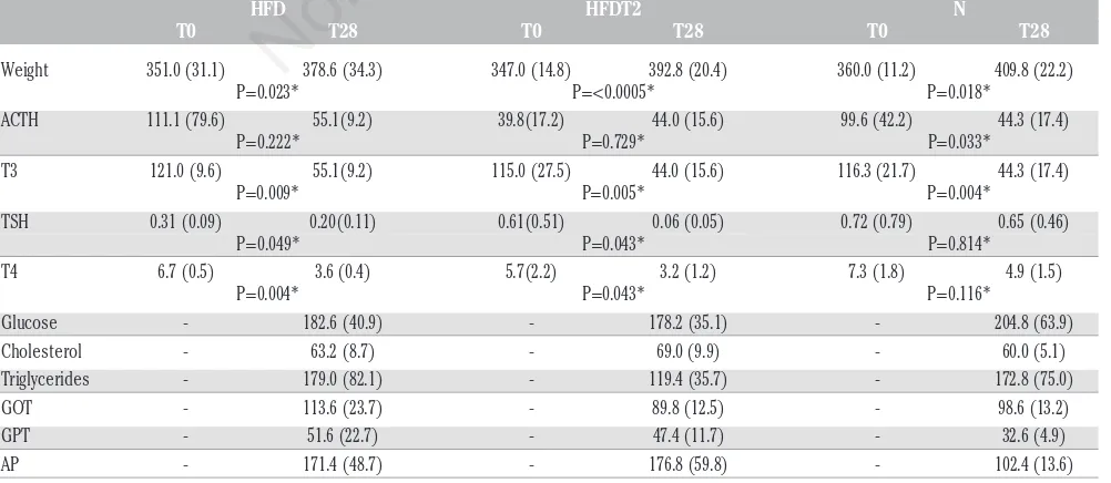
Table 1. Weight, metabolic and hormonal data of three groups.
HFD HFDT2 N
T0 T28 T0 T28 T0 T28
Weight 351.0 (31.1) 378.6 (34.3) 347.0 (14.8) 392.8 (20.4) 360.0 (11.2) 409.8 (22.2) P=0.023* P=<0.0005* P=0.018*
ACTH 111.1 (79.6) 55.1(9.2) 39.8(17.2) 44.0 (15.6) 99.6 (42.2) 44.3 (17.4) P=0.222* P=0.729* P=0.033*
T3 121.0 (9.6) 55.1(9.2) 115.0 (27.5) 44.0 (15.6) 116.3 (21.7) 44.3 (17.4) P=0.009* P=0.005* P=0.004*
TSH 0.31 (0.09) 0.20(0.11) 0.61(0.51) 0.06 (0.05) 0.72 (0.79) 0.65 (0.46) P=0.049* P=0.043* P=0.814*
T4 6.7 (0.5) 3.6 (0.4) 5.7(2.2) 3.2 (1.2) 7.3 (1.8) 4.9 (1.5) P=0.004* P=0.043* P=0.116*
Glucose - 182.6 (40.9) - 178.2 (35.1) - 204.8 (63.9) Cholesterol - 63.2 (8.7) - 69.0 (9.9) - 60.0 (5.1) Triglycerides - 179.0 (82.1) - 119.4 (35.7) - 172.8 (75.0) GOT - 113.6 (23.7) - 89.8 (12.5) - 98.6 (13.2) GPT - 51.6 (22.7) - 47.4 (11.7) - 32.6 (4.9) AP - 171.4 (48.7) - 176.8 (59.8) - 102.4 (13.6) HFD, hight fat diet group; HFDT2, fat diet group treated with T2; N, control group; T0, day 0 of the test; T28, day 28 of the test; ACTH, adrenocorticotropic hormone; T3, triiodothyronine; TSH, thyroid-stimulating hor-mone; T4, thyroxine; GOT, glutamic oxaloacetic transaminase; GPT, glutamic pyruvic transaminase; AP, alkaline phosphatase. Values are expressed as mean (standard deviation). *T28 vsT0.
Non-commercial
paired sample Student’s t-test. Data were analyzed by IBM SPSS Software 22 version (IBM Corp., Armonk, NY, USA). All P values were two-sided and P<0.05 was considered statistically significant.
Results
The BW of the animals was recorded at the beginning of the treat-ment (day 0) and on day 28. No significant differences in the mean BW among the three groups of animals were observed (Table 1). However, a significant increase in BW was highlighted for each rat between day 0 and day 28 (Table 1). Furthermore, no significant differences in the serum levels of glucose or cholesterol were noted among the three groups on day 28 when compared to those measured on day 0 (Table 1). Still, the serum levels of triglycerides decreased in HFDT2 animals as compared to the other groups; this reduction did not reach the statistical significance. Among the biochemical parameters of liver function exam-ined, only the plasma levels of AP resulted significantly more elevated (P=0.041) in the HFD and HFDT2 groups as compared to control group on day 28 while, again, no significant difference was observed for glu-tamic oxaloacetic transaminase and gluglu-tamic pyruvic transaminase. Interestingly, the mean plasma levels of T3 resulted in all the three groups significantly lower on day 28 than those measured on day 0 (Table 1). On the other hand, a similar trend was observed in the case of T4 plasma levels (Table 1). However, in this case, stastistically signif-icant difference was observed only for the HFD group (P=0.004). Finally, no significant difference in the plasma levels of T3 and T4 was highlight among the three groups of animals between day 0 and day 28. Although no significant difference in TSH plasma concentrations was observed among the different groups of animals, in the HFDT2 groups the circu-lating levels of this parameter on day 28 were significantly lower as
com-pared to control group (P=0.013). In all the animals TSH levels were lower on day 28 as compared to day 0. However these differences result-ed statistically significant for the HFD group (P=0.049) e HFDT2 group (P=0.043). At the histological evaluation the rats of group B (standard diet) showed no signs of steatosis (Figure 1), while rats of the HFD group (high fat diet) showed a fatty medium entities (Figure 2), which was not highlighted in the control group and in HFDT2 group (Figures 1 and 3).
The histological sections of the liver of the animals of the group HFD, examined at 100×200× magnification showed the presence of optically empty vacuoles in the cytoplasm of hepatocytes. In some cases this phenomenon determined the relocation of the nuclei in a periph-eral position. Although cell damage was small, it was widespread and classified as fatty liver score 2.
Discussion
Recent studies have shown the beneficial effects of 3,5-T2 in pre-venting liver steatosis in animals fed a high-fat diet. Experimental evi-dence indicates that the beneficial effects exerted by 3,5-T2 are non-genomic but rather mediated by metabolic pathways mainly at the mitochondrial level. Our investigations were aimed at assessing the suppressive effects of this molecule on the pituitary-thyroid axis.7In
line with the recent findings of Padron and colleagues,6our data show
that the administration of 3,5-diiodo-L-thyronine to rats three times a week up to 4 weeks leads to a reduction in BW gain despite the decreased level of T4 and T3 serum levels. On the other hand, Lanni
and colleagues8have shown that the administration of 25 mg/100 g of
3,5-T2 to rats may reduce BW gain and adiposity while no change in serum T4 levels was observed in animals fed a diet rich in fat and
treat-Figure 3. Histological section of liver tissue from 3,5-diiodo-L-thyronine high fat diet fed rats. No sign of steatosis is present (score 0). A) 100× magnification; B) 200× magnification.
Non-commercial
ed with 3,5-T2. This phenomenon was not observed in control animals. In addition, it was also shown that 3,5-T2 increased the rate of fatty acid oxidation in skeletal muscle. In addition to the chronic effects of 3,5-T2, a non-genomic acute and most likely increased mitochondrial
activity in rat liver has also been described.2An increased consumption
of hepatic oxygen and oxidative activity in the liver of rats fed a high-fat diet, and the recovery of energy expenditure in hypothyroid rats
have been also described.9-11The metabolic effects induced by 3,5-T2
could, in part, explain the decrease in BW gain and in that of retroperi-toneal fat observed in aged rats treated with this agent. In addition to the beneficial effects of 3,5-T2 on BW increase and adiposity, rats treat-ed with 3,5-T2 also showtreat-ed an improvement in glucose tolerance, more than 10%, compared to control animals. This result suggests that 3,5-T2 appears to improve glucose tolerance, directly or indirectly by decreasing adiposity in the treated animals. On the other hand, these findings are consistent with the observation that 3,5-T2, in addition to other effects on the liver, prevents insulin resistance in skeletal muscle of rats fed a high-fat diet and increases the expression of GLUT4 by insulin-induced phosphorylation of Akt. 3,5-T2 also was shown to increase nuclear sirtuin 1 expression and to decreases lipogenic
genes.12,13In agreement with previous data demonstrating that 3,5-T2
exerts important metabolic effects,2experimental findings highlight a
significant increase in oxygen consumption in rats treated with 50
mg/100 g of 3,5-T2 as compared to controls. Interestingly these animals
had lower, serum levels of T3 and T4. These hormones are the main regulators of energy metabolism. Accordingly, 3,5-T2 may increase the rate of mitochondrial fatty acid oxidation and thermogenesis in the rat
skeletal muscle, β-oxidation of lipids,8mitochondrial oxygen
consump-tion10and also the RMR.14Thus, it is conceivable to hypothesize that
3,5-T2 may function as a stimulator of RMR and may increase oxygen consumption. TSH serum levels were low. In animals treated with 3,5-T2 this phenomenon did not appear to be related to the reduced serum
levels of T4 and T3. However, Antonelli and colleagues3have recently
highlighted evident changes in the levels of thyroid hormones in serum in two euthyroid subjects treated for 3 weeks with a daily dose of 300
mg of 3,5-T2. On the other hand, Horst and colleagues9and Giammanco
and colleagues15 have shown that 3,5-T2 reduces the secretion of TSH
from the pituitary fragments of rat stimulated by T2.15These authors
also showed a reduction in T4 serum levels after 90 days of treatment
with 25 mg of 3,5-T2/100 g, these data are consistent with the results
obtained by our studies.15Furthermore, other observations highlighted
that a single dose of 3,5-T2 reduced serum concentrations of the β
sub-unit of pituitary TSH.16Thyroid hormone receptor β(TRβ) is the main
mediator of the negative feedback of thyroid hormone.17The results of
Ball and colleagues16showing that TRβ2 binds 3,5-T2 with higher
affin-ity than the other TR isoforms may well explain the effectiveness of 3,5-T2 in suppressing the secretion of TSH. Recently, it has been also reported that in humans that 3,5-T2 binds and activates the B isoform
of human TRβ, showing once again that this metabolite exerts
genom-ic effects.18Consequently the reduced serum levels of TSH in rats
treat-ed with 3,5-T2 may explain the rtreat-eduction in their thyroid deiodinase D1. On the other hand, the activity of deiodinase D2 is increased both in the hypothalamus in the pituitary of rats treated with the 3,5-T2, despite the thyreomimetic effects of 3,5-T2. It has been shown that lev-els of D2 protein and its activities are regulated by post-translational mechanisms, in particular by ubiquitination processes, that seem to be
mainly driven by T4.19Since T4 levels were decreased in rats treated
with 3,5-T2, this could explain the increase in Q2 in these animals.19
Taken together, the results show that the 3,5-T2 significantly down reg-ulated thyroid function. In addition to the decreased levels of thyroid hormones, the inhibitory effect of 3,5-T2 on thyroid function appears, at least partly, to be due to reduced serum levels of TSH because it is the main stimulant of the thyroid gland. The expression of the TSH
receptor (TSHR) and TSHR mRNA were increased in the thyroid of rats treated with 3,5-T2. It has been reported that TSH exerts inhibitory
effects on the activity of the promoter of the gene for TSHR.20
Therefore, it is likely that thyroid TSHR expression is higher in rats treated with the 3,5-T2 as TSH serum levels were significantly reduced. Furthermore, the expression of NOX4 is inhibited, while that of DUOX2
is up-regulated, just as previously shown in other models.21,22 The
chronic administration of 3,5-T2 reduces the increase in BW and retroperitoneal fat mass and increases RMR. These effects do not appear to be correlated with the decreased levels of thyroid hormone. The reduction in thyroid hormone levels may be secondary to the reduction in serum levels of TSH, which leads to a reduced activity and expression of sodium-iodide symporter (NIS), thyroid D1, and thyroid
peroxidase (TPO).6These new data support the hypothesis that 3,5-T2
causes exogenous TSH suppression. Thus, 3,5-T2 may be used as a non-thyreomimetic pharmacological agent in the treatment of hypothy-roidism. On the other hand, in hyperthyroidism, TSH circulating levels are typically <0.1 mU/L or even lower than 0.05 mU/L.
Conclusions
Until recently 3,5-T2 has been considered as a hormone with little or no physiological effects, due to its low plasma and its short half-life. On the contrary, recent findings show that this molecule may increase energy expenditure and may also have anti-steatotic activity without producing thyrotoxic effects and according to some authors without affecting the secretion of TSH. The present study carried out on Wistar
rats further confirms this effect. However, at a dose level of 70 mg/100
g BW, the molecule suppresses TSH secretion from hypophysis. The reduction of the thyroid hormone levels that might be secondary to the decreased TSH serum levels, may lead to the reduced activity and expression of NIS, thyroid D1, and TPO. These new data support the idea that exogenous 3,5-T2 causes TSH suppression. On the other hand, a molecule that may increase energy expenditure endowed with protective effect on the liver represents an important therapeutic goal for a more effective treatment of obesity and non-alcoholic fatty liver disease. To this aim, some 3,5-T2 agonists have been synthesized. However, their effects on energy expenditure on the liver and on the hypothalamic-pituitary-adrenal axis still remain to be better defined.
References
1. Jonas W, Lietzow J, Wohlgemuth F, et al. 3,5-Diiodo-L-thyronine (3,5-t2) exerts thyromimetic effects on hypothalamus-pituitary-thyroid axis, body composition, and energy metabolism in male diet-induced obese mice. Endocrinology 2015;156:389-99. 2. Lanni A, Moreno M, Lombardi A, Goglia F. Calorigen effect of
diiodothyronines in the rat. J Physiol 1996;494:831-7.
3. Antonelli A, Fallahi P, Ferrari SM, et al. 3,5-diiodo-L-thyronine increases resting metabolic rate and reduces body weight without undesirable side effects. J Biol Reg Homeos Ag 2011;25:655-60. 4. Lanni A, Moreno M, Cioffi M, Goglia F. Effect of
3,3’-diiodothyro-nine and 3,5-diiodothyro3,3’-diiodothyro-nine on rat liver oxidative capacity. Mol Cell Endocrinol 1992;86:143-8.
5. Cioffi F, Zambad SP, Chhipa L, et al. TRC150094, a novel functional analog iodothyronines, reduces adiposity by increasing energy expenditure and fatty acid oxidation in rats receiving a high-fat diet. Fed Am Soc Exp Biol 2010;24:3451-61.
6. Padron AS, Neto RA, Pantaleão TU, et al. Administration of
3,5-Non-commercial
diiodothyronine (3,5-T2) causes central hypothyroidism and stim-ulates thyroid-sensitive tissues. J Endocrinol 2014;221:415-27. 7. Silvestri E, Cioffi F, Glinni D, et al. Pathways affected by
3,5-diiodo-L-thyronine in liver of high fat-fed rats: evidence from two-dimen-sional electrophoresis, blue-native PAGE, and mass spectrometry. Mol Bio Syst 2010;6:2256-71.
8. Lanni A, Moreno M, Lombardi A, et al. 3,5-Diiodo-L-thyronine pow-erfully reduces adiposity in rats by increasing the burning of fats. Fed Am Soc Exp Biol 2005;19:1552-4.
9. Horst C, Harneit A, Seitz HJ, Rokos H. 3,5-Di-iodo-L-thyronine
sup-presses TSH in rats in vivoand in rat pituitary fragments in vitro.
J Endocrinol 1995;145:291-7.
10. Mollica MP, Lionetti L, Moreno M, et al. 3,5-diiodo-L-thyronine, by modulating mithocondrial function, reverses hepatic fat accumula-tion in rats fed a higt-fat diet. J Hepatol 2009;51:363-70.
11. Cimmino M, Mion F, Goglia F, et al. Demostration of in vivo
meta-bolic effects of 3,5-diiodothyronine. J Endocrinol 1996;149:319-25. 12. Moreno M, Silvetri E, De Matteis R, et al. 3,5-Diiodo-L-thyronine
prevents high-fat-diet-induced insulin resistance in rat skeletal muscle through metabolic and structural adaptations. Fed Am Soc Exp Biol 2011;25:3312-24.
13. De Lange P, Cioffi F, Silvestri E, et al. (Healthy) ageing: focus on iodothyronines. Int J Mol Sci 2013;14:13873-92.
14. Moreno M, Lombardi A, Beneduce L, et al. Are the effects of T3 on resting metabolic rate in euthyroid rats entirely caused by T3 itself? Endocrinology 2002;143:504-10.
15. Giammanco M, Cassata G, Cicero L, et al. 3,5-diiodo-L-thyronine
induced modification in pituitary thyroid axis in rats fed higt fat diet. A preliminary report. In: Giammanco M, ed. Proceedings of the 86th SIBS National Congress, 24-25 October 2013, Palermo, Italy. J Biol Res 2015;87:5161.
16. Ball SG, Sokolov J, Chin WW. 3,5-Diiodo-l-thyronine (T2) has
selec-tive thyromimetic e Vects in vivoand in vitro. J Mol Endocrinol
1997;19:137-47.
17. Williams GR, Bassett JHD. Deiodinases: the balance of thyroid hor-mone local control of thyroid horhor-mone action: role of type 2 deiod-inase. J Endocrinol 2011;209:261-72.
18. Mendoza A, Navarrete-Ramirez P, Hernandez-Puga G, et al. 3,5-T2 is an alternative ligand for the thyroid hormone receptor b1. Endocrinology 2013;154:2948-58.
19. Gereben B, Goncalves C, Harney JW, et al. Selective proteolysis of human type 2 deiodinase: a novel ubiquitin-proteasomal mediated mechanism for regulation of hormone activation. Mol Endocrinol 2000;14:1697-708.
20. Ikuyama S, Ohe K, Takayanagi R, et al. Cloning and characteriza-tion of the 4.2 kb region of the rat thyrotropin receptor promoter. Endocr J 1997;44:247-56.
21. Milenkovic M, De Deken X, Jin L, et al. Duox expression and relat-ed H2O2 measurement in mouse thyroid: onset in embryonic development and regulation by TSH in adult. J Endocrinol 2007;192:615-26.
22. Weyemi U, Caillou B, Talbot M, et al. Intracellular expression of reactive oxygen species-generating NADPH oxidase NOX4 in nor-mal and cancer thyroid tissues. Endocr-Relat Cancer 2010;17:27-37.
.](https://thumb-us.123doks.com/thumbv2/123dok_us/1207412.1624056/2.595.64.546.430.703/figure-histological-section-tissue-control-steatosis-evident-magnification.webp)