LASER TREATMENT OF BLADDER CANCER
Thesis submitted for the degree of
Doctor of Medicine
in the
University of London
by
Alvan John Pope
T.D., B.Sc., M.B., B.S., F.R.C.S., F.R.C.S.Ed
Academic Unit, Institute of Urology
University College and Middlesex School of Medicine
The National Medical Laser Centre
Faculty of Clinical Sciences, University College London
ProQuest Number: 10106859
All rights reserved
INFORMATION TO ALL USERS
The quality of this reproduction is dependent upon the quality of the copy submitted. In the unlikely event that the author did not send a complete manuscript and there are missing pages, these will be noted. Also, if material had to be removed,
a note will indicate the deletion.
uest.
ProQuest 10106859
Published by ProQuest LLC(2016). Copyright of the Dissertation is held by the Author. All rights reserved.
This work is protected against unauthorized copying under Title 17, United States Code. Microform Edition © ProQuest LLC.
ProQuest LLC
789 East Eisenhower Parkway P.O. Box 1346
ABSTRACT
Lasers techniques are able to produce precise and sometimes unique tissue effects. These promise an improvement over the conventional techniques for treating both superficial and early muscle invasive bladder cancer.
Photodynamic Therapy (PDT) is an experimental treatment that shows great promise for treating superficial bladder cancer, especially resistant carcinoma in situ (Cis). Some clinical studies though have reported serious side effects, mainly in producing an irreversible functional impairment in many bladders due to fibrosis. This thesis presents a study of the effect of changes in dosimetry variables on the normal rat bladder using a new photosensitiser, aluminium chlorosulphonated phthalocyanine. The uptake of this drug into the different layers of the bladder wall has also been investigated using sensitive fluorescence microscopy techniques. The maximum concentration gradient of photosensitiser between the superficial and the deep layers of the bladder wall was reached after 24 h following ' administration and was increased by the photobleaching observed at low sensitiser concentrations. Morphological and functional changes (bladder capacity and compliance) were also studied and it was found that if PDT damage was restricted to the superficial layers of the bladder, the resulting functional disturbance was less severe and recovered more fully than when the muscle layers were also involved. At low concentrations of photo sensitiser a selective, superficial necrosis was achieved across a wide range of light doses. If these experimental results can be achieved in clinical practice then PDT should provide an effective and bladder preserving treatment for Cis without the complications that have been seen previously.
The possible role of the flashlamp pulsed-dye laser for PDT was studied using cultured human bladder carcinoma cells (MGH-Ul) sensitised with dihaematoporphyrin ether. It was found that this clinical laser was of a comparable efficacy to the more complex systems currently used for PDT.
PREFACE
The title of this thesis implies that it will aim to cover the whole field of
laser therapy as applied to bladder cancer. This will comprise both
experimental and some clinical work with a review of relevant literature.
Section 1 outlines the pathology and current treatment of bladder cancer
and how laser techniques might complement or improve on conventional
therapy. The principles of laser action and the several different types of
lasers available are described. At present there are only 2 types of laser
applications; either the thermal destruction of bladder tumours or a photo
chemical technique not requiring heat but using a photosensitising drug to
produce tissue damage which is known as photodynamic therapy (PDT).
Section 2 deals with PDT in detail and describes our experimental work
in this field. This comprises both in vitro studies on a human bladder tumour cell line and in vivo studies on the effect of PDT on bladder function which is the area in which most clinical problems have been
encountered. It had been hoped to carry out a clinical PDT study, but due
to delay in acquiring the necessary regulatory approval for the photo
sensitiser drug this has not proved possible.
Section 3 covers the use of thermal lasers to ablate bladder tumours.
This is by far the commonest urological application of the
neodymium:YAG laser and where the bulk of clinical experience has been
gained. The experimental work presented in this section looks at the
morphology of laser lesions in normal pig bladder, and compares them to
conventional diathermy, as an accurate assessment of the expected tissue
effect is necessary to match treatment dosimetry variables to tumour
pathology. A small study in which tumour recurrences in patients have
been treated without anaesthetic using a NdiYAG laser in conjunction with
TABLE OF CONTENTS
Page
Statem ent of originality ... 9
List of figures and tables ... 10
A cknow ledgem ents ... 15
SECTION 1 BACKGROUND C hapter 1 1.1 1.2 1.2.1 1.2.2. 1.3 1.3.1 1.3.2 1.4 1.4.1 1.4.2 1.5 1.5.1 1.5.2 1.5.3 1.5.4 1.6 Chapter 2 2.1 2.2 2.2.1 2.2.2 2.3 2.3.1 2.4 Bladder Cancer Introduction ... 18
Superficial papillary bladder cancer ... 20
Treatment ... 20
The pTl 0 3 bladder tumour ... 21
Carcinoma in situ ... 23
Incidence ... 23
Treatment ... 24
Invasive bladder cancer ... 25
Radical treatment ... 26
Local excision ... 27
Laser treatment of bladder cancer ... 27
Introduction ... 27
Historical background ... 28
Photodynamic therapy ... 29
Neodymium: YAG laser coagulation ... 31
Discussion ... 32
Lasers: principles and effects Introduction ... 36
Lasers for photodynamic therapy ... 38
Copper vapour pumped dye laser ... 39
Flashlamp pumped dye laser ... 41
Neodymium: YAG laser ... 42
Delivery systems ... 45
Chapter 3 H istorical review of Photodynam ic Therapy
3.1 Discovery of photodynamic action ... 53
3.2 Development of porphyrin photosensitisers ... 54
3.3 Tumour localisation of porphyrins ... 55
3.3.1 Bladder tumour localisation with HpD ... 57
3.4 Clinical studies of photodynamic therapy ... 58
3.4.1 Early work ... 58
3.4.2 PDT in Urology... 59
3.4.3 Overview of PDT in other specialties ... 70
3.5 The way forward ... 73
C hapter 4 4.1 4.1.1 4.1.2 4.1.3 4.1.4 4.1.5 4.2 4.2.1 4.2.2 4.2.3 4.2.4 4.3 4.3.1 4.3.2 4.3.3 The n atu re of Photodynamic Therapy Principles of photodynamic action ... 76
Introduction ... 76
Singlet oxygen ... 78
Cellular effects ... 79
Vascular effects ... 80
Role of oxygen ... 82
Photosensitisers ... 84
Newer photosensitisers ... 84
Ideal properties of a photosensitiser ... 85
Porphyrin photosensitisers ... 87
Phthalocyanine photosensitisers ... 88
Selectivity of photodynamic effect ... 92
Selectivity of PDT in the bladder ... 94
Fluorescence ... 96
SECTION 2 EXPERIMENTAL STUDIES ON PDT
C hapter 5 The photodynamic action of a pulsed dye laser on human bladder cancer cells sensitised with DHE
5.1 Introduction ... 103
5.2 Materials and methods ... 106
5.2.1 Cell culture techniques ... 106
5.2.2 Experiment design ... 112
5.3 Results ... 116
5.3.1 Direct toxicity and the effect of white light ... 116
5.3.2 The effect of pulsed green laser light ... 119
5.4 Discussion ... 124
5.4.1 Saturation fluence ... 127
5.4.2 Comparison with CW lasers ... 129
5.4.3 Conclusions ... 130
C hapter 6 Uptake and microscopic distribution of alum inium sulphonated phthalocyanine in norm al ra t bladder 6.1 Introduction ... 133
6.2 Materials and methods ... 135
6.2.1 Photosensitiser ... 135
6.2.2 Rat bladder model ... 136
6.2.3 Preparation of specimens ... 139
6.2.4 Fluorescence detection ... 140
6.2.5 Light exposure for photobleaching studies ... 144
6.2.6 Pharmacokinetic studies ... 145
6.3 Results ... 146
6.3.1 Microscopic tissue distribution of AlSPc ... 147
6.3.2 Photobleaching ... 154
6.3.3 Tissue distribution of AlPcS2 by chemical extraction ... 154
6.4 Discussion ... 158
6.4.1 Timing of light exposure ... 159
6.4.2 Intravenous or intravesical administration? ... 160
C hapter 7 7.1 7.2 7.2.1 7.2.2 7.2.3 7.3 7.3.1 7.3.2 7.3.3 7.3.4 7.4 7.4.1 7.4.2
Morphological changes in the norm al ra t bladder following photodynam ic therapy with
phthalocyanine photosensitisation
Introduction ... 166
Materials and methods ... 169
Phototherapy ... 169
Experimental groups ... 178
Histological studies ... 180
Results ... 181
Whole animal effects ... 181
Macroscopic changes ... 183
Microscopic changes ... 185
Intravesical administration of AlSPc ... 189
Discussion ... 193
Improving the selectivity of PDT ... 198
Conclusions ... 200
C hapter 8 8.1 8.2 8.2.1 8.2.2 8.2.3 8.3 8.3.1 8.3.2 8.4 8.4.1 8.4.2 Functional changes in the norm al ra t bladder following photodynam ic therapy with phthalocyanine photosensitisation Introduction ... 203
Materials and methods ... 204
Animals, photosensitiser and phototherapy ... 204
Evaluation of anaesthetic technique ... 205
Assessment of bladder function ... 207
Results ... 213
Bladder compliance ... 213
Bladder capacity ... 221
Discussion ... 226
Correlation of functional and morphological changes ... 229
SECTION 3 NEODYMIUM:YAG LASER THERAPY
C hapter 9 The use of the Nd:YAG laser for the treatm ent of bladder cancer
9.1 Introduction ... 235
9.2 Superficial bladder cancer ... 235
9.2.1 Clinical results ... 236
9.2.2 Convenience and economy ... 239
9.2.3 Complications ... 240
9.2.4 Patient selection and tumour staging ... 241
9.3 Invasive bladder cancer ... 242
9.3.1 Introduction ... 242
9.3.2 Clinical results ... 243
9.4 Morphology of experimental laser lesions ... 246
9.4.1 Materials and methods ... 248
9.4.2 Results ... 252
9.5 Discussion ... 261
9.5.1 Experimental studies ... 261
9.5.2 Conclusions ... 266
C hapter 10 Nd:YAG laser coagulation of superficial bladder tum ours with flexible cystoscopy on outpatients 10.1 Introduction ... 270
10.1.1 Flexible cystoscopy ... 270
10.2 Patients and methods ... 272
10.2.1 Patients and instruments ... 272
10.2.2 Organisation of the Laser Unit ... 274
10.2.3 Laser technique ... 274
10.3 Results ... 281
10.4 Discussion ... 285
SECTION 4 SUMMARY
C hapter 11 G eneral Conclusions
11.1 Introduction... 296
11.2 Photodynamic therapy ... 297
11.2.1 Further work ... 303
11.3 Nd:YAG laser coagulation ... 306
11.3.1 Further work ... 308
11.4 Laser treatment - an integrated approach ... 309
11.4.1 The role for a Laser Unit ... 311
APPENDICES 1 Cell culture data (from chapter 5) ... 314
2 Fluorescence data (from chapter 6) ... 325
3 Bladder compliance and capacity data (from chapter 8) ... 330
4 Publications and presentations ... 340
5 Abbreviations ... 342
BIBLIOGRAPHY ... 343
STATEMENT OF ORIGINALITY
The work presented in this thesis has been carried out by myself and has not been entered for a higher degree or award of this or any other University. The major element of this work involves original concepts and observations as to the effect of photodynamic therapy on the normal bladder and the means by which this might be assessed with the help of an animal model. No similar work has been published or carried out by any other group to my knowledge and 1 hope that it will make a contribution to the scientific understanding and clinical practice of photodynamic therapy for bladder cancer.
Fig. Fig. Fig, Fig. Fig. Fig. Fig,
LIST OF FIGURES AND TABLES
Page
1.1 Staging of bladder cancer ... 19
2.1 Diagram of a copper vapour laser ... 40
2.2 Diagram of a flashlamp pumped dye laser ... 41
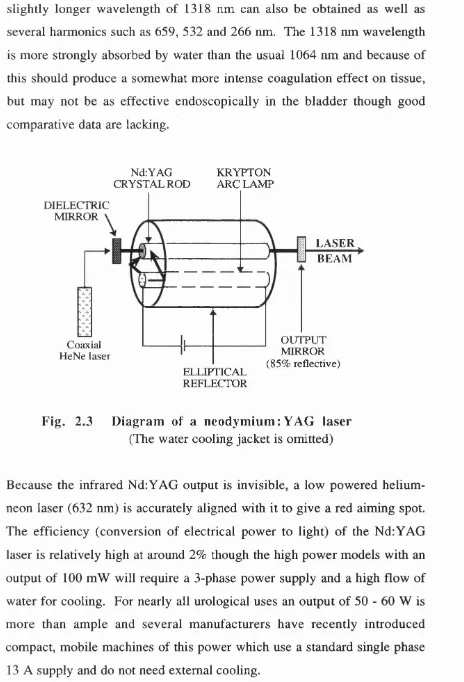
2.3 Diagram of a neodymium:YAG laser ... 43
2.4 Laser cystoscope ... 44
2.5 Laser resectoscope ... 44
2.6 Types of laser light interaction with skin ... 46
Fig. 2.7 Variation of thermal damage with tissue depth ... 49
Fig. 2.8 The effect of wavelength on tissue penetration ... 49
Table 3.1 Results of PDT in bladder carcinoma ... 63
Fig. 3.1 Absorption spectra for HpD and AlSPc ... 69
Fig. 4.1 Mechanism of photodynamic action ... 77
Table 4.1 Comparison of photochemical properties of HpD and AlSPc ... 86
% 4.2 The planar structures of AlSPc and Hp ... 89
5.1 Stained dishes showing cell kill ... 114
5.2 The Candela flashlamp pumped dye laser ... 114
5.3 Effect of exposure to white light for 30 min on MGH-Ul cells sensitised with DHE ... 117
5 .4 Effect of exposure to white light on MGH-U1 cells sensitised with either DHE or AlSPc ... 118
5.5 The effect of exposure to pulsed green laser light on unsensitised MGH-Ul cells ... 119
5.6 Survival of cells sensitised with DHE exposed to laser light (504 nm) at varying power densities ... 120
Fig. 5.8 Effect of the duration of light exposure time
on cell survival ... 122
Fig. 5 .9 The effect of varying the pulse repetition rate ... 123
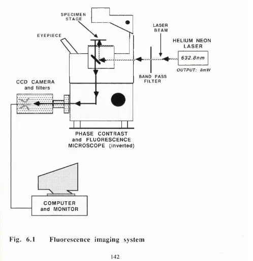
Fig. 6.1 Fluorescence imaging system ... 142
Fig. 6.2 CCD camera attached to fluorescence microscope ... 143
Figs. 6.3 Line or box integration for measurement ... 143
Fig. 6.4 Fluorescence image 1 h after sensitisation ... 148
Figs. 6.5 Fluorescence at 1 h in vascular endothelium ... 148
Figs. 6.6 24 h after sensitisation with 5 mg/kg AlSPc ... 149
Figs. 6.7 Fluorescence at 72 h after sensitisation ... 150
Fig. 6.8 Fluorescence after IV sensitisation ... 151
Fig. 6.9 Intravesical administration of AlSPc mixture ... 153
Fig. 6.10 Intravesical administration of AlPcS2 ... 153
Fig. 6.11 Concentration of AlPcS2 in whole bladder and kidney compared with plasma levels ... 155
F ig s .6.12 Photobleaching effect of 80 J/cm^ red light ... 156
Fig. 6.13 Photobleaching after 20 J/cm^ red light ... 157
Fig. 6.14 Effect of cessation of blood flow on photobleaching ... 157
Fig. 7.1 Copper vapour pumped dye laser ... 170
Fig. 7.2 Laser fibre within diffuser cannula ... 170
Fig. 7.3 Diagram of bladder illumination ... 171
Fig. 7.4 Laparotomy to expose bladder and to illustrate illumination from diffused laser fibre ... 173
Fig. 7.5 Shielding of adjacent bowel from transmitted light .... 173
Fig. 7.6 Irradiance measurement from a diffused fibre ... 176
Fig. 7.7 Irradiance measurement from an intact bladder ... 176
Fig. 7.9
T able 7.1
F ig s .7.10
F ig s .7.11
T able 7.2
F ig. 7.12
F ig. 7.13
F ig. 7.14
Fig. 7.15
Figs .7.16
T able 8.1
F ig. 8.1
Fig. 8.2
Fig. 8.3
Fig. 8 .4
Figs. 8.5 Fig. 8.6 Fig. 8.7 Fig. 8.8 Fig. 8.9 Fig. 8.10 Fig. 8.11 Fig. 8.12
Plot of fibre irradiance (m vivo) ... 179
Percentage incidence of morbidity and mortality in the treatment groups ... 182
Necrosis produced by high doses of PDT ... 184
Transmural acute inflammation of the bladder ... 186
Effect of AlSPc and light dose on PDT necrosis in the superficial and deep layers of the rat bladder w all.. 187
Photodynamic damage limited to the superficial layers of the bladder ... 190
Healing with complete regeneration of bladder wall architecture after 21 days ... 190
HVG stained section 3 months after PDT ... 191
PDT after intravesical administration of AlSPc ... 191
PDT after intravesical AlPcS2 ... 192
Response to stimuli during anaesthesia ... 207
Apparatus for cystometry ... 209
Bladder pressure measuring system ... 210
Repeat CMG’s performed without laparotomy ... 210
Pressure / volume curve in normal rat bladder ... 213
Filling pressures in rat bladder after PDT ... 215
Change in compliance ratio (controls) ... 216
Change in compliance ratio (0.5 mg/kg AJSPc) .. 217
Change in compliance ratio (1 mg/kg AlSPc) ... 218
Change in compliance ratio (1.5 mg/kg AJSPc) .. 219
Changes in mean compliance ratio after PDT ... 220
Changes in rat bladder capacity (controls) ... 221
Fig. 8.13 Changes in rat bladder capacity (1 mg/kg AlSPc) ... 223
Fig. 8.14 Changes in rat bladder capacity (1.5 mg/kg AlSPc) .... 224
Fig. 8.15 Changes in rat bladder capacity after PDT ... 225
T able 8.2 Effect of initial muscle damage from PDT on the recovery of bladder function ... 230
T able 9.1 Recurrences after laser treatment of superficial bladder tumours ... 237
T able 9.2 Recurrences after laser treatment of invasive bladder cancer ... 244
Fig. 9.1 Comparative schematic representation of lesions produced by diathermy and Nd:YAG laser ... 247
Fig. 9.2 Laser cystoscope inserted via a cystotomy in the dome of the bladder ... 250
Fig. 9.3 HeNe aiming beam seen through the bladder wall ... 250
T able 9.3 Depth of necrosis produced by laser ... 253
T able 9.4 Depth of necrosis produced by diathermy ... 254
T able 9.5 Mean depth of laser and diathermy coagulation ... 255
Fig. 9.4 Superficial coagulation from resectoscope loop ... 257
Fig. 9.5 Diathermy electrode 20 W for 4 s ... 258
Fig. 9.6 Nd: YAG laser coagulation 20 W for 4 s ... 258
Fig. 9.7 Diathermy electrode 40 W for 4 s ... 259
Fig. 9.8 Nd: YAG laser coagulation 40 W for 4 s ... 259
Fig. 9.9 Diathermy electrode 60 W for 4 s ... 260
Fig. 9.10 Nd: YAG laser coagulation 60 W for 4 s ... 260
F ig . 10.1 The Nd: YAG laser used in this study ... 273
F ig s .10.2 Effect of tangential incidence on a sizeable tumour with a laser fibre and a diathermy loop ... 276
F ig. 10.4 400 micron fibre with a retractable outer sheath to
aid insertion through flexible endoscopes ... 278
F ig. 10.5 Recurve T manoeuvre for bladder neck lesions ... 279
F ig s .10.6 Reduction in the maximum deflection of a flexible
cystoscope caused by 600 and 400 micron fibres .... 280
F ig. 10.7 Relationship between the number of tumours treated
and the laser energy delivered ... 282
ACKNOW LEDGEMENTS
The work presented in this thesis was carried out in the National Medical
Laser Centre under the guidance of its director Professor S G Bown except
for the cell culture work which was performed at the Institute of Urology
under the supervision of Dr J R Masters. I am most grateful to them both,
especially to Professor Bown who has been a continual source of education,
inspiration and direction and whose high standards and enthusiasm for laser
research have ensured a thriving department embracing many specialties.
Several colleagues have assisted me to some extent in this work, Mr T A
McNicholas with the minipig dosimetry work (chapter 9), Miss J Bedwell
for the extraction assays of phthalocyanine (chapter 6) and particularly Dr
A J MacRobert of Imperial College for his considerable help and patience
in running the CCD camera system (chapter 6). Dr T Mills, senior medical
physicist, kept the lasers running most of the time and managed to remain
cheerful when they didn’t as well as being a great source of technical
advice. Mr C Keast and colleagues. Biological Services, University College
provided the animal facilities and the histological processing was largely
done by the Imperial Cancer Research Fund Histopathology Unit.
My thanks are also due to Mr H Barr, Dr S J Harland and to Dr M C
Parkinson for allowing me to use her photographic microscope. Mr J E A
Wickham, Director of the Academic Unit, Institute of Urology has been a
valuable source of encouragement and I am most grateful for the facilities
provided by the Institute particularly from the department of Medical
Illustration who printed many of the photographs.
I am indebted to the Sir Jules Thom Charitable Trust who generously
awarded me a 2 year fellowship (1988/9) for research into photodynamic
therapy during which time the bulk of this experimental work was
completed. Finally my dear wife Rachel has survived my long-running
preoccupation with writing this thesis with her considerable good humour
SECTION 1
Chapter 1
BLADDER CANCER
1.1 Introduction ... 18
1 .2 Superficial papillary bladder cancer ... 20
1.2.1 Treatment ... 20
1.2.1.1 Prognostic indicators ... 20
1.2.1.2 Intravesical chemotherapy ... 21
1.2.2 The pTl G3 bladder tumour ... 21
1.3 C arcinom a in situ ... 23
1.3.1 Incidence ... 23
1.3.2 Treatment ... 24
1.4 Invasive bladder cancer ... 25
1.4.1 Radical treatment ... 26
1.4.2 Local excision ... 27
1.5 Laser treatm ent of bladder cancer ... 27
1.5.1 Introduction ... 27
1.5.2 Historical background ... 28
1.5.3 Photodynamic therapy ... 29
1.5.4 Neodymium:YAG laser coagulation ... 31
1.1 INTRODUCTION
Bladder carcinoma is the commonest urological malignancy requiring
treatment (prostate cancer is more common in males but frequently occult)
and the commonest one to cause death. In 1985, 9500 new cases were
registered in the U.K. (Cancer Statistics, 1990), and in 1988 there were
4800 deaths (Mortality Statistics, 1990). It is 2.6 times more common in
males and maximal around the 7th decade. Smoking increases the risk of
developing the disease from 2 -6 times. The aetiological role of industrial
carcinogens, particularly certain aniline dyes and chemicals such as 13-
naphthalenes which were used in the rubber industry has been recognised
for years and their use is now generally banned. Nearly all bladder
tumours are transitional cell carcinomas arising from the urothelium and
all the trials reviewed here relate to these. Other histological types are
rare and often advanced at presentation, they will be discussed briefly later.
As with other carcinomas, bladder tumours are classified by grade (G1 - 3)
and stage (Ta-T4). Grade 1 (Gl) indicates a well-differentiated tumour,
G2 moderate and G3 a poorly differentiated tumour. Grade is the single
most important prognostic feature though high grade tumours also have a
positive correlation with more advanced stage.
Staging of the local tumour indicates the depth to which it has extended into
the bladder wall (fig. 1.1). Superficial tumours comprise carcinoma in situ
(Cis) and Ta which are confined to the epithelium, and T1 - into the cores
of the papillae or lamina propria. Muscle invasive cancers may be T2 (into
the muscle but not palpable bimanually) or T3 (subdivided rather
artificially into T3a - invading more than half-way through the muscle
layer, and T3b which is through the bladder wall but not invading adjacent
structures). T4 tumours invade other organs such as the uterus or rectum.
gynaecological organs). When a tumour has been pathologically staged
either a lowercase “p” (biopsy), or an uppercase “P” (cystectomy specimen)
is applied though many authors seem to interchange these or drop the “T” -
e.g. Tl, pTl, PTl, PI can all refer to the same superficial tumour (UICC,
1987)
à
A - mucosa B - lamina propria C - superficial muscle D - deep muscle E - prostate gland
Fig. 1.1 Staging of bladder cancer
This chapter summarises the conventional treatment for bladder cancer and
how much laser techniques can offer at present, or promise for the future,
1.2 SUPERFICIAL PAPILLARY BLADDER CANCER
The diagnosis and treatment of patients with superficial (Ta,Tl) papillary
bladder cancer comprise a significant proportion of a general urologist’s
workload. Around 70% of all superficial tumours are stage Ta, almost all
of which are G l or 0 2 and are most unlikely either to progress locally and
become invasive or to metastasise (Abel et al., 1988). Once diagnosed,
however, patients are usually advised to undergo long term surveillance by
regular cystoscopy as many will require repeated treatment of recurrences.
1 .2 .1 T reatm ent
Standard treatment is by transurethral resection (TUR) or diathermy
fulguration and, in general, only when recurrences become particularly
frequent or numerous do other adjuvant therapies such as intravesical
chemotherapy become applicable. New treatment modalities that may
reduce tumour recurrences in these patients would therefore have little
survival benefit except in the small number of patients who present with
superficial cancer which is destined to become invasive.
1 .2 .1.1 Prognostic indicators
The most useful of these for indicating a potentially more aggressive nature
are high tumour grade, stage, size, multiplicity and rate of recurrence of
new tumours, and the presence of Cis (Heney et al., 1983). A patient with
0 2 or 0 3 superficial recurrences has a high chance of developing an
invasive tumour whereas the only categories of patients with 01 disease
who have a real risk of developing subsequent invasion are those with
either multiple recurrences (greater than 5 at each cystoscopy), or those
who develop disease in the upper tracts or prostatic ducts. Other
prognostic factors include DNA analysis by flow cytometry as tumours
However although these techniques may have a role in routine clinical
practice in the future it is doubtful whether at present they provide any
more useful information over histological grade and stage alone.
1 .2 .1 .2 Intravesical chem otherapy
The first intention in managing a patient with superficial bladder cancer
must be to surgically resect or fulgurate all visible tumour. Patients who
show the risk factors for developing invasive disease listed above, and those
who persist in developing frequent recurrences are candidates for some
form of intravesical therapy in the hope of normalising the bladder
urothelium, or at least reducing subsequent recurrences and hopefully
preventing progression. Treatment may be either adjuvant (prophylactic)
with the aim of preventing recurrence, or therapeutic to treat established
disease. Agents commonly used include epodyl (no longer available),
thiotepa, mitomycin C, adriamycin or Bacillus Calmette-Guérin (BCG).
There have been many trials over the years and although results vary
considerably, it would appear that the chief benefit seems to be in slightly
reduced numbers of recurrences and prolonged interval to first recurrence
rather than in altering the natural history of superficial bladder cancer (i.e.
reducing the progression rate) or giving any long term survival advantage
(Herr et al., 1987).
1 .2 .2 The p T lG 3 bladder tum our
Around 60% of invasive transitional cell tumours arise in patients with pTl
G3 disease yet these constitute less than 20% of all bladder tumours (Smith
et al., 1986). Furthermore these tumours are often associated with
coexistent carcinoma in situ (Cis) which is a further high risk factor for
progression (see below). Five year local recurrence rates after TUR alone
bladder (Jakse et al., 1987). The risk of an invasive tumour developing in
such a patient within 2-5 years has been estimated at around 40% overall
from series comprising some 180 patients, and 60% in those who get
recurrent superficial tumours (Birch and Harland, 1989).
Intravesical chemotherapy - Because of this risk most clinicians would
give adjuvant intravesical therapy to patients with high grade superficial
tumours though, as mentioned above, no controlled study has yet been able
to demonstrate that this will reduce the progression to invasive disease in
the long term (Riibben et al., 1988). Time to first recurrence is delayed
and the number of recurrences are reduced in the first 1-2 years and
although it would see reasonable to expect that if recurrence rates are
reduced so might progression rates, unfortunately this does not seem to be
the case. A study of 60 patients with superficial G3 tumours treated with
adjuvant intravesical chemotherapy had a similar rate of progression (40%)
to that in a series treated by TUR alone (Smith et al., 1986).
Radiotherapy - Radiotherapy, most commonly used for muscle-invasive
bladder cancer, has been shown of benefit in the treatment of high grade
superficial tumours. Most series quote freedom from progression in
around 50% after 2 -5 years follow-up though those who do not achieve an
initial complete response do very badly (Quilty and Duncan, 1986). There
also seems little benefit in irradiation after failed intravesical chemotherapy
both in terms of eradicating disease and in producing severe side-effects
(Sawczuk etal., 1988).
Surgery - There are several studies supporting the view that the best
survival rates are obtained when cystectomy is carried out early. Stockle et
al. (1987) reported a 90% 5-year survival in 55 patients whose bladders
were removed upon diagnosis compared with only a 62% 5-year survival,
until further recurrences had occurred. However early surgery inevitably
overtreats the 15 - 20% of patients who will be cured by a single TUR alone
and, as most urologists are loath to perform possibly unnecessary
cystectomies, this is an unusual course of action, at least in the U.K.
Perhaps with the increasing refinement and availability of reconstructive
surgery this will begin to change.
1.3 CARCINOMA IN SITU
The term carcinoma in situ (Cis) covers a wide spectrum of disease from a
flat localised area in an otherwise normal bladder which may remain
relatively static and asymptomatic for years, to a generalised involvement
of the epithelium producing severe symptoms (malignant cystitis) and
having a high risk (60-80%) of rapid progression to muscle invasive
disease by which stage the patient is invariably doomed (Utz, 1970). The
histological appearances in Cis are of high grade (G3) malignant cells
confined to the epithelial layer (UICC, 1981). Their propensity for
exfoliation can lead to confusion in small biopsies which may be completely
denuded of epithelium, although these patients will always have positive
urine cytology. Dysplasia which is a benign condition suggesting urothelial
instability may lead on to Cis, though severe dysplasia is morphologically
indistinguishable from Cis and therefore synonymous with it.
1.3.1 Incidence
Primary Cis (unassociated with any exophytic bladder tumour) is a rare
condition with a strong male predominance. Reported incidences vary
considerably from as low as 0.3% of all bladder tumours in one large
induced by industrial carcinogens (Glashan, 1989). Secondary Cis in
association with other transitional cell carcinomas is more common,
increasing with the stage of the primary tumour, so that it is found in
association with about 20% of T l tumours and 33% of T2 tumours
(Glashan, 1989). However secondary Cis is probably under-reported and
will often be missed unless random biopsies of normal looking as well as
the suspicious areas of bladder mucosa are taken.
1 .3 .2 T reatm ent
As there is so much variability in the natural course of a patient found to
have Cis, a single treatment protocol is inappropriate. Although there is a
high chance of recurrence of Cis and an overall risk of some 60% of
progression to invasive cancer without adjuvant therapy (Utz et al., 1980),
it is reasonable just to treat conservatively by fulguration, or even by
observation alone, those patients with small areas of flat asymptomatic Cis.
Patients with the more aggressive widespread forms of the disease which
appear raised and angry-looking at cystoscopy, invariably have symptoms
of bladder irritability, reduced capacity and pain which may be most
disabling and in themselves an indication for cystectomy.
Intravesical agents - The common chemotherapeutic agents such as
Thiotepa (Farrow et al., 1977), Mitomycin C (Heney, 1985), and
Adriamycin (Jakse et al., 1984) will achieve a complete response rate in at
least 50% of patients with Cis. Bacillus Calmette-Guérin (BCG) seems
slightly more effective with initial normalisation of the bladder epithelium
in 60-70% of patients (Haff et al., 1985). Herr et al. (1986) still had a
complete remission in 50% of 47 patients at 5 years (68% initial response).
Systemic chemotherapy - Encouraging responses of extensive Cis have
proving more effective than intravesical mitomycin C, also led to the rapid
resolution of symptoms of malignant cystitis and reversed prostatic duct
involvement (Jenkins et al., 1988a). Although this experience was in only
17 patients none of the 75% who responded developed disease progression
during follow-up of more than 3 years. However major irritative
symptoms were common, for which 2 patients required cystectomy.
R adiotherapy - Most evidence suggests that there is no place for
radiotherapy in the management of primary Cis (Riddle et al., 1976;
Whitmore and Prout, 1982).
Radical surgery - There is little doubt that early cystectomy produces
the best chance of survival from carcinoma in situ (Riddle et aL, 1976;
Farrow et al., 1977). The need is to identify those patients who need
surgery before progression occurs.
The most satisfactory current approach is to locally fulgurate all visible
tumour at the initial cystoscopy and then to repeat this with random
biopsies a few weeks later. Residual disease or positive cytology should be
treated with no more than 2 courses of BCG, and cystectomy performed
without undue delay on those whose disease persists or later relapses.
1.4 INVASIVE BLADDER CANCER
Invasion of the muscle layers of the bladder wall by tumour is an ominous
prognostic sign with an overall 5 year survival rate less than 50%. Most
invasive tumours present as such (about 25% of all new bladder cancers)
with only a small minority developing in patients with a preceding history
of superficial carcinoma. The great majority are transitional cell
1.4.1 Radical Treatm ent
The standard treatment in the UK of non-metastatic transitional cell
carcinoma which is invading muscle consists of radical cystectomy with
urinary diversion or radiotherapy, either alone or in combination. There
still exists some diversity of opinion as to the best management of primary,
potentially curable, invasive bladder cancer largely because there have been
few large, randomised prospective trials of the various treatment options
available. The Institute of Urology and Royal Marsden study of T3
tumours for instance, which commenced in the 1960's, took 10 years to
recruit 200 patients (Bloom et aL, 1982).
Radiotherapy prior to radical cystectomy offers the potential of reducing
pelvic recurrence and several series have shown some benefit from this
combination over surgery alone. Others though have not found any added
benefit from preoperative radiotherapy (Skinner and Lieskovsky, 1984).
The lower morbidity of modem radiotherapy compared with cystectomy
has led many centres to adopt radiotherapy as their primary treatment of
muscle invasive bladder cancer. This will result in a complete regression
in about 50% of cases though in only 60% of these will the response be
durable (Quilty et aL, 1986). Cystectomy can then be reserved for those
who fail local control and have no evidence of métastasés. A recent series
of 182 patients with T2 or T3 bladder cancer showed an overall corrected
5 year survival rate of 40% following radical radiotherapy as the primary
treatment, with salvage cystectomy in suitable cases (18%) (Jenkins et aL,
1988b). Adjuvant chemotherapy has given encouraging initial response
rates of 55 - 80% but no clear survival advantage over radiotherapy alone
has so far emerged in randomised trials (Raghavan 1988). A current MRC
trial is underway to assess the effect of combination chemotherapy before
1.4 .2 Local excision
Partial cystectomy has been commonly employed for the rarer non
transitional-cell carcinomas, particularly urachal tumours. There is also
sometimes a role for partial cystectomy for favourably placed solitary
invasive tumours in frail patients, or for those tumours arising in a
diverticulum. In general, though, results are disappointing with a high
incidence of local recurrence and métastasés (Gill et aL, 1989).
Radical TUR whereby the full thickness of the bladder wall is resected has
been shown to give good results in the hands of enthusiasts. Clearly though
this is only suitable for relatively small tumours in extraperitoneal sites and
there is a worry that the inevitable extravasation may lead to extravesical
recurrence. For this reason combination with chemotherapy seems
advisable (Hall etaL, 1984).
1.5 LASER TREATMENT OF BLADDER CANCER
1.5.1 In tro d u ctio n
There has been enormous interest in the possibility that lasers could add to
the current management of bladder cancer. Lasers are not magic, healing
rays as they have often been portrayed but they can produce unique,
precise and predictable effects on tissue which may complement existing
techniques and treatments. The ability to transmit powerful light energy
along fine quartz or silica fibres has opened up the whole field of
therapeutic endoscopy to laser techniques. Most interest has focussed on
the treatment of malignant disease of which bladder cancer is but an
example. Clearly to be worthwhile a new technique must offer real
advantages over existing methods, if not in overall improved prognosis
Most work has centred around the thermal effects of the neodymium:
yttrium aluminium garnet (Nd:YAG) laser or the photosensitising abilities
of certain drugs activated by specific wavelengths of laser light. The
various types of urological lasers will be discussed in detail in chapter 2,
but for now it is useful to reflect more generally on the introduction of
laser techniques to urology.
1.5.2 H istorical background
The first workers to apply laser energy to the bladder were Parsons and
co-workers (1966), who exposed an opened canine bladder to 694 nm light
from a pulsed ruby laser, using energy levels of 10-15 J. The histological
appearances after 3-10 days showed minor damage confined to the mucosa
with a sharp transition from neighbouring normal tissue. They suggested
that this new energy modality might destroy bladder cancer endoscopically,
provided a suitable “light pipe” could be found. This breakthrough did not
come until the development of the flexible quartz fibre (Nath et aL, 1973)
and the argon ion laser.
Several urologists have studied the argon ion laser (Staehler et aL, 1976).
It is not ideal for urological use as it is low powered, poorly haemostatic
and although readily transmitted by quartz glass fibres, only penetrates
tissue for 1-2 mm. The blue/green argon laser light is well absorbed by
haemoglobin and whilst effective for small bladder tumours (Smith and
Dixon, 1984), it has in clinical practice been supplanted in this role by the
more powerful, compact and reliable neodymium:YAG laser (section
2.2.1). The argon laser can be used to pump (excite) a dye laser and this
configuration is still the standard light source for photodynamic therapy
(Benson, 1988). Even in this field though the argon dye laser is likely to
Müssiggang and Katsaros (1971) were the first to describe the use of the
NdiYAG laser, for urological purposes. They realised its advantage over
the carbon dioxide laser in being transmissible via flexible glass fibres, and
designed a prototype laser cystoscope using bundles of thin glass fibres to
transmit the energy. Unfortunately glass caused significant transmission
losses, and the resultant heat generated quickly melted the adhesive used to
hold the bundles together unless the whole apparatus was water cooled.
They studied the effect of the pulsed Nd:YAG laser on bladder, prostate
and renal tissue as well as calculi. In the bladder they were able to produce
coagulation to a depth of about 1 mm using a power of 25 W. This damage
was sharply demarcated from the surrounding normal tissue. Staehler and
Hofstetter (1979) produced full thickness lesions (2 mm) in rabbit bladder
with a power of 40 W for 2 s, in comparison to the more shallow lesions
made by an argon laser.
1.5 .3 Photodynam ic Therapy
Photodynamic therapy (PDT) is an attractive approach to the treatment of
superficial bladder cancer, especially resistant Cis. The mechanism of PDT
will be described in detail in chapter 4 but in brief involves the
administration of a photosensitising drug which is retained with some
selectivity in malignant tissue (Benson et aL, 1982). Light of a specific
wavelength corresponding to an absorption peak of the photosensitiser is
then used to illuminate the bladder mucosa. This light is best produced by
a laser and transmitted via a diffusing fibre placed centrally within the
bladder at cystoscopy. Illumination activates the photosensitising drug to
produce local necrosis of tumour, probably mediated via singlet oxygen,
with ideally little adverse effect on adjacent normal tissue. Because PDT is
carried out on an outpatient basis though all workers so far have used
general or epidural anaesthesia because of the relatively lengthy
illumination times required (Stamp et al., 1990). Treatment of superficial
bladder cancer with PDT should still be regarded as an experimental
therapy whilst applications to tumours in solid “urological” organs such as
the prostate are still in the pre-clinical phase.
It is in the patient with widespread Cis that PDT is most likely to be of
value. It has the advantage of being a single treatment and although there
are insufficient clinical data available to assess durability of response it
should be possible to retreat patients who relapse without additional
toxicity. Although the limitations of extravesical field changes apply to
bladder PDT as well as intravesical chemotherapy it would be technically
possible with currently available delivery systems to also treat the prostatic
urethra and lower ureter directly.
The bladder seems ideally suited to PDT as it is readily accessible and as
the entire mucosa can be treated simultaneously, areas of occult dysplasia
and Cis do not have to be precisely defined. The published clinical data are
discussed in detail in chapter 3, but they generally show that PDT is most
effective at treating Cis, less so for superficial papillary disease, and
ineffective for invasive tumour. Although good results have been achieved
in terms of the eradication of superficial bladder cancer resistant to all
conventional modes of treatment short of cystectomy, in around 65% of
patients treated (Benson, 1986; Shumaker and Hetzel, 1987); this has been
at the cost of considerable morbidity in most patients. Those serious side
effects reported include severe irreversible bladder shrinkage, sometimes
sufficient to require cystectomy for intolerable symptoms (Nseyo et aL,
1985a), and perhaps more worrying, a high incidence of upper tract
1.5.4 Neodymium:YAG laser coagulation
The NdiYAG laser energy is transmitted efficiently along modem optical
fibres and through water and has been used by urologists for more than 10
years. Its deep tissue penetration and excellent haemostatic properties
make it the laser of choice for thermal coagulation of bladder cancer. It is
with the treatment of small, superficial tumours that most expertise has
been gained, notably from European centres such as Oslo (Beisland and
Seland, 1986), and Hofstetter's groups in Munich and Liibeck (Hofstetter,
1987). As a result of these studies several advantages have been advanced
for the Nd:YAG laser compared with standard electrocoagulation,
noticeably that of improved efficacy in terms of controlling the disease
with some authors reporting a much lower recurrence rate after laser
coagulation compared to that seen following electrocautery (Hofstetter et
aL, 1981; Malloy et aL, 1984). However there have been few prospective
trials between laser and diathermy and these all have major shortcomings,
as will be discussed in chapter 9. One of the most beneficial aspects of
laser coagulation is that it is fairly painless and should enable, in
conjunction with flexible cystoscopy, the treatment of the great majority of
bladder cancer patients as out-patients without anaesthetic (Fowler, 1987).
There have also been some 7 studies, comprising around 150 patients,
investigating Nd:YAG coagulation as a means of local control of invasive
bladder tumours and generally showing promising results for T2 tumours
Some authors quote recurrence rates of less than 10% (Staehler et aL,
1985; McPhee et aL, 1988). On the other hand laser coagulation seems to
give much poorer local control of T3 tumours with recurrence rates
generally in excess of 50% (Smith, 1986b; McPhee etaL, 1988).
With the current rapid advances in instrument technology it also seems
be treatable endoscopically, either via a percutaneous tract or more
attractively via retrograde ureterorenoscopy. Ureteric tumours are
already being successfully coagulated by laser with less risk of traumatic
perforation or subsequent stricture than after diathermy (Schmeller and
Pensel, 1989).
1.6 DISCUSSION
The role for laser treatment in bladder cancer is still evolving. Often the
adoption of a new technique has as much to do with the personality of the
enthusiast as with any clear evidence for its superiority over existing
methods. Such has been the case with Nd:YAG laser coagulation of
bladder tumours. The great vigour that accompanies the emergence of a
new treatment such as PDT may lead to its wide application in a somewhat
haphazard way resulting inevitably in some poor results which then bring
the whole concept into disrepute. The right balance will be impossible to
achieve without careful experimentation and logical clinical trials.
Widespread bladder Cis or high grade multifocal papillary carcinoma
frequently herald the onset of a lethal invasive bladder cancer. Standard
intravesical chemotherapy is unlikely to produce a durable response in
more than 50% of patients. Early cystectomy often seems an excessively
radical step for such superficial disease with the result that surgery may be
delayed too long in non-responders, until the disease is incurable. An
effective new treatment which allowed the patient to retain his natural
bladder would therefore be very valuable and it is for this reason that PDT
has aroused our interest.
There seems little doubt that PDT can destroy bladder tumours, but before
bladder cancer there needs to be much more pre-clinical research and
careful clinical trials. This is because the rapid clinical introduction of
PDT in the bladder whilst it was still an under-researched treatment, led to
major side-effects and there is a current need to revert to the sort of
preclinical studies described in this thesis. These should investigate the
effects on normal bladder as well as on tumour areas, to identify ways of
reducing the inevitable side effects to acceptable levels. As these seem to
be the result of necrosis followed by fibrosis in the muscle layer of the
bladder, i.e. the result of damage to normal tissue rather than to tumour,
these experiments can be most expeditiously performed on normal bladder.
This is fortunate as an animal model of superficial bladder cancer and in
particular of Cis, that closely approaches the human condition is not
available.
The major part of the experimental work in this thesis (chapters 6, 7 and 8)
seeks to explore the morphological and functional effects of PDT on
normal rat bladder and the subsequent recovery of bladder function during
healing. Although it is recognised that such work may not be directly
transferable to the clinical situation nevertheless it is hoped that general
principles may be established that will aid our further understanding of this
exciting technique.
The place of thermal, i.e. Nd:YAG laser, coagulation in the treatment of
superficial bladder cancer is by no means clear. The early laser pioneers
treated large numbers of patients and claimed a significant reduction in the
overall recurrence rate after laser coagulation (Hofstetter et aL, 1981).
However these studies were not properly randomised against standard
therapies and most subsequent authors have not been able to substantiate
their claims, though there may be some advantage over diathermy in terms
An effective local treatment for early muscle-invasive bladder cancer
would be a worthwhile advance but the extent of laser necrosis produced
must be matched to the dimensions of the tumour being treated under
conditions where one can be sure of safe healing. Therefore not only must
the laser clinician know the extent of clinical disease (and here the more
widespread use of improved imaging techniques should be useful), but he
must also know the biological effect of the dosimetry parameters to be
applied in treatment. Some studies of the morphology of Nd:YAG lesions
in the pig bladder and a comparison of laser with diathermy coagulation
are presented in chapter 9.
We seem therefore in the not uncommon situation with new technologies in
that enough work has been done to show that a treatment is feasible but,
after the initial novelty has worn off, little careful work has been done to
define its proper place in the therapeutic armamentarium. In these days of
restricted in-patient beds and the need for improved efficiency we should
perhaps be looking at laser treatment to aid our service to patients by
enabling a reduction in the need for repeated in-patient admissions and
general anaesthetics. The prognosis of superficial low grade papillary
bladder cancer is unlikely to be improved by Nd:YAG laser coagulation
compared with diathermy coagulation as the incidence of new tumours
seems unaltered. The scope for improvement is likely to be in providing
more efficient treatment for the great majority of cases as outpatients
without anaesthetic. If this is feasible, and there is only 1 study suggesting
it may be (Fowler, 1987), then the economics of this approach, balancing
the high cost of a laser against savings in bed occupancy and theatre time,
need to be addressed. This was the justification for embarking on the study
Chapter 2
LASERS: PRINCIPLES AND EFFECTS
2.1 Introduction ... 36
2.2 Lasers for photodynamic therapy ... 38 2.2.1 Copper vapour pumped dye laser ... 39 2.2.2 Flashlamp pumped dye laser ... 41
2.3 The neodymium:YAG laser ... 42
2.3.1 Delivery systems ... 45
2.1 INTRODUCTION
Light is an essential part of photodynamic therapy which requires the
production of a specific wavelength depending on the absorption spectrum
of the photosensitiser used. For external illumination of small areas the
filtered beam from an incandescent light source is usually adequate but it
has become customary to use a laser light source as this offers several
advantages. Laser light can be produced at precisely the wavelength and
power required and being so pure should contribute towards a consistent
biological effect, essential for the evaluation of this technique. Lasers
become essential for PDT when internal organs are to be treated as it is not
possible to effectively focus other forms of light down the fine optical
fibres necessary for endoscopic or interstitial use.
High energy lasers producing thermal effects (e.g. coagulation or cutting),
or photomechanical effects (e.g. lithotripsy) also have a place in urological
practice. Of these the thermal action of the Nd:YAG laser is the most
widely used and is the instrument employed for some of the work in this
thesis. It is necessary, therefore, to discuss briefly how a laser works and
to outline the characteristics of the relevant laser systems. This is not
intended to be a comprehensive account of urological laser practice for
which the reader is referred elsewhere (McNicholas, 1990).
The production of laser light requires the stimulated emission of photons in
a controlled manner from a medium excited by an external energy source.
When an atom absorbs energy it becomes excited but will soon
spontaneously return to its stable ground state by releasing this absorbed
energy in the form of a photon. Normally more atoms are in the ground
state than in an excited state but if sufficient energy is applied to ensure that
released from spontaneous decay can strike an already excited atom in its
path to release another photon identical to the first in terms of wavelength,
direction of travel, amplitude and phase. This is the process of stimulated
emission of radiation, first predicted in theory by Einstein over 70 years
ago, and which results in an amplified cascade of these identical photons
hence the acronym Light Amplification by the Stimulated Emission of
Radiation - LASER.
The light is produced within an optical cavity configured in such a way that
two parallel mirrors on opposite faces cause the internal reflection of those
photons travelling exactly perpendicular to the mirror face whilst any
others are lost. This means that the stimulated photons will also be
travelling in the same direction, resulting in a narrow, precisely coherent
beam. One of these mirrors is made partially reflective so that some of the
light is emitted as the laser output which can be focussed into a small spot
or transmitted via a flexible fibre. If light is prevented from escaping (e.g.
by rotating one mirror) whilst the laser medium is being energised, then a
very large population inversion builds up and upon restoring the exit path
all this energy is released as a very high energy, short duration pulse; this
is known as Q(quality)-switching. Other types of pulsed lasers producing
their output somewhat differently by energising the laser medium using
repeated electrical or optical pulses which results in a pulsed laser output.
Specific lasers derive their names from the laser medium used which may
be solid (e.g. ruby, neodymiumiyttrium aluminium garnet - NdrYAG,
holmium:YAG, or alexandrite), liquid (various coloured organic dyes
depending on the output wavelength required) or gaseous (e.g. argon ion,
carbon dioxide or metal vapour). New variants are continually coming
onto the market, though these rarely represent a real advance and will not
2 .2 LASERS FOR PHOTODYNAMIC THERAPY
All photosensitisers have an absorption spectrum with a peak in the red
part of the visible spectrum. The exact wavelength depends on the
sensitiser though for the most commonly used (haematoporphyrin
derivative - HpD, and dihaematoporphyrin ether/ester - DHE) it is at 630
nm. Dye lasers use liquid organic dyes as the lasing medium which have a
fluorescence spectrum spread over about 50 nm. The particular dye used
depends on the wavelength band required and final fine tuning is achieved
with a filter. The dye laser has to be excited by another laser which is
termed “pumping”. In theory any laser can be used for this job as long as
it produces an output wavelength shorter than that desired from the dye,
though in practice the efficiency is greatly reduced if they get too similar.
At present, the laser sources most often used to pump a dye laser are the
argon ion or copper vapour lasers. The gold vapour laser emits at 628 nm
which is exactly right to excite HpD and DHE, but would not be suitable
for the newer photosensitisers that generally absorb at a higher wavelength.
Other possibilities include pumping dye lasers with excimer or frequency-
doubled NdiYAG lasers (e.g. the KTP laser - potassium titranyl phosphate).
The laser output is capable of being focussed onto a very small spot (100 -
200 )x) which enables it to be coupled into the end of an optical fibre and
transmitted to the target tissue. Fibres are made from a core of quartz
glass or fused silica enclosed in a cladding layer of lower refractive index
material to minimise transmission losses. This is then covered by a
protective plastic sheath (usually PTFE - polytetrafluoroethylene). The
laser light emerges from the distal end of the fibre as a slightly divergent
cone so, in the absence of any focussing devices, the power density on the
In the work described in this thesis a copper vapour pumped dye laser
system was used for the in vivo experiments, with aluminium sulphonated
phthalocyanine (AlSPc) as the photosensitiser. A flashlamp-pumped dye
laser was used for the in vitro work with DHE as the photosensitiser. Both
of these are pulsed lasers but it should be recognised that the most
commonly used laser system for PDT in most centres is still the continuous
wave argon ion pumped dye laser.
2.2.1 Copper vapour pum ped dye laser
The lasing medium is gaseous copper vaporised in a vacuum within a
ceramic tube by repeated high voltage pulses from a thyratron (10-20 kV).
This produces a population inversion within the copper atoms and a lasing
action is produced yielding two wavelengths, at 511 nm (green) and 578
nm (yellow). The copper vapour laser only operates in a high frequency
pulsed mode at a pulse repetition rate of 10 kHz and pulse duration of 30 -
50 nanoseconds (fig. 2.1).
The copper vapour laser is coupled to a dye laser so that its output passes
into the dye chamber. The dye which is continuously circulating is pumped
as a fine jet set at an angle (Brewster’s angle) across the optical path of the
laser beam resulting in a population inversion within the dye molecules.
The output beam from the dye laser is passed through a biréfringent filter,
which can be rotated around an axis perpendicular to the laser beam. For
each position of the filter only a single wavelength of light can oscillate
within the dye cavity and hence fine tuning of the output wavelength is
achieved. The output band of the dye laser depends on the dye used. To
get an output at 630 nm suitable for HpD or DHE, Rhodamine B is the dye
used, whereas an output at 675 nm necessary for AlSPc was achieved by
THYRATRON
Buffer gas flow Buffer gas
flow
Quartz window
(set at Brewster's angle) Quartz window
(set at Brewster's angle)
1 r
Reflective
mirror “ LASER
BEAM
i i
Insulation
Electrode COPPER CHARGE
Electrode
OUTPUT MIRROR Vacuum Ceramic
envelope tube
Fig. 2.1 Diagram of a copper vapour laser
The output is coupled to a dye laser (not show n)
The output powers achievable with the copper laser system can be greater
than 5 W, rather more than from the argon dye laser as it is more efficient.
This is not so important for most small animal work but would become
vital when for instance performing whole bladder illumination clinically.
This laser has also been more reliable in our experience than the argon dye
and is simpler and cheaper to maintain. One disadvantage though is that
the copper tube takes around 1.5 hours to reach operating temperature and
peak lasing effect so this effectively rules out morning activities! Initially
there was some concern as to whether the pulsed output would produce a
different photodynamic effect to continuous wave light but it now seems
that for all practical purposes this can be considered a continuous wave
2 . 2 .2 Flashlamp pumped dye laser
This laser uses a high intensity light source to excite the laser medium. A gas discharge flashlamp produces intense pulses o f light, rather than continuous light, which are focussed onto a narrow dye channel by an elliptical reflector (fig. 2.2). These m icrosecond light pulses produce a population inversion within the dye channel. The pulse rate is many orders slower than that of the copper vapour laser (5 - 20 Hz) and large volumes o f dye are pumped though the circulating system, which results in large pulse energies and peak pulse powers.
The wavelength emitted can be varied depending on the particular dye used: cresol violet will produce a wavelength o f 675 nm (red) whereas coumarin dye produces a green light output (504 nm).
DYE RESERVOIR
OUTPUT MIRROR REFLECTIVE
REAR MIRROR
Dye channel LASER
BEAM
Gas discharge flashlamp
ELLIPTICAL REFLECTOR
Fig. 2.2 Diagram of a flashlamp pumped pulsed-dye laser
In very close proximity to tissue the intense light from a flashlamp dye
laser will disrupt electron bonds, forming a tissue “plasma” and clinically
its most important application is to fragment urinary and biliary calculi. If
the laser fibre tip is some distance from tissue this shockwave effect is lost
but it was thought interesting to see if the intense pulses of light would
produce a photodynamic action and this is the basis of work described in
chapter 5. The laser used was a prototype (Candela MDL-IP, Candela
Corp., Natick, M.A., USA), which needed a lot more maintenance than the
current clinical machines though generally it was reliable with a stable
output and needed little warm-up time. It also required 3-phase electrical
supply and external water supply for cooling the flashlamp.
2.3 THE NEODYMIUM: Y AG LASER
The Nd:YAG laser has proved most effective as a thermal coagulator and
has been used successfully to treat lesions from the external genitalia to the
renal pelvis. Good transmission through water, deep tissue penetration and
excellent haemostatic properties make it the laser of choice for coagulating
bladder tumours. It is therefore with the Nd:YAG that most expertise has
been gained, notably from European groups such as Hofstetter’s who have
treated over 1000 patients since 1976 (Hofstetter, 1987), and whom should
be credited with the introduction of laser surgery to urology.
The laser medium is a synthetic crystal of yttrium aluminium garnet
(YAG) doped with a small concentration of neodymium atoms (Nd). This
is pumped by light from a high intensity krypton lamp which is focussed
onto the crystal rod (fig. 2.3). Lasing is achieved between long-lived high
energy states and short-lived, low energy states of the excited neodymium