ARTICLE
Genomic Imbalances in Neonates With Birth Defects:
High Detection Rates by Using Chromosomal
Microarray Analysis
Xin-Yan Lu, MDa,b, Mai T. Phung, MDb,c, Chad A. Shaw, PhDa, Kim Pham, BSa, Sarah E. Neil, BSa, Ankita Patel, PhDa, Trilochan Sahoo, MDa, Carlos A. Bacino, MDa,b, Pawel Stankiewicz, MD, PhDa, Sung-Hae Lee Kang, PhDa, Seema Lalani, MDa,b, A. Craig Chinault, PhDa,
James R. Lupski, MD, PhDa,b, Sau W. Cheung, PhDa, Arthur L. Beaudet, MDa,b
Departments ofaMolecular and Human Genetics andbPediatrics, Baylor College of Medicine, Houston, Texas;cDepartment of Neonatal Medical Services, Winnie Palmer Hospital for Women and Babies, Orlando, Florida
The authors have indicated they have no financial relationships relevant to this article to disclose.
What’s Known on This Subject
Birth defects are the leading cause of neonatal morbidity and mortality. Genomic imbalances are believed to be the cause in a significant number of birth defects; however, submicroscopic genomic imbalances are not identifiable using the con-ventional clinical cytogenetic analysis.
What This Study Adds
Molecular cytogenetic studies using chromosomal microarray analysis (CMA) in neo-nates with birth defects detects many more genomic imbalances than conventional cytogenetics. Furthermore, the improved resolution provided by CMA enables more informed decision making and medical management in a timely manner.
ABSTRACT
OBJECTIVES.Our aim was to determine the frequency of genomic imbalances in neo-nates with birth defects by using targeted array-based comparative genomic hybrid-ization, also known as chromosomal microarray analysis.
METHODS.Between March 2006 and September 2007, 638 neonates with various birth defects were referred for chromosomal microarray analysis. Three consecutive chro-mosomal microarray analysis versions were used: bacterial artificial chromosome– based versions V5 and V6 and bacterial artificial chromosome emulated oligonucle-otide– based version V6 Oligo. Each version had targeted but increasingly extensive genomic coverage and interrogated ⬎150 disease loci with enhanced coverage in genomic rearrangement–prone pericentromeric and subtelomeric regions.
RESULTS.Overall, 109 (17.1%) patients were identified with clinically significant ab-normalities with detection rates of 13.7%, 16.6%, and 19.9% on V5, V6, and V6 Oligo, respectively. The majority of these abnormalities would not be defined by using karyotype analysis. The clinically significant detection rates by use of chromo-somal microarray analysis for various clinical indications were 66.7% for “possible chromosomal abnormality” ⫾ “others” (other clinical indications), 33.3% for am-biguous genitalia ⫾ others, 27.1% for dysmorphic features ⫹multiple congenital anomalies⫾others, 24.6% for dysmorphic features⫾others, 21.8% for congenital heart disease⫾others, 17.9% for multiple congenital anomalies⫾others, and 9.5% for the patients referred for others that were different from the groups defined. In all, 16 (2.5%) patients had chromosomal aneuploidies, and 81 (12.7%) patients had segmental aneusomies including common microdeletion or microduplication syn-dromes and other genomic disorders. Chromosomal mosaicism was found in 12 (1.9%) neonates.
CONCLUSIONS.Chromosomal microarray analysis is a valuable clinical diagnostic tool that allows precise and rapid identification of genomic imbalances and mosaic abnormalities as the cause of birth defects in neonates. Chromosomal microarray analysis allows for timely molecular diagnoses and detects many more clinically relevant genomic abnormalities than conventional cytogenetic studies, enabling more informed decision-making and management and appropriate assessment of recurrence risk.Pediatrics2008;122:1310–1318
I
N THE UNITEDStates, birth defects are the leading cause of neonatal morbidity and mortality1 and occur in ⬃3% of all newborns.2Genetic factors have been long recognized to be an important cause for syndromic and nonsyndromic congenitalwww.pediatrics.org/cgi/doi/10.1542/ peds.2008-0297
doi:10.1542/peds.2008-0297
The Department of Molecular and Human Genetics at Baylor College of Medicine, where the authors are faculty members, offers extensive genetic laboratory testing and derives revenue from this activity. Baylor College of Medicine currently uses oligonucleotide arrays manufactured by Agilent Technologies.
Key Words
sporadic birth defects, array comparative genomic hybridization, copy-number variation, molecular cytogenetic analysis, and mosaicism
Abbreviations
GTG banding—trypsin-Giemsa banding FISH—fluorescence in situ hybridization DG/VCFS—DiGeorge/velocardiofacial syndrome
aCGH—array comparative genomic hybridization
CNV— copy-number variation BAC— bacterial artificial chromosome CMA— chromosomal microarray analysis DF— dysmorphic feature
MCA—multiple congenital anomalies CHD— congenital heart disease PWS/AS—Prader-Willi/Angelman syndrome
OMIM—Online Mendelian Inheritance in Man
Accepted for publication Jul 14, 2008 Address correspondence to Sau W. Cheung, PhD, Baylor College of Medicine, Department of Molecular and Human Genetics, One Baylor Plaza, NAB 2015, Houston, TX 77030. E-mail: [email protected]
anomalies.3Previous cytogenetic investigations demon-strated that constitutional chromosomal abnormalities affect ⬃0.5% of live-born infants4,5and contribute to a significant percentage of birth defects.6 Chromosomal abnormalities are found in⬃25% of deaths in newborns with congenital anomalies.7 Common chromosomal aneuploidies, such as monosomy X and trisomy for chro-mosomes 21, 18, and 13, have traditionally been diag-nosed during the neonatal period by chromosome stud-ies by using conventional cytogenetic methods, trypsin-Giemsa staining (GTG banding) analysis. Improved cytogenetic methods, such as fluorescence in situ hybrid-ization (FISH) by using chromosome- or locus-specific probes, enable the diagnosis of the common microdeletion syndromes such as DiGeorge/velocardiofacial syndrome (DG/VCFS),8 Wolf-Hirschhorn syndrome (4p⫺),9 and Cri-du-chat syndrome (5p⫺)10in patients with dysmor-phic features and/or congenital anomalies; however, FISH assays using genomic locus-specific probes are ap-plicable only for patients with a strong clinical suspicion of a specific genetic defect. This is often challenging in neonates with birth defects because their clinical presen-tations may be atypical, they may have nonspecific phe-notypic features shared by several different genetic dis-orders, or they may lack specific syndromic features that appear only at a later age. Syndrome recognition can also be challenging in premature children because some clinical features may not yet be evident or may be ob-scured by other pathologies seen in these newborns.
Despite advances in prenatal diagnosis and perina-tal management, birth defects remain a major cause of neonatal morbidity and mortality,11 and the genetic cause of the majority of birth defects remains un-known.12 Submicroscopic genomic rearrangements may be the cause in a significant proportion of birth defects, and many such cryptic genomic imbalances are not identifiable by using conventional cytogenetic methods because of their limited coverage (eg, FISH) and resolution (eg, GTG banding) for genomic analy-sis.13 Precise determinations of a diagnosis for the child’s pattern of birth defects is critical because a diagnosis may determine medical and surgical man-agement as well as help to provide accurate informa-tion to families regarding prognosis and recurrence risks. Thus, newer molecular cytogenetic diagnostic tools with improved genomic coverage and resolution are needed to increase the detection rate for genomic imbalances, especially in neonates with major birth defects and seemingly normal constitutional chromo-somes.
Array comparative genomic hybridization (aCGH)14 has proved to be such a powerful innovative tool for high-resolution genome analysis and for the detection of gains or losses (ie, genomic imbalances) resulting in copy-number variations (CNVs). CNVs are segments of deletion or duplication in the genome that may represent rare or common benign variants (benign CNVs) or pathologic variants (eg, deletions in DG/ VCFS or Williams-Beuren syndrome). The use of aCGH for genomic analysis detects both benign and pathologic CNVs, and there is an important need to
better understand benign CNVs in the normal popu-lation15,16or in the various ethnic populations17and to distinguish the pathologic CNV that can be responsible for the human constitutional disorders18and autism.19 Two well-established aCGH platforms have been de-veloped and clinically implemented: the large insert bacterial artificial chromosome (BAC)-based array20 and the oligonucleotide-based array.21–23 Both whole-genome arrays that interrogate the entire whole-genome and targeted arrays focused on specific genomic regions of clinical interest24have had a major impact on postna-tal evaluation25–29 and have the potential for use in prenatal diagnosis.30–32 aCGH also has enabled the identification of novel disease genes,33 discovery of new microdeletion syndromes,34,35 and detection of low-level mosaicism that is undetected by conven-tional chromosome analysis.36,37This has significantly increased the identification of genomic imbalances (eg, pathogenic CNV) as the potential cause in patients with dysmorphic features (DFs), multiple congenital anomalies (MCA), or mental retardation.13,27,38–41
To our knowledge, to date, no studies have specifi-cally addressed CNVs by using aCGH in the neonatal population. We used a targeted aCGH platform, termed chromosomal microarray analysis (CMA), for 638 con-secutive neonates who presented with various birth de-fects.
METHODS
Neonatal Patient Samples
aCGH
Genomic DNA isolated from peripheral blood obtained from the 638 neonatal patients was analyzed on 1 of the 3 different CMA versions that interrogated ⬎150 genomic disorder loci, with expanded coverage in peri-centromeric and subtelomeric regions, depending on the time of sample submission (Table 2). The first 197 sam-ples were analyzed on BAC V5, which consisted of 853 BAC clones.27There were 175 samples in the second set analyzed on BAC V6, which consisted of 1475 BAC clones and with greater backbone coverage of the ge-nome with inclusion of 1 clone per band at 650 cytoge-netic banding resolution.22The most recent group of the patients consisted of 266 neonates analyzed using V6 Oligo, which consisted of 42 640 oligonucleotides, with the average of 20 to 40 oligonucleotides corresponding to each V6 BAC clone genomic locus.22CMA procedures, data analyses, and confirmation of abnormalities by
us-ing partial karyotype analysis and/or FISH were per-formed as previously described.22,25,27
RESULTS
CMA Resolution and Clinically Significant CNV-Detection Rates CNVs were classified as clinically significant, likely be-nign, or of uncertain significance as follows. Clinically significant abnormalities included aneuploidy, detection of well-characterized deletion/duplication syndromes, deletion/duplication ⬎3 Mb in size or cytogenetically visible, and de novo deletions or duplications⬍1 Mb of which most were⬎0.5 Mb in size. Parental studies were performed or recommended for the patients for whom we reported clinically significant CMA findings (except for the patients with common trisomies or monosomy X) to determine whether the CMA findings represent de novo or familial events. Deletions or duplications that were not diagnostic of well-characterized syndromes were classified as “likely benign” when the variant was well documented to occur in the normal population on the basis of public databases (http://projects.tcag.ca/vari-ation) or internal experience or when the variant was also present in a healthy parent. In cases in which pa-rental samples were unavailable and the variants were ⬍1 Mb in size and not cytogenetically visible, deletions were considered to be CNVs of uncertain significance. That parental samples were unavailable in such cases leads to an underestimation of the clinically significant abnormalities.27The expectation is that parental studies will eventually be performed in most of these cases.
The detection rates of clinically significant CNVs or genetic aberrations in this study were 13.7% (27 of 197) for BAC V5, 16.6% (29 of 175) for BAC V6, and 19.9% (53 of 266) for V6 Oligo (see Table 2). A consistent increase in detection rate was observed for each ad-vanced version of the arrays; especially in the 266 neo-nates who were studied by use of the most recent oligo-nucleotide array with expanded genomic coverage. In addition, 62 (9.6%) patients were found to have CNVs (see Table 2), including 2 likely benign groups of 13 (2.0%) with CNVs that were also reported to be present in the normal population and 22 (3.4%) with familial
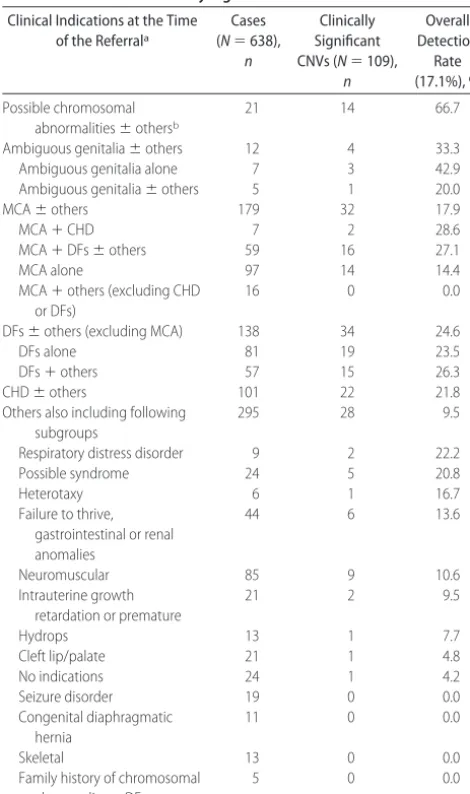
TABLE 1 Clinical Indications at the Time of Referral and Detection Rates for Clinically Significant CNV
Clinical Indications at the Time of the Referrala
Cases (N⫽638),
n
Clinically Significant CNVs (N⫽109),
n Overall Detection Rate (17.1%), % Possible chromosomal abnormalities⫾othersb
21 14 66.7
Ambiguous genitalia⫾others 12 4 33.3
Ambiguous genitalia alone 7 3 42.9
Ambiguous genitalia⫾others 5 1 20.0
MCA⫾others 179 32 17.9
MCA⫹CHD 7 2 28.6
MCA⫹DFs⫾others 59 16 27.1
MCA alone 97 14 14.4
MCA⫹others (excluding CHD or DFs)
16 0 0.0
DFs⫾others (excluding MCA) 138 34 24.6
DFs alone 81 19 23.5
DFs⫹others 57 15 26.3
CHD⫾others 101 22 21.8
Others also including following subgroups
295 28 9.5
Respiratory distress disorder 9 2 22.2
Possible syndrome 24 5 20.8
Heterotaxy 6 1 16.7
Failure to thrive, gastrointestinal or renal anomalies
44 6 13.6
Neuromuscular 85 9 10.6
Intrauterine growth retardation or premature
21 2 9.5
Hydrops 13 1 7.7
Cleft lip/palate 21 1 4.8
No indications 24 1 4.2
Seizure disorder 19 0 0.0
Congenital diaphragmatic hernia
11 0 0.0
Skeletal 13 0 0.0
Family history of chromosomal abnormality or DFs
5 0 0.0
aPatients who hadⱖ2 clinical indications were included inⱖ2 groups in this table. bBroad spectrum of birth defects.
TABLE 2 CNVs Detected by CMA in 638 Neonates With Birth Defects
Parameter March 2006 BAC V5 November 2006 BAC V6a March 2007 V6 Oligo
Coverage of BACs/Oligos,n 853 1475 1475; 44 K
Genomic disorders,n ⬎75 ⬎150 ⬎150
Subtelomeric coverage, Mb 10 10 10
Pericentric coverage Yes Yes Yes
Backbone coverage Yes Expanded Expanded
Neonates studied,n 197 175 266
Clinically significant CNVs,n 27 29 53
Clinically significant CMA detection rate, %
13.7 16.6 19.9
Benign or unknown CNVs,n 18 20 24
Benign or unknown CNVs detection rate, %
9.1 11.4 9.0
variants present in phenotypically normal parents; 27 (4.2%) were classified as being of uncertain significance and awaiting parental studies (data not shown). The detection rates of the “likely benign” group decreased from 9.1% (18 of 197) and 11.4% (20 of 175) to 9.0% (24 of 266) on the 3 sequential versions of arrays (see Table 2), through systematic removal of interrogating clones or oligonucleotides from genomic regions that consistently detected seemingly benign variants. This restricts or reduces the need to perform parental studies on variants that ultimately prove to be benign. These results also reflect our learning curve and experience in designing high-resolution targeted clinical diagnostic ar-rays through better BAC and oligonucleotide probe se-lection.
Clinically Significant Abnormality-Detection Rate and Clinical Indications
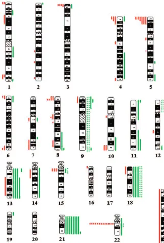
Of a total of 638 neonates, CMA revealed 109 (17.1%) patients with clinically significant chromosomal aberra-tions (Fig 1, Table 1, and Supplemental Table 3, which is published as supporting information on www.pediatrics. org/content/full/122/6/1310); all of these abnormalities were verified by a secondary independent laboratory test: FISH and/or high-resolution retrospective partial karyo-type analyses. The clinical information available for each individual neonatal patient is limited. As a general measure of clinical presentation, we used the clinical indication as determined by the clinician referring to our diagnostic lab-oratory.
In the group of 21 patients who were suspected
clin-FIGURE 1
ically to have chromosomal abnormalities⫾others (eg, DFs, cleft palate, MCA), the CMA clinically significant detection rate was the highest at 66.7% (n ⫽ 14; see Table 1). CMA identified trisomies 21 (6), 18 (2), and 13 (1); all of these were concordant with the clinical indi-cation for the concurrent karyotype analysis. In addi-tion, a derivative chromosome 6 was identified in a patient who was suspected to have trisomy 18; 2 patients were found to have a deletion in Prader-Willi/Angelman syndrome (PWS/AS) critical region, 1 with a deletion in chromosome 8q and 1 with 45,X, which was consistent with a diagnosis of Turner Syndrome.
In 12 patients with ambiguous genitalia⫾other in-dications (see Table 1), CMA detected abnormalities in 4 (33.3%) patients, including 2 with a mosaic 45,X karyo-type with the presence of the Y material, 1 with a com-plex chromosome 5 rearrangement, and 1 with an in-terstitial deletion in the subtelomeric region of the long arm of chromosome 10.
A total of 179 patients were referred for MCA ⫾ others (eg, DFs, CHD, club feet, broad thumbs), and CMA identified significant chromosomal aberrations in 32 cases. All of the CMA findings were summarized in 4 subgroups (see Table 1). For 7 patients who were re-ferred for MCA and CHD, CMA revealed that 2 (28.6%) patients had clinically significant CNVs involving subte-lomeric imbalances in chromosome 1q (1) and 3p (1). A total of 59 patients were referred for MCA ⫹ DFs ⫾ others (eg, polydactyly, failure to thrive; see Table 2), CMA detected 16 (27.1%) with clinically significant genomic imbalance including 2 mosaic tetrasomy 12p, 1 trisomy 21, 1 trisomy 22, 1 deletion in PWS/AS critical region, a complex chromosome 8 rearrangement, and other genomic imbalances involving chromosome 1 (4), chromosome 4 (3), chromosome 11 (2), and 10qter (1).The identified nonmosaic trisomy 22 in 1 patient was later verified by metaphase FISH analysis. This infant was admitted to the NICU secondary to DFs and MCA, where she died at the age of 35 days. She received a diagnosis of CHD with major abnormalities involving double-outlet right ventricle and pulmonary vein steno-sis. No autopsy was performed, thereby limiting the clinical information available. The identified aneuploidy and early death are clearly compatible with a severe phenotype often seen in this rarely reported live-born trisomy. In 113 patients who were referred for MCA alone or MCA⫹others (eg, hydrocephalus, limb anom-alies, but excluding CHD or DFs), CMA detected signif-icant CNVs in 14 patients (see Table 1). Most of these aberrations involved subtelomeric (6) and pericentro-meric (3) imbalances; 3 had microdeletions in DG/VCFS critical region, and 2 had mosaicism for inv dup (15) (1) and inv dup (22) (1).
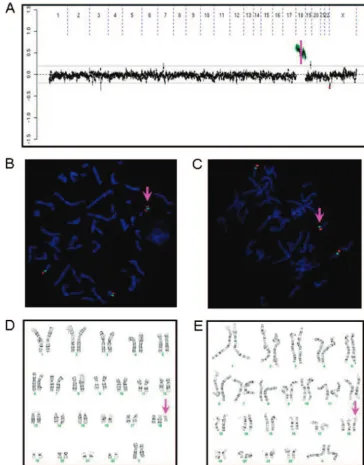
CMA detected 34 (24.6%) patients with clinically significant abnormalities in 138 patients who were re-ferred for DFs alone or DFs ⫹ others (eg, cleft palate, hypotonia, but excluding MCA; see Table 1). Of these, 14 were found to have subtelomeric imbalances, 4 were revealed to have microdeletions involving DG/VCFS (2), Williams-Beuren syndrome (1), and PWS/AS (1) critical regions; 11 were observed to have aneuploidies
includ-ing trisomy 21 (4), trisomy 13 (2), trisomy 18 (1), 45,X (1), mosaic trisomy 9 (2) and mosaic trisomy 18 (1); the remaining 5 had genomic imbalances that involved other rare disease regions. It is interesting that in the mosaic trisomy 18, the CMA ratio plot was suggestive of copy-number differences between the p and q arms of chromosome 18; this result was concordant with the concurrent karyotype analysis of 2 cell lines that con-sisted of 47,XX,⫹18[27]/47,XX,⫹i(18)(p10)[3] (Fig 2).
Among 101 patients who were referred for CHD ⫾ others (see Table 1), CMA identified 22 (21.8%) patients with significant chromosomal aberrations. Eight patients with a common 3-Mb deletion in 22q11.2 had clinical features compatible with a diagnosis of DG/VCFS; 1 pa-tient had an interstitial deletion at 1p36, and the remain-ing patients had subtelomeric imbalances in chromo-somes 3p, 6p, 8p, and 11q.
In 295 patients who were referred for other birth defects (see Table 1), the average clinically significant detection rate was 9.5% (28 of 295). In a small group of 9 patients who were referred for respiratory distress syndrome, CMA detected 2 (22.2%) of them with genomic imbalance, including 1 mosaic tetrasomy 12p, that can be associated with the Pallister-Killian syn-drome, and another 1 involving an unbalanced translo-cation between chromosomes 14 and 18. In 24 patients who were suspected for a possible syndrome, CMA clin-ically significant detection rate was 20.8%.
In summary, of the 109 (17.1% of the 638 neonates referred for testing) patients in which clinically signifi-cant genomic imbalances or pathogenic CNVs were de-tected by CMA (see Fig 1), 16 (14.7%) had numerical anomalies including trisomy 21 (8), trisomy 18 (3), trisomy 13 (3), trisomy 22 (1), and monosomy X (1). The remaining 93 (85.3%) patients had genomic imbal-ances that may not be detected by standard cytogenetic studies, including 37 (33.9%) with common microdele-tions or microduplicamicrodele-tions involving 22q11.2 (13), 5p15.2 (6), 3p26.3 (4), 4p16.3 (4), and other chromo-somal regions (10); 44 (40.4%) with genomic imbal-ances at relatively rare disease loci; and 12 (11.0%) with chromosomal mosaicism.
DISCUSSION
could not be defined by GTG-banded karyotype (see Supplemental Table 3). In fact, the 85.3% (93 of 109) of genomic imbalances detected by CMA were not simply aneuploidies and might have been missed by conven-tional clinical cytogenetic analysis. Although many could have been diagnosed by locus-specific FISH tests or subtelomeric FISH, ordering locus-specific FISH re-quires a presumptive clinical diagnosis. CMA provides a more efficient and reliable method for detecting all of these cases with a single test that enables a “genomic” screen.
More than half of the patients (see Table 1) were referred for MCA ⫾ others, DFs ⫾ others, or CHD ⫾
others. CMA detected the most abnormalities (28.6% and 27.1%) in neonates with MCA⫹CHD and MCA⫹ DFs, respectively. CMA detection rates for DFs⫾others and CHD ⫾ others referrals were 24.6% and 21.8%, respectively. These data suggest that the neonates with these phenotypes are mostly likely to have higher frequencies of genomic imbalances or pathogenic CNVs. In the neonates who were referred with MCA alone, the detection rate was relatively lower at 14.4% (see Table 1).
CMA detected the highest abnormal rate for neonates who were referred with a clinical indication of suspected chromosomal abnormalities. Previous cytogenetic
stud-FIGURE 2
ies showed that the average chromosomal aberration incidence was 21.6% in a total of 6183 pediatric patients who were suspected to have chromosomal abnormali-ties.46–49Remarkably, by using CMA,⬃66.7% (see Table 1) of the neonates who were referred for possible chro-mosomal aberrations, a threefold increase from previous cytogenetic studies, were found to have genomic imbal-ances. The significantly improved detection of chromo-some aberrations substantiates the power of CMA as a high-resolution molecular cytogenetic diagnostic tool.
Many submicroscopic genomic rearrangements have been associated with well-defined clinical syndromes, such as contiguous gene microdeletion/microduplication disorders.50–52 CMA tests hundreds or thousands of genomic loci for CNVs (gains or losses) simultaneously and thus greatly improves the detection rate for identi-fying microdeletion and microduplication syn-dromes.27,53At least 37 (5.8%) patients were identified as having microdeletion or microduplication syndromes that involved the common chromosomal regions in 22q11.2, 5p15.4, and 4p16.3. Among these, the com-mon 3-Mb deletion in DG/VCFS critical region at 22q11.2 (Online Mendelian Inheritance in Man [OMIM] No. 188400 and 192430) was the most prevalent, found in 13 (2.0%) of the referred neonates. In 1 neonate with an apparently balanced inversion of chromosome 2 (not included in this series), CMA revealed a reciprocal mi-croduplication in DG/VCFS critical region at 22q11.2 that was consistent with the clinical diagnosis of 22q11.2 microduplication syndrome (OMIM No. 608363). Both syndromes present with CHD and have other overlap-ping clinical features.54 Often, a significant number of duplication patients were first referred for a FISH test for suspected DG/VCFS. Many patients with FISH-negative results were later found to have the 22q11.2 microdu-plication syndromes by CMA27or interphase FISH anal-ysis.55 Recent work that applied clinical arrays docu-mented a new genomic disorder that is associated with deletion just distal to the common DG/VCFS locus.56 Common subtelomeric rearrangements were identified in 3 chromosomal regions in the distal 3p, 4p, and 5p. Monosomy 4p and 5p have been well defined to be associated with Wolf-Hirschhorn syndrome (OMIM No.
194190) and Cri-du-chat syndrome (OMIM No.
123450); however, the clinical manifestation of micro-duplications in the distal 4p as well as the deletions in the distal region of 3p57,58are not very well defined, and they could represent new genomic syndromes.
CMA in this study also demonstrated an increased detection rate for identifying chromosomal mosaicism. CMA for 12 patients, mostly referred for DFs and MCA, were suggestive for mosaicism. Five (38.5%) patients had low-level mosaicism with aberrations that were not detected in retrospective chromosomal metaphase anal-yses, including tetrasomy 12p (3) and trisomy 9 (2). Tetrasomy 12p has been associated with Pallister-Killian syndrome, and skin biopsy for conventional cytogenetic studies is generally recommended when clinically indi-cated; however, none of these 3 neonates whom we reported with mosaic tetrasomy 12p in this study was suspected for this diagnosis, including 1 patient who
was reported to have a 45,X karyotype before CMA and was known to manifest clinically DFs and MCA findings that were not consistent with a Turner syndrome phe-notype. Interphase FISH analysis on blood smears was needed to verify mosaicism in these cases. As elucidated in previous studies,36,37CMA on DNA from multiple cell lineages in whole blood improves the ability to detect mosaicism compared with conventional karyotype anal-ysis, which examines only the stimulated T lymphocytes that respond to mitogens. Chromosomal low-level mo-saicism has been shown in this study to exist in neonates with unexplained birth detects such as MCA, DFs, and CHD and seemingly normal chromosomes by karyotype analysis, and, therefore aCGH/CMA is highly recom-mended for these neonates for additional evaluation of the possible chromosomal mosaicism.
To our knowledge, this is the largest study to date to use aCGH to detect the frequencies of genomic imbal-ances in a large series of neonates with birth defects and has important implications for the evaluation of congen-ital anomalies with unknown genetic cause. Limitations of this study include the relatively modest sample size (638) and that neonates with birth defects in this study were not characterized on the basis of the coding of birth defects because of limited clinical information.12,59 We propose that additional investigations use CMA in large neonatal sample cohorts, such as the DNA sample col-lection of the National Birth Defects Prevention Study (NBDPS),60 to identify the potential genomic imbal-ances and disease gene(s) that underlie specific birth defects.
CONCLUSIONS
This study demonstrates that CMA can effectively diag-nose birth defects that are caused by submicroscopic genomic imbalances in the neonatal period and, there-fore, assist the clinician in diagnosis, early neonatal in-tervention, and genetic counseling. Importantly, the CMA was ⬃3 times as likely to detect an abnormality than routine clinical chromosome analysis in a neonate who was referred with a clinical suspicion of a chromo-somal syndrome. Better understanding of the causes of birth defects can potentially enable effective prenatal detection and potential avenues for therapy on the basis of mitigating the clinical consequences of the dosage-sensitive gene(s) whose CNV is responsible for disease.63 We recommend the consideration of using CMA in lieu of routine cytogenetic testing to evaluate neonates with birth defects of unknown cause.
ACKNOWLEDGMENTS
This work is supported in part by Baylor College of Medicine Mental Retardation Developmental Disabilities Research Center grant HD024064 –20.
We thank all of the referring physicians, patients, and families for cooperation. We also thank all of our col-leagues and co-workers from the CMA laboratory, Array Production Laboratory, Cytogenetic/FISH Laboratory, and DNA Diagnostic Laboratory at Baylor College of Medicine for contributions and support.
REFERENCES
1. Mathews TJ, MacDorman MF. Infant mortality statistics from the 2003 period linked birth/infant death data set.Natl Vital Stat Rep.2006;54(16):1–29
2. Canfield MA, Honein MA, Yuskiv N, et al. National estimates and race/ethnic-specific variation of selected birth defects in the United States, 1999 –2001.Birth Defects Res A Clin Mol Tera-tol.2006;76(11):747–756
3. Yoon PW, Rasmussen SA, Lynberg MC, et al. The National Birth Defects Prevention Study. Public Health Rep. 2001; 116(suppl 1):32– 40
4. Hamerton JL, Canning N, Ray M, Smith S. A cytogenetic survey of 14,069 newborn infants: I—incidence of chromo-some abnormalities.Clin Genet.1975;8(4):223–243
5. Evans JA, de von FR, Greenberg C, Ramsay S, Hamerton JL. A cytogenetic survey of 14,069 newborn infants: IV—further follow-up on the children with sex chromosome anomalies. Birth Defects Orig Artic Ser.1982;18(4):169 –184
6. Seashore MR. Chromosomal abnormalities in the newborn period.Semin Perinatol.1993;17(5):312–317
7. Smith A, Bannatyne P, Russell P, Ellwood D, den Dulk G. Cytogenetic studies in perinatal death.Aust N Z J Obstet Gynae-col.1990;30(3):206 –210
8. Larson RS, Butler MG. Use of fluorescence in situ hybridization (FISH) in the diagnosis of DiGeorge sequence and related dis-eases.Diagn Mol Pathol.1995;4(4):274 –278
9. Gandelman KY, Gibson L, Meyn MS, Yang-Feng TL. Molecular definition of the smallest region of deletion overlap in the Wolf-Hirschhorn syndrome. Am J Hum Genet. 1992;51(3): 571–578
10. Gersh M, Grady D, Rojas K, et al. Development of diagnostic tools for the analysis of 5p deletions using interphase FISH. Cytogenet Cell Genet.1997;77(3– 4):246 –251
11. Liu S, Joseph KS, Wen SW. Trends in fetal and infant deaths
caused by congenital anomalies. Semin Perinatol.2002;26(4): 268 –276
12. Rasmussen SA, Moore CA. Effective coding in birth defects surveillance.Teratology.2001;64(suppl 1):S3–S7
13. Ming JE, Geiger E, James AC, et al. Rapid detection of submi-croscopic chromosomal rearrangements in children with mul-tiple congenital anomalies using high density oligonucleotide arrays.Hum Mutat.2006;27(5):467– 473
14. Pinkel D, Segraves R, Sudar D, et al. High resolution analysis of DNA copy number variation using comparative genomic hy-bridization to microarrays.Nat Genet.1998;20(2):207–211 15. Sebat J, Lakshmi B, Troge J, et al. Large-scale copy number
polymorphism in the human genome.Science.2004;305(5683): 525–528
16. Iafrate AJ, Feuk L, Rivera MN, et al. Detection of large-scale variation in the human genome. Nat Genet. 2004;36(9): 949 –951
17. White SJ, Vissers LE, Geurts van KA, et al. Variation of CNV distribution in five different ethnic populations.Cytogenet Ge-nome Res.2007;118(1):19 –30
18. Lee C, Iafrate AJ, Brothman AR. Copy number variations and clinical cytogenetic diagnosis of constitutional disorders. Nat Genet.2007;39(7 suppl):S48 –S54
19. Sebat J, Lakshmi B, Malhotra D, et al. Strong association of de novo copy number mutations with autism. Science. 2007; 316(5823):445– 449
20. Cai WW, Mao JH, Chow CW, et al. Genome-wide detection of chromosomal imbalances in tumors using BAC microarrays. Nat Biotechnol.2002;20(4):393–396
21. Rouillard JM, Herbert CJ, Zuker M. OligoArray: genome-scale oligonucleotide design for microarrays. Bioinformatics. 2002; 18(3):486 – 487
22. Ou Z, Kang SH, Shaw CA, et al. Bacterial artificial chromo-some-emulation oligonucleotide arrays for targeted clinical ar-ray-comparative genomic hybridization analyses. Genet Med. 2008;10(4):278 –289
23. Friedman JM, Baross A, Delaney AD, et al. Oligonucleotide microarray analysis of genomic imbalance in children with mental retardation.Am J Hum Genet.2006;79(3):500 –513 24. Aradhya S, Cherry AM. Array-based comparative genomic
hybridization: clinical contexts for targeted and whole-genome designs.Genet Med.2007;9(9):553–559
25. Cheung SW, Shaw CA, Yu W, et al. Development and valida-tion of a CGH microarray for clinical cytogenetic diagnosis. Genet Med.2005;7(6):422– 432
26. Shaffer LG, Bejjani BA. Medical applications of array CGH and the transformation of clinical cytogenetics. Cytogenet Genome Res.2006;115(3– 4):303–309
27. Lu X, Shaw CA, Patel A, et al. Clinical implementation of chromosomal microarray analysis: summary of 2513 postnatal cases.PLoS ONE.2007;2(3):e327
28. Sharp AJ, Hansen S, Selzer RR, et al. Discovery of previously unidentified genomic disorders from the duplication architec-ture of the human genome.Nat Genet.2006;38(9):1038 –1042 29. Menten B, Maas N, Thienpont B, et al. Emerging patterns of cryptic chromosomal imbalance in patients with idiopathic mental retardation and multiple congenital anomalies: a new series of 140 patients and review of published reports.J Med Genet.2006;43(8):625– 633
30. Sahoo T, Cheung SW, Ward P, et al. Prenatal diagnosis of chromosomal abnormalities using array-based comparative genomic hybridization.Genet Med.2006;8(11):719 –727 31. Van den Veyver I, Beaudet AL. Comparative genomic
hybrid-ization and prenatal diagnosis.Curr Opin Obstet Gynecol.2006; 18(2):185–191
of unbalanced chromosomal rearrangements by array CGH. J Med Genet.2006;43(4):353–361
33. Vissers LE, Veltman JA, van Kessel AG, Brunner HG. Identifi-cation of disease genes by whole genome CGH arrays.Hum Mol Genet.2005;14(Spec No. 2):R215–R223
34. Lupski JR. Genome structural variation and sporadic disease traits.Nat Genet.2006;38(9):974 –976
35. Ballif BC, Hornor SA, Jenkins E, et al. Discovery of a previously unrecognized microdeletion syndrome of 16p11.2–p12.2.Nat Genet.2007;39(9):1071–1073
36. Ballif BC, Rorem EA, Sundin K, et al. Detection of low-level mosaicism by array CGH in routine diagnostic specimens.Am J Med Genet A.2006;140(24):2757–2767
37. Cheung SW, Shaw CA, Scott DA, et al. Microarray-based CGH detects chromosomal mosaicism not revealed by conventional cytogenetics.Am J Med Genet A.2007;143A(15):1679 –1686 38. Bar-Shira A, Rosner G, Rosner S, Goldstein M, Orr-Urtreger A.
Array-based comparative genome hybridization in clinical ge-netics.Pediatr Res.2006;60(3):353–358
39. Caselli R, Mencarelli MA, Papa FT, et al. A 2.6 Mb deletion of 6q24.3–25.1 in a patient with growth failure, cardiac septal defect, thin upperlip and asymmetric dysmorphic ears. Eur J Med Genet.2007;50(4):315–321
40. Stankiewicz P, Beaudet AL. Use of array CGH in the evaluation of dysmorphology, malformations, developmental delay, and idiopathic mental retardation.Curr Opin Genet Dev.2007;17(3): 182–192
41. Shaw-Smith C, Redon R, Rickman L, et al. Microarray based comparative genomic hybridisation (array-CGH) detects sub-microscopic chromosomal deletions and duplications in pa-tients with learning disability/mental retardation and dysmor-phic features.J Med Genet.2004;41(4):241–248
42. Schoumans J, Ruivenkamp C, Holmberg E, et al. Detection of chromosomal imbalances in children with idiopathic mental retardation by array based comparative genomic hybridisation (array-CGH).J Med Genet.2005;42(9):699 –705
43. Aradhya S, Manning MA, Splendore A, Cherry AM. Whole-genome array-CGH identifies novel contiguous gene deletions and duplications associated with developmental delay, mental retardation, and dysmorphic features.Am J Med Genet A.2007; 143A(13):1431–1441
44. de Vries BB, Pfundt R, Leisink M, et al. Diagnostic genome profiling in mental retardation.Am J Hum Genet.2005;77(4): 606 – 616
45. Krepischi-Santos AC, Vianna-Morgante AM, Jehee FS, et al. Whole-genome array-CGH screening in undiagnosed syn-dromic patients: old syndromes revisited and new alterations. Cytogenet Genome Res.2006;115(3– 4):254 –261
46. Shah VC, Murthy DS, Murthy SK. Cytogenetic studies in a population suspected to have chromosomal abnormalities. In-dian J Pediatr.1990;57(2):235–243
47. Kenue RK, Raj AK, Harris PF, el-Bualy MS. Cytogenetic anal-ysis of children suspected of chromosomal abnormalities.J Trop Pediatr.1995;41(2):77– 80
48. Kim SS, Jung SC, Kim HJ, Moon HR, Lee JS. Chromosome abnormalities in a referred population for suspected chromo-somal aberrations: a report of 4117 cases. J Korean Med Sci. 1999;14(4):373–376
49. Goud MT, Al-Harassi SM, Al-Khalili SA, et al. Incidence of chromosome abnormalities in the Sultanate of Oman.Saudi Med J.2005;26(12):1951–1957
50. Lupski JR. Genomic disorders: structural features of the ge-nome can lead to DNA rearrangements and human disease traits.Trends Genet.1998;14(10):417– 422
51. Stankiewicz P, Lupski JR. Genome architecture, rearrange-ments and genomic disorders.Trends Genet.2002;18(2):74 – 82 52. Lupski JR, Stankiewicz P, ed.Genomic Disorders: The Genomic
Basis of Disease. Totowa, NJ: Humana Press; 2006:1– 427 53. Berg JS, Brunetti-Pierri N, Peters SU, et al. Speech delay and
autism spectrum behaviors are frequently associated with du-plication of the 7q11.23 Williams-Beuren syndrome region. Genet Med.2007;9(7):427– 441
54. Ou Z, Berg JS, Yonath H, et al. Microduplications of 22q11.2 are frequently inherited and are associated with variable phe-notypes.Genet Med.2008;10(4):267–277
55. Ensenauer RE, Adeyinka A, Flynn HC, et al. Microduplication 22q11.2, an emerging syndrome: clinical, cytogenetic, and mo-lecular analysis of thirteen patients.Am J Hum Genet. 2003; 73(5):1027–1040
56. Ben-Shachar S, Ou Z, Shaw CA, et al. 22q11.2 distal deletion: a recurrent genomic disorder distinct from DiGeorge syndrome and velocardiofacial syndrome.Am J Hum Genet.2008;82(1): 214 –221
57. Dijkhuizen T, van ET, van der Vlies P, et al. FISH and array-CGH analysis of a complex chromosome 3 aberration suggests that loss ofCNTN4andCRBNcontributes to mental retardation in 3pter deletions.Am J Med Genet A.2006;140(22):2482–2487 58. McCullough BJ, Adams JC, Shilling DJ, et al. 3p-syndrome defines a hearing loss locus in 3p25.3.Hear Res.2007;224(1–2): 51– 60
59. Rasmussen SA, Olney RS, Holmes LB, et al. Guidelines for case classification for the National Birth Defects Prevention Study. Birth Defects Res A Clin Mol Teratol.2003;67(3):193–201 60. Rasmussen SA, Lammer EJ, Shaw GM, et al. Integration of
DNA sample collection into a multi-site birth defects case-control study.Teratology.2002;66(4):177–184
61. De Gregori M, Ciccone R, Magini P, et al. Cryptic deletions are a common finding in “balanced” reciprocal and complex chro-mosome rearrangements: a study of 59 patients.J Med Genet. 2007;44(12):750 –762
62. Higgins AW, Alkuraya FS, Bosco AF, et al. Characterization of apparently balanced chromosomal rearrangements from the developmental genome anatomy project. Am J Hum Genet. 2008;82(3):712–722
DOI: 10.1542/peds.2008-0297
2008;122;1310
Pediatrics
Lalani, A. Craig Chinault, James R. Lupski, Sau W. Cheung and Arthur L. Beaudet
Trilochan Sahoo, Carlos A. Bacino, Pawel Stankiewicz, Sung-Hae Lee Kang, Seema
Xin-Yan Lu, Mai T. Phung, Chad A. Shaw, Kim Pham, Sarah E. Neil, Ankita Patel,
Using Chromosomal Microarray Analysis
Genomic Imbalances in Neonates With Birth Defects: High Detection Rates by
Services
Updated Information &
http://pediatrics.aappublications.org/content/122/6/1310
including high resolution figures, can be found at:
References
http://pediatrics.aappublications.org/content/122/6/1310#BIBL
This article cites 62 articles, 7 of which you can access for free at:
Subspecialty Collections
http://www.aappublications.org/cgi/collection/genetics_sub
Genetics
following collection(s):
This article, along with others on similar topics, appears in the
Permissions & Licensing
http://www.aappublications.org/site/misc/Permissions.xhtml
in its entirety can be found online at:
Information about reproducing this article in parts (figures, tables) or
Reprints
http://www.aappublications.org/site/misc/reprints.xhtml
DOI: 10.1542/peds.2008-0297
2008;122;1310
Pediatrics
Lalani, A. Craig Chinault, James R. Lupski, Sau W. Cheung and Arthur L. Beaudet
Trilochan Sahoo, Carlos A. Bacino, Pawel Stankiewicz, Sung-Hae Lee Kang, Seema
Xin-Yan Lu, Mai T. Phung, Chad A. Shaw, Kim Pham, Sarah E. Neil, Ankita Patel,
Using Chromosomal Microarray Analysis
Genomic Imbalances in Neonates With Birth Defects: High Detection Rates by
http://pediatrics.aappublications.org/content/122/6/1310
located on the World Wide Web at:
The online version of this article, along with updated information and services, is
http://pediatrics.aappublications.org/content/suppl/2008/12/01/122.6.1310.DC1
Data Supplement at:
by the American Academy of Pediatrics. All rights reserved. Print ISSN: 1073-0397.