Congenital Transmission of Visceral Leishmaniasis (Kala Azar) From an
Asymptomatic Mother to Her Child
Christoph K. Meinecke, MD*; Justus Schottelius, PhD‡; Linda Oskam, PhD§; and Bernhard Fleischer, MD‡
ABSTRACT. In this article, we report the case of a 16-month-old German boy who was admitted to the Chil-dren’s Hospital of Stuttgart with a 4-week history of intermittent fever, decreased appetite, weakness, fatigue, and difficulty sleeping. He was healthy at birth and remained so for the first 15 months of his life. On admis-sion, physical examination showed enlarged cervical, ax-illary, and inguinal lymph nodes, as well as hepato-splenomegaly. Laboratory data revealed pancytopenia, elevated liver function tests, and hypergammaglobuline-mia. Blood, stool, and urine culture results were negative. Viral infections and rheumatologic and autoimmune dis-orders were ruled out, but a positive titer forLeishmania
antibodies was noted. In a liver and bone marrow biopsy, the amastigote form of the parasite could not be seen in cells. The promastigote form of Leishmania was found and the diagnosis of visceral leishmaniasis was made by combining the cultures of both the liver and the bone marrow biopsy material in 5 mL 0.9% saline on brain heart infusion agar, supplemented with defibrinated rab-bit blood and incubated at 25 to 26°C for 5 days. The parasite was identified by Southern blot analysis as
Leishmania infantum.
Specific therapy with the antimonial compound so-dium stibogluconate with a dose of 20 mg/kg body weight was begun immediately. Within 4 days, the pa-tient became afebrile. The side effects of treatment, in-cluding erosive gastritis, cholelithiasis, worsening hepa-tosplenomegaly, elevation of liver enzymes, pancreatitis, and electrocardiogram abnormalities, necessitated the discontinuation of treatment after 17 days. On discharge 4 weeks later, the patient was stabilized and afebrile with a normal spleen, normal complete blood count, normal gammaglobulins, and decreasing antibody titers to Leish-mania. During the next 24 months, the patient experi-enced intermittent episodes of abdominal pain, de-creased appetite, recurrent arthralgia, and myalgia. But at his last examination in January 1998, he was well; all symptoms mentioned above had disappeared.
Because the child had never left Germany, nonvector transmission was suspected and household contacts were examined. His mother was the only one who had a pos-itive antibody titer against Leishmania donovani com-plex. She had traveled several times to endemic Mediter-ranean areas (Portugal, Malta, and Corse) before giving birth to the boy. But she had never been symptomatic for visceral leishmaniasis. Her bone marrow, spleen, and liver biopsy results were within normal limits. Culture
results and polymerase chain reaction of this material were negative. A Montenegro skin test result was posi-tive, indicating a previous infection with Leishmania. Western blot analysis showed specific recognition by maternal antibodies of antigens ofLeishmania cultured from the boy’s tissue.
Visceral leishmaniasis is endemic to several tropical and subtropical countries, but also to the Mediterranean region. It is transmitted by the sand fly (Phlebotomus,
Lutzomyia). Occasional nonvector transmissions also have been reported through blood transfusions, sexual intercourse, organ transplants, excrements of dogs, and sporadically outside endemic areas. Only 8 cases of con-genital acquired disease have been described before 1995, when our case occurred.
In our patient, additional evaluation showed that the asymptomatic mother must have had a subclinical infec-tion withLeishmaniathat was reactivated by pregnancy, and then congenitally transmitted to the child. Visceral leishmaniasis has to be considered in children with fe-ver, pancytopenia, and splenomegaly, even if the child has not been to an endemic area and even if there is no evidence of the disease in his environment, because leishmaniasis can be transmitted congenitally from an asymptomatic mother to her child.Pediatrics1999;104(5). URL: http://www.pediatrics.org/cgi/content/full/104/5/ e65;visceral leishmaniasis in infants, kala azar, congeni-tal transmission, nonvector transmission.
ABBREVIATIONS. EIA, enzyme-linked immunosorbent assay; IFA, immunofluorescence assay; CF, complement fixation test; ECG, electrocardiogram; PCR, polymerase chain reaction.
V
isceral leishmaniasis is endemic to several
tropical and subtropical countries but also to
the Mediterranean region. It is transmitted by
the sand fly (
Phlebotomus
,
Lutzomyia
). Occasional
nonvector transmissions also have been reported
through blood transfusions, sexual intercourse,
or-gan transplants, excrements of dogs, and
sporadi-cally outside endemic areas. Only 8 cases of
congen-ital acquired disease have been described before
1995.
1– 8In all these case reports, the mothers were
symptomatic for the disease. In this article, we report
the case of a boy who acquired the disease
congeni-tally by transmission from his asymptomatic mother.
METHODS
All diagnostic procedures and the treatment of the boy were conducted at the Children’s Hospital of Stuttgart in Germany. The serologic tests onLeishmania donovani species included enzyme-linked immunosorbent assay (EIA; cutoff values: weak positive $10 antibody units and strong positive$30 antibody units), im-munofluorescence assay (IFA; cutoff values: weak positive titers $1:20 and strong positive titers$1:80), and complement fixation
From the *Children’s Hospital of Stuttgart (Olgahospital), Stuttgart, many; ‡Bernhard Nocht Institute for Tropical Medicine, Hamburg, Ger-many; and the §Department of Biomedical Research, Royal Tropical Institute, Amsterdam, the Netherlands.
Received for publication Oct 21, 1998; accepted May 17, 1999.
tests (CFs; cutoff values: weak positive titers $1:4 and strong positive titers$1:16). The cultivation of liver and bone marrow biopsy material was performed in 5 mL 0.9% saline on brain heart infusion agar, supplemented with defibrinated rabbit blood (Difco, Detroit, MI) and incubated at 25 to 26°C for 5 days. Then the promastigote form ofLeishmaniawas detected by microscopy. The serologic tests, the cultivation, the microscopic examinations, and the Western blot analysis of the mother’s serum against Leishmaniaantigens from the child were performed at the Bern-hard Nocht Institute for Tropical Medicine in Hamburg, Germany. The identification of the promastigotes was performed by South-ern blot analysis, using probe pDK209at the Royal Tropical
Insti-tute, Department of Biomedical Research in Amsterdam, the Neth-erlands.
CASE REPORT
In February 1995, a 16-month-old boy from a small village in southwestern Germany was admitted to the Children’s Hospital of Stuttgart with a 4-week history of intermittent fever up to 40°C, decreased appetite, weakness, fatigue, and difficulty sleeping. He was born to a 31-year-old prima gravida, prima para at 40 weeks’ gestation by vacuum extraction with a birth weight of 3720 g and a length of 53 cm. The mother’s pregnancy was complicated by a febrile gastroenteritis, but the newborn was healthy, and the child remained healthy for the first year of his life. At 15 months of age, he developed fever, enlarged cervical lymph nodes, poor appetite, and a transient icterus with elevated liver function tests. A pre-sumptuous diagnosis of infectious mononucleosis was made. Be-cause the symptoms persisted for.3 weeks, however, he was readmitted to the hospital for additional evaluation and treatment. Physical examination on admission showed a 16-month-old febrile male in mild distress. There were no signs of meningitis, and ear, nose, and throat examination results were normal, with normal lung and heart auscultation. The examination was remarkable for enlarged cervical, axillary, and inguinal lymph nodes (338 cm, 132 cm, and 234 cm, respectively), as well as hepatospleno-megaly (4 and 6 cm below the costal margin, respectively). Labo-ratory data revealed a normal erythrocyte sedimentation rate (10
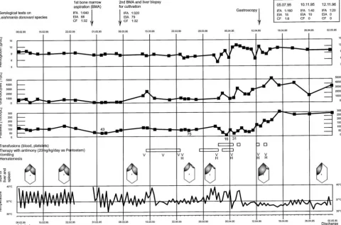
mm per hour), pancytopenia (hemoglobin 7.8 g/dL, leukocytope-nia with an absolute neutrophil count of 100 –200/mL, platelets 40 000/mL), elevated liver function tests (serum glutamic oxaloa-cetic transaminase 153 U/L, serum glutamic pyruvate transami-nase 184 U/L, lactic acid dehydrogetransami-nase 467 U/L), with normal bilirubin levels, and hypergammaglobulinemia (immunoglobulin G 2577 mg/dL). The results of a chest radiograph were within normal limits. Abdominal sonography verified the hepatospleno-megaly. Cardiac echography showed a mild pericardial effusion. The results of three blood cultures were negative. Urine analysis and stool and urine culture results were also negative. Serologic studies showed no evidence of brucellosis, leptospirosis, or Ep-stein-Barr virus infection. Human immunodeficiency virus infec-tion, rheumatologic, and autoimmune disorders were ruled out. A bone scan was negative, and a bone marrow aspirate showed no evidence of malignancy, only a proliferation of lymphocytes and macrophages with increased hemophagocytosis. Because of the severity of the illness, an antibiotic treatment was initiated empir-ically, without any clinical improvement. After 7 weeks of inter-mittent fever, a positive titer forLeishmaniaantibodies (L donovani complex, IFA 1:640, EIA 68, CF 1:32) was noted. The results were confirmed on a second specimen. In a repeat liver and bone marrow biopsy, the amastigote form of the parasite could not be seen in cells. Only by combined culture of both the liver and the second bone marrow biopsy material was the promastigote form ofLeishmania found and the diagnosis of visceral leishmaniasis made. The parasite was identified as Leishmania infantumusing Southern blot analysis.
Specific therapy with the antimonial compound sodium stibo-gluconate (Pentostam; Wellcome Foundation, London, UK) at a dose of 20 mg/kg body weight was begun immediately after the biopsies were taken, given once daily intravenously (Fig 1). Within 4 days, the patient became afebrile but developed hemate-mesis, elevation of liver enzymes, and electrocardiogram (ECG) abnormalities, necessitating discontinuation of treatment. After the status of the patient improved, treatment was restarted. The side effects of the treatment, including erosive gastritis, cholelithi-asis, worsening hepatosplenomegaly, and pancreatitis
tated the discontinuation of treatment after 17 days. The patient stabilized and was discharged from the hospital 4 weeks after treatment with Pentostam. On discharge, he was afebrile with a normal spleen, normal complete blood count and gammaglobu-lins, and decreasing antibody titers toLeishmania. During the next 24 months, the patient experienced intermittent episodes of ab-dominal pain, decreased appetite, recurrent arthralgia, and myal-gia. His hepatomegaly slowly resolved, however, and there was no recurrence of splenomegaly or fever. His physical examination remained unremarkable and the antibody titers toLeishmaniawere low (in November 1996, IFA 1:20, EIA negative, CF negative). No objective signs of rheumatologic, immunologic, or neurological diseases were found during detailed investigations in March 1997 at the University Children’s Hospital of Tu¨bingen. Antibody titers remained low, polymerase chain reaction (PCR) was negative, and bone marrow aspirate was normal. At his last examination in January 1998, the boy was well and all of the symptoms that were mentioned above had disappeared.
Because the child had never left Germany, transmission by the sand fly could be excluded. Household contacts (including the family’s dog) were examined. His mother was the only one who had a positive antibody titer againstL donovanicomplex (IFA 1:40, EIA 16, CF 1:4). The mother had traveled to several endemic Mediterranean countries 2, 5, and 6 years before giving birth to the boy (Table 1). She had never had any symptoms compatible with visceral leishmaniasis. Her bone marrow, spleen, and liver biop-sies were within normal limits. Culture and PCR of this material were negative. A Montenegro skin test result (test for cutaneous delayed hypersensitivity response to a killed promastigote prep-aration called leishmanin) was positive, indicating a previous infection withLeishmania. Western blot analysis showed specific recognition by maternal antibodies of antigens ofLeishmania cul-tured from the boy’s tissue. Neither the mother nor the child had ever received a blood transfusion.
DISCUSSION
Infections with the protozoan parasite
Leishmania
can lead to three different forms of disease:
cutane-ous, mucocutanecutane-ous, and visceral leishmaniasis.
Vis-ceral leishmaniasis, also called kala azar, is usually
caused by
L donovani
,
L infantum
, or
L chagasi
and
rarely by
L tropica
or
L mexicana
. These are protozoan
parasites that are generally transmitted by sand flies
and then disseminate in the body of their host by
infecting macrophages in multiple organs, but
par-ticularly in the spleen, liver, bone marrow, and
lymph nodes. The clinical incubation period ranges
typically from 6 weeks to 6 months but can vary from
10 days to
.
10 years. The course of the disease is
identical in children and adults. It may begin either
suddenly with high fevers, vomiting, diarrhea, and
coughing, or insidiously with irregular daily
increas-ing fever, poor appetite, weight loss, lassitude, and
pallor. Later characteristic findings are
splenomeg-aly, fever up to 41°C with a pattern of double daily
spikes (however, this pattern is seen rarely),
recur-rent respiratory and intestinal infections,
pancytope-nia, and hyperglobulinemia. Hepatomegaly,
lymph-adenopathy, and general bleeding diathesis may
follow. Untreated, the disease is fatal in 90% of cases
after 1 to 3 years. Diagnosis should be confirmed
either by microscopic identification of the amastigote
or promastigote form of the parasite in liver, spleen,
or bone marrow biopsies, or by detection of the
desoxyribonucleic acid of
Leishmania
by PCR in
blood or biopsy material. The mainstay treatment is
with pentavalent antimony (20 mg/kg intravenously
in the form of sodium stibogluconate once daily for
28 days), which reduces mortality from 90% to 1 to
15%. Side effects may occur, including nausea,
vom-iting, jaundice, ECG abnormalities, elevation of liver
enzymes, pancreatitis, worsening of hepatomegaly,
bleeding diathesis, and thrombocytopenia. Other
ac-cepted drug regimens include meglumine
anti-monate (Glucantime, 60 –100 mg/kg intramuscularly
once daily for 14 –28 days). The daily intramuscular
injection may cause painful local inflammation.
Tox-icity is similar to sodium stibogluconate but side
effects include renal failure and sudden death.
10Al-lopurinol (15 mg/kg per day) in combination with
antimonials may reduce the rate of relapse.
Pentam-idine (2– 4 mg/kg daily intramuscularly or
intrave-nously for 15 days or every second day for 30 days)
and amphotericin B (1 mg/kg intravenously on
al-ternate days giving a total dose of 30 –35 mg/kg)
show much more toxicity and are used only as
sec-ond-line regimens. Liposomal amphotericin B
(Am-Bisome) is more expensive but less toxic and more
efficient than conventional amphotericin B. Recent
studies in children suggest that even in comparison
with pentavalent antimonials, it may provide better
results, shorter courses of treatment, lower costs of
hospitalization, and fewer side effects. Two
alterna-tive regimens are recommended: a daily dose of 3
mg/kg for 10 days or a daily dose of 3 mg/kg only
on days 0, 1, 2, 3, 4, and 10.
11,12Congenital visceral leishmaniasis was described
first in 1926 by Low and Cooke.
1Seven more case
reports had been published until 1995, when our case
occurred.
2– 8One more case report has been
pub-lished since
13(Table 2). The course of the disease
seems to be identical in congenital transmitted and
otherwise acquired kala azar. Most of the children
developed the disease in the first year of life.
How-ever, in congenital cases the route of transmission
remains unclear. Most likely the infection occurs
dur-ing labor via blood exchange from the mother to the
child. Transplacental transmission during pregnancy
before birth is rather unprobable, because no
para-sites were found in the organs of an aborted fetus of
5 months’ gestational age who was born to a
30-year-old infected mother in Sudan while the placenta
showed large numbers of amastigotes.
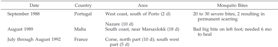
7TABLE 1. Mother’s Travel History to Endemic Mediterranean Countries
Date Country Area Mosquito Bites
September 1988 Portugal West coast, south of Porto (2 d) 20 to 30 severe bites, 2 resulting in permanent scarring
Nazare (10 d)
August 1989 Malta South coast, near Marsaxlokk (18 d) Bad big bite on left foot; needed 6 mo to heal
Each year 500 000 new infections with visceral
leishmaniasis occur. According to the World Health
Organization, the ratio of subclinical to clinical
infec-tions is 5:1.
14A study from Kenya suggested that
asymptomatic persons can be a reservoir of
Leishma-nia
parasites for expended periods.
15Patients can
develop leishmaniasis years and even decades after
traveling to an endemic area, if they become
immu-nosuppressed.
16During pregnancy, a shift from
cell-mediated to humoral immunity has been described
in mice as well as in humans.
17Therefore, females
may have a higher susceptibility to leishmaniasis
during pregnancy, as has been shown recently in
mice.
18This also may suggest that pregnancy can
trigger (re)activation of leishmaniasis.
In our patient, visceral leishmaniasis started
insid-iously. On hospital admission, the patient presented
with intermittent fever, lymphadenopathy,
hepato-splenomegaly, and pancytopenia. Bacterial
infec-tions, autoimmune disorders, and malignancies were
ruled out. An unusual infection was suspected.
Al-though the patient had no travel history to any
en-demic country, a laboratory diagnosis of visceral
leishmaniasis was made. The diagnosis was
con-firmed by culture. The promastigote parasite could
be identified as
L infantum.
(PCR was not available at
our hospital and institute at that time).
Additional evaluation showed that the
asymptom-atic mother was the only potential carrier in the
environment of the child who showed antibody titers
against
Leishmania
. Therefore, she must have had a
subclinical infection that could have been reactivated
by pregnancy, which was then transmitted
congeni-tally to the child. The flu-like symptoms that the
mother described in early pregnancy might represent
nonspecific signs of reactivation. The mother’s
posi-tive Montenegro skin test result and the negaposi-tive
culture result for
Leishmania
suggest that the
para-sites persisted after infection and eventually
reap-peared during pregnancy to induce parasitemia but
after pregnancy were suppressed in the mother by
cellular immunity. She most likely contracted the
disease years before this pregnancy during her
jour-neys to several endemic Mediterranean areas:
Portu-gal, Malta, and Corse. The prevalence of visceral
leishmaniasis in this areas is not known, but the
latest available reports show
;
70, 8, and 3 new cases
each year, respectively.
14Leishmania infantum
is
en-demic to the whole Mediterranean region.
During therapy with pentavalent antimony
(Pen-tostam), our patient developed hematemesis and
gastrointestinal bleeding, which may be part of the
disease. There is evidence about a toxic effect of
Pentostam on the platelets
19that could have
aggra-vated the bleeding. Most likely, the elevation of liver
enzymes and ECG changes were also side effects.
Temporary discontinuation of treatment is necessary
when these side effects occur. We do not know
whether treatment with liposomal amphotericin B
might have minimized the complications in our case.
Future studies are needed to evaluate the advantages
of this medication.
Three years after discharge from the hospital, our
patient is a normally developed 4-year-old boy. He
has a younger brother who is now 2 years old. The
brother never showed any signs of leishmaniasis and
is seronegative for the disease.
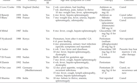
TABLE 2. Reported Cases of Congenital Kala Azar
Author Year Country Mother
III
Age and Symptoms of Child Therapy Outcome
1 Low/Cooke 1926 England (India) Yes 2 wk: convulsion, bad feeding Antimon as Cured 7 wk: diarrhoea, pain, failure to thrive Stibosan (75
10 mo: weight loss, fever, splenomegaly mg/d)
2 Banerji 1955 India Yes 6 mo: fever, hepato-splenomegaly Antimon as Cured (after
3 Blanc 1984 France Yes 7 mo: weight loss, fever, icterus, hepato- Stibatin 1 recurrence) splenomegaly, adenopathy Glucantime
(glucamine antimonate) 500 mg/d
Cured
4 Mittal 1987 India Yes 8 mo: fever, cough, hepato-splenomegaly Glucantime 100 mg/kg/d for 12 d
Cured
5 Nyakundi 1988 Kenya Yes Premature, born after 6 months’ GA Pentostam Not reported
6 d: poor feeding (sodium
4 mo: pneumonia, urinary tract infection (specific symptoms not reported)
stibogluconate) 20 mg/kg/d
6 Yadav 1989 India Yes 6 wk: 3 mo: fever and diarrhoea Glucantime 100 Parasite free bone 10 mo: fever, hepato-splenomegaly,
dystrophy
mg/kg/d for 2 wk
marrow 2 wk after therapy
7 El-Toum 1992 Sudan Yes Birth: small for gestation age Pentostam Died
7 mo: fever, cough, hepato-splenomegaly 8 Elamin 1992 Sudan Yes 4 wk: fever, hepato-splenomegaly,
dystrophy
Pentostam Died
9 Our case 1995 Germany No 12 mo: poor appetite, weight loss, Pentostam 20 Cured, no
(Mediterranean) recurrent infections mg/kg/d for recurrence until
15 mo: fever, cough, lymph-adenopathy, 17 d now icterus, hepato-splenomegaly
10 Sharma 1996 India No 18 mo: failure to thrive, anemia, splenomegaly
Not reported Cured
CONCLUSION
If an infant presents with fever, pancytopenia, and
splenomegaly, leishmaniasis has to be considered if
the child is in an endemic area. Our report shows
that visceral leishmaniasis has to be considered even
if the child has not been to an endemic area and even
if there is no evidence of the disease in his
environ-ment, because leishmaniasis can be transmitted
con-genitally from an asymptomatic mother to her child.
Beyond this, in endemic areas, congenital
transmis-sion may occur much more often than is known.
Therefore, more detailed investigations on this
ques-tion may be warranted in endemic areas.
REFERENCES
1. Low GC, Cooke WE. A congenial infection of kala azar.Lancet. 1926;ii: 1209 –1211
2. Banerji D. Possible congenial infection of kala-azar.J Indian Med Assoc. 1955;24:433– 435
3. Blanc C, Robert A. Cinquie`me observation de kala-azar conge´nital. English translation: Fifth observation of congenital Kala Azar.La Presse Me´dicale. 1984;13:1751
4. Mittal V, Sehgal S, Yadav TP, Singh VK. Congenital transmission of kala-azar.J Commun Dis. 1987;19:184 –185
5. Nyakundi PM, Muigai R, Were JBO, Oster CN, Gachihi GS, Kirigi G. Congenital visceral leishmaniasis: case report.Trans R Soc Trop Med Hyg. 1988;82:564
6. Yadav TP, Gupta H, Satteya U, Kumar R, Mittal V. Congenital kala-azar.Ann Trop Med Parasitol. 1989;83:535–537
7. El-Toum IA, Zijlstra EE, Ali MS, et al. Congenital kala-azar and leish-maniasis in the placenta.Am J Trop Med Hyg. 1992;46:57– 62
8. Elamin A, Omer MIA. Visceral leishmaniasis in a 6-week-old infant: possible congenital transmission.Trop Doct. 1992;22:133–135
9. Van Eys GJJM, Guizani I, Ligthart GS, Dellagi K. A nuclear DNA probe for the identification of strains within the Leishmania donovani com-plex.Exp Prasitol. 1991;72:459 – 463
10. Bouree P, Belec L. Leishmaniasis: report of 33 cases and a review of the literature.Comp Immun Microbiol Infect Dis. 1993;16:251–265
11. Davidson RN, Di Martino L, Gradoni L, et al. Liposomal amphotericin B (AmBisome) in Mediterranean visceral Leishmaniasis: a multi-centre trial.Q J Med.1994;87:75– 81
12. Gradoni L, Bryceson A, Desjeux P. Treatment of Mediterranean visceral leishmaniasis.Bull WHO. 1995;73:191–197
13. Sharma R, Bahl L, Goel A, et al. Congenital kala-azar: a case report.
J Commun Dis. 1996;28:59 – 61
14. World Health Organization/Organisation Mondiale de la Sante´. Lutte contre les leishmanioses. Rapport d9un comite´ OMS d9experts. Serie de rapports techniques 793. English translation: The fight against leish-maniasis. Report of a WHO committee experts. Technical genes no. 793. Geneva, Switzerland: World Health Organization; 1990
15. Scha¨fer KU, Schoone GJ, Gachihi GS, Mueller AS, Kager PA, Meredith SE. Visceral leishmaniasis: use of the polymerase chain reaction in an epidemiological study in Baringo District, Kenya.Trans R Soc Med Hyg. 1995;89:492– 495
16. Grogl M, Daugirda JL, Hoover DL, Magill AJ, Berman JD. Survivability and infectivity of viscerotropicLeishmania tropicafrom Operation Desert Storm participants in human blood products maintained under blood bank conditions.Am J Trop Med Hyg. 1993;49:308 –315
17. Wegmann TG, Lin H, Guilbert LJ, Mosmann TR. Bidirectional cytokine interactions in the maternal-fetal relationship: is successful pregnancy a TH2 phenomenon?Immunol Today. 1993;14:353–356
18. Krishnan L, Guilbert LJ, Russel AS, Wegmann TG, Mosmann TR, Be-losevic M. Pregnancy impairs resistance of C57BL/6 mice toLeishmania majorinfection and causes decreased antigen-specific IFN-gresponses and increased production of T helper 2 cytokines.J Immunol. 1996;156: 644 – 652