University of Pennsylvania
ScholarlyCommons
Publicly Accessible Penn Dissertations
1-1-2015
The Role of Celf2 in the Signal Induced Alternative
Splicing of Lef1 Exon 6 in T Cells
Sandya Ajith
University of Pennsylvania, [email protected]
Follow this and additional works at:
http://repository.upenn.edu/edissertations
Part of the
Biochemistry Commons
This paper is posted at ScholarlyCommons.http://repository.upenn.edu/edissertations/1581 For more information, please [email protected].
Recommended Citation
Ajith, Sandya, "The Role of Celf2 in the Signal Induced Alternative Splicing of Lef1 Exon 6 in T Cells" (2015).Publicly Accessible Penn Dissertations. 1581.
The Role of Celf2 in the Signal Induced Alternative Splicing of Lef1 Exon 6
in T Cells
Abstract
Alternative splicing is the process by which an exon is preferentially included or excluded from an mRNA
transcript. Recent global sequencing studies have shown that >95% of the transcriptome undergoes some
form of alternative splicing. Such regulation often alters protein isoform expression, as is especially apparent in
T cells of the immune system that change their expression of RNA and protein according to signaling cues.
The focus of this thesis is on one alternative exon in the pre-mRNA of transcription factor LEF1 and its
regulation by the splicing factor CELF2. LEF1 is crucial for T cell function as it upregulates the expression of
TCRα. Upon signal induction in T-cells, CELF2 promotes the inclusion of exon 6 in LEF1 (LEF1-E6) in the
final mRNA transcript. This increase in LEF-E6 inclusion generates an isoform of LEF1 that is preferentially
active in promoting transcription of TCRα. CELF2 regulates LEF1-E6 inclusion upon stimulation by
increasing its binding to two conserved elements (USE60 and DSE120) in the upstream and downstream
introns flanking exon 6. My goal is to understand how the increase of binding of CELF2 to the USE60 and
DSE120 upon stimulation results in an increase in LEF1-E6 inclusion. Using a combination of in vivo
minigene assays, in vitro splicing assays and UV-crosslinking assays I correlate the binding of CELF2 to the
function of the USE60 and DSE120. I show that the USE60 and DSE120 do not work synergistically to
enhance inclusion but function antagonistic to each other. The USE60 is a repressor of splicing while the
DSE120 is an enhancer. In order to achieve an increase in exon 6 inclusion only upon stimulation, CELF2
binding is highly regulated between the USE60 and DSE120. In unstimulated T cells, binding is biased
towards the repressive USE60 and upon stimulation the increase in CELF2 binding happens purely on the
activating DSE120. This bolus of CELF2 binding on the DSE120 upon stimulation leads to an increase in
exon 6 inclusion. These studies reveal a model where binding of CELF2 to the DSE120 is inhibited in
unstimulated cells and this inhibition is relieved upon stimulation.
Degree Type
Dissertation
Degree Name
Doctor of Philosophy (PhD)
Graduate Group
Biochemistry & Molecular Biophysics
First Advisor
Kristen Lynch
Keywords
alternative splicing, CELF2, LEF1, T cells
Subject Categories
Biochemistry
THE ROLE OF CELF2 IN THE SIGNAL INDUCED ALTERNATIVE SPLICING OF
LEF1 EXON 6 IN T CELLS
Sandya Ajith
A DISSERTATION
In
Biochemistry and Molecular Biophysics
Presented to the Faculties of the University of Pennsylvania
in
Partial Fulfillment of the Requirements for the
Degree of Doctor of Philosophy
2015
Supervisor of Dissertation
Signature_______________________
Kristen W. Lynch, Professor of Biochemistry and Biophysics
Graduate Group Chairperson
Signature_______________________
Kim A. Sharp, Associate Professor of Biochemistry and Biophysics
Dissertation Committee
Gregory D. Van Duyne, Professor of Biochemistry and Biophysics
James Shorter, Associate Professor of Biochemistry and Biophysics
Arjun Raj, Assistant Professor of Bioengineering
Russ P. Carstens, Associate Professor of Medicine
Yoseph Barash, Assistant Professor of Genetics
Tara Davis, Assistant Professor of Biochemistry and Molecular Biology, Drexel University College
ii
DEDICATION
I would like to dedicate this thesis to my late father, K.C Ajith. Thank you for teaching me to be
eternally curious, believing that I could achieve anything and watching the discovery channel with
iii
ACKNOWLEDGEMENTS
I’d like to thank my advisor Kristen Lynch for her consistent patience, wisdom and support through
this journey. Thanks to her I know the importance of always celebrating the victories, no matter
how small, even when the task ahead is daunting.
I’d like to thank my wonderful thesis committee for their guidance and invaluable wisdom through
the progression of my thesis.
I’d like to thank Ben Black for hiring me out of undergrad and starting me down this path of
discovery. Thank you for believing in my abilities and teaching me how to use Illustrator.
I’d like to thank current and past members of the Lynch Lab for making my time here unforgettable.
Thank you for indulging my obsession with making crossword solving a group activity.
I’d like to thank Nicole Martinez for her unwavering friendship and support. Thank you for journeying
with me to streets far and wide in search for new food trucks to try.
I’d like to thank Lee Solomon for being my lab husband. Thank you for motivating me to stay later
in lab than I had originally planned and walking back home from lab with me in the pouring rain.
Thank for all the conversations about Farscape, video games and Twin Peaks while we waited for
experiments to finish. There is no Shepard without Vakarian.
I’d like to thank Bruce Berry for being my quarterback through this journey. You always calmed me
down when I felt hopeless and gave me the best advice when I was in a bind.
I’d like to thank Samantha Falk for being my partner in crime. Between the blog, tennis, dream
moods, coffee, quizzo and random chats during the day, you have been my source of creativity,
stress relief and fun.
I’d like to thank my kindred spirit Nikolina Sekulic for reminding me of the bigger picture when things
got too stressful.
I’d like to thank Chris Yarosh for being the younger brother I never had and for always getting me
iv
I’d like to thank Michael Mallory for being the best bench-mate a scientist could have. I hope to
always strive to live by the motto we set.
I’d like thank Erin Wohler and the quizzo team for always making Mondays better.
I’d like to thank Sam Getchell and Kyle Harpole for always making me laugh and inspiring me with
your awesome brains.
I’d like to thank Zach Smoak for being the Zach to my hobbles and Evan Smoak for always being
able to put things in perspective for me.
I’d to thank my mother Amrita and my brother Sandeep for their constant support and encouraging
words.
I’d like to thank my wife Stephanie for always believing in me even when I don’t believe in myself.
Thank you for knowing what I am capable of and challenging me to new heights. Thank you for
v
ABSTRACT
THE ROLE OF CELF2 IN THE SIGNAL INDUCED ALTERNATIVE SPLICING OF
LEF1 EXON 6 IN T CELLS
Sandya Ajith
Kristen Lynch
Alternative splicing is the process by which an exon is preferentially included or excluded from an
mRNA transcript. Recent global sequencing studies have shown that >95% of the transcriptome
undergoes some form of alternative splicing. Such regulation often alters protein isoform
expression, as is especially apparent in T cells of the immune system that change their expression
of RNA and protein according to signaling cues. The focus of this thesis is on one alternative exon
in the pre-mRNA of transcription factor LEF1 and its regulation by the splicing factor CELF2. LEF1
is crucial for T cell function as it upregulates the expression of TCRα. Upon signal induction in
T-cells, CELF2 promotes the inclusion of exon 6 in LEF1 (LEF1-E6) in the final mRNA transcript. This
increase in LEF-E6 inclusion generates an isoform of LEF1 that is preferentially active in promoting
transcription of TCRα. CELF2 regulates LEF1-E6 inclusion upon stimulation by increasing its
binding to two conserved elements (USE60 and DSE120) in the upstream and downstream introns
flanking exon 6. My goal is to understand how the increase of binding of CELF2 to the USE60 and
DSE120 upon stimulation results in an increase in LEF1-E6 inclusion. Using a combination of in
vivo minigene assays, in vitro splicing assays and UV-crosslinking assays I correlate the binding of
CELF2 to the function of the USE60 and DSE120. I show that the USE60 and DSE120 do not work
synergistically to enhance inclusion but function antagonistic to each other. The USE60 is a
repressor of splicing while the DSE120 is an enhancer. In order to achieve an increase in exon 6
inclusion only upon stimulation, CELF2 binding is highly regulated between the USE60 and
vi
stimulation the increase in CELF2 binding happens purely on the activating DSE120. This bolus of
CELF2 binding on the DSE120 upon stimulation leads to an increase in exon 6 inclusion. These
studies reveal a model where binding of CELF2 to the DSE120 is inhibited in unstimulated cells
vii
TABLE OF CONTENTS
DEDICATION ... ii
ACKNOWLEDGEMENTS ... iii
ABSTRACT ... v
LIST OF FIGURES ... ix
CHAPTER 1: INTRODUCTION ... 1
Alternative splicing and spliceosome assembly ... 1
Regulation of alternative splicing via
cis
- and
trans
-acting factors ... 2
Alternative Splicing in the Immune System ... 5
CELF2 is a regulator of alternative splicing ... 6
LEF1 exon 6 and its effect on TCRα expression ... 9
The regulation of LEF1-E6 by CELF2 in T cells ...10
CHAPTER 2: THE USE60 AND DSE120 ARE SUFFICIENT FOR SIGNAL INDUCED
ENHANCEMENT OF LEF-E6 INCLUSION ...12
Introduction: ...12
Results: ...13
Discussion: ...17
CHAPTER 3: CELF2 BINDING TO THE USE60 AND DSE120 WITHIN THE CONTEXT
OF LEF1 EXON 6 ...20
Introduction: ...20
Results: ...20
Discussion: ...24
CHAPTER 4: FUNCTION OF THE USE60 AND DSE120 ...27
Introduction: ...27
Results: ...27
Discussion: ...35
CHAPTER 5: POST-TRANSLATIONAL MODIFICATION OF CELF2 IN
UNSTIMULATED AND STIMULATED T CELLS ...38
Introduction ...38
viii
Discussion: ...51
CHAPTER 6: COMBINATORIAL CONTROL OF LEF1-E6 ALTERNATIVE SPLICING .53
Introduction: ...53
Results: ...54
Discussion: ...61
CHAPTER 7: CONCLUSIONS AND FUTURE DIRECTIONS ...64
CHAPTER 8: MATERIALS AND METHODS ...70
Minigenes and RNA ...70
Nuclear extract and recombinant proteins ...70
Cell culture ...71
RT-PCR ...71
Western blotting ...71
in vitro
splicing assay ...71
RNA electro-mobility shift assays (EMSA) ...72
UV Crosslinking ...72
APPENDICES ...73
ix
LIST OF FIGURES
Figure 1.1 Assembly of the major spliceosome components during pre-mRNA……….3
Figure 1.2 cis and trans regulatory elements regulate every individual alternative splicing event………..4
Figure 1.3 Domain structure of CELF2 protein……….7
Figure 1.4 Regulation of LEF1-E6 inclusion upon PMA stimulation in JSL1 cells………….…...10
Figure 1.5 USE60, DSE120, CELF2 are required for enhancement of LEF1-E6 inclusion upon T cell stimulation………...…11
Figure 2.1 Downstream Connecting Sequence (DCS) is not required for enhancement of LEF1-E6………...14
Figure 2.2 Conserved nucleotides in DCS are not required for enhancement of LEF1-E6 Inclusion………..…….16
Figure 2.3 Location of the DSE120 downstream of LEF1-E6 is crucial for signal induced regulation………..17
Figure 3.1 The USE60 and DSE120 bind CELF2 in a stimulation dependent manner…………21
Figure 3.2 his-CELF2 binds USE60 and DSE120 with high affinity………23
Figure 3.3 Increase of CELF2 binding upon stimulation occurs predominantly on the DSE120 not the USE60………..25
Figure 4.1 DSE120 activates splicing and the USE60 represses splicing in vitro………28
Figure 4.2 Deletion or replacement of the USE60 results in slicing activation………..29
Figure 4.3 DSE120 activates splicing and the USE60 represses splicing in vivo……….30
Figure 4.4 Knockdown of CELF2 in JSL1 cells has opposite effects on LEF1-E6 inclusion in unstimulated and stimulated cells………31
x
Figure 4.6 Differential binding of US-CELF2 and S-CELF2, purified in the absence of
RNase with USE60 and DSE120………..34
Figure 4.7 Differential binding of US-CELF2 and S-CELF2, purified in the presence of RNase with USE60 and DSE120……….36
Figure 5.1 Post-translation modifications on CELF1 and CELF2 described in the literature……….39
Figure 5.2 Identification of PTMs on US and S-CELF2 using mass spectrometry…………...41
Figure 5.3 The population of modified FLAG-CELF2 isolated from US and S cells as well as endogenous CELF2 in US and S JSL1 nuclear extract is small………49
Figure 5.4 Post-translational modification of US and S-CELF2………..50
Figure 6.1 Proteins that bind the region of LEF1-E6……….………55
Figure 6.2 hnRNP H binds the region around LEF1-E6……….……..57
Figure 6.3 hnRNP C binding to the region around LEF1-E6……….….….59
Figure 7.1 Model for the enhancement of LEF1-E6 inclusion upon T cell stimulation by the differential binding of CELF2 to the USE60 and DSE120………..…..66
1
CHAPTER 1: INTRODUCTION
Alternative splicing and spliceosome assembly
The central dogma of biology describes a linear progression of events that starts with DNA
(genes) that is transcribed into messenger RNA (mRNA) that is translated into protein. This 1:1:1
association between the three states means that the proteomes of humans and the nematode C.
Elegans should be relatively equal with ~20,000 protein for 20,000 RNA for ~20,000 protein-coding
genes. However, global studies of RNA and protein have shown that diversity in both these realms
goes well beyond the 1:1:1 ratio. It is this diversity that accounts for how although humans have a
similar number of genes as worms - humans are much more biologically complex having greater
proteomic and cellular diversity3.
A major mechanism by which this diversity is achieved is at the level of RNA during a
process called splicing. Splicing is a co-/post-transcriptional process by which the introns in a
pre-mRNA transcript are excised and the exons, which contain the protein coding information, are
ligated together to form a mature transcript. However, various versions of a transcript can be
created by regulating not only intron removal but exon fate as well. During the process of splicing,
through a system of regulatory sequences and proteins, certain exons can be preferentially
included or excluded, leading to many isoforms of a protein from the same coding gene. This is
called alternative splicing and global sequencing studies have shown that this process occurs in
>95% of coding genes4,5
The ability to create and regulate the expression of functionally diverse isoforms makes
alternative splicing a powerful tool used by the cell to dictate its internal and external environment
in response to various stimuli. The mechanisms underlying how an exon is alternatively spliced in
response to developmental signals are based on a network of regulatory proteins that control the
splicing machinery. The chemistry of splicing is undertaken by the spliceosome, a RNA-protein
macro-molecular machine whose final catalytic conformation is achieved on the pre-mRNA
2
The spliceosome is composed of 5 distinct RNAs - U1-2 and U4-6 - which associate with
~145 proteins to form ribonuclear protein complexes (snRNPs)6,7. These snRNPs interact with varying sequences on the intron and exon - particularly the 5’ and 3’ Splice Sites (ss),
Poly-Pyrimidine Track (PPT) and the Branch Point Sequence (BPS). Besides the splice site consensus
sequences, the BPS has a conserved adenine whose 2’OH performs the first nucleophilic attack
and is therefore crucial to the catalytic activity of the final spliceosomal complex. The earliest step
in the process is the formation of the E (early) complex. It involves the binding of U1 snRNP to the
5’ss, U2AF heterodimer (U2AF 35 and 65) to the PPT and 3’ss. This is then converted into the A
complex by the ATP-dependent addition of U2 snRNP to the 3’ss. This is followed by the
recruitment of the remaining snRNPs U4-U6 (tri-snRNP) to form the B complex. Finally, after a
series of re-arrangements the splicing competent C-complex (catalytic complex) is formed with the
release of U1 and U4 snRNP6,8–10. (Figure 1.1). The catalytic complex is now capable of enabling the nucleophilic attacks required for intron excision and lariat release.
Regulation of alternative splicing via cis- and trans-acting factors
It is important to note that the interactions that drive spliceosome assembly are largely
weak, such that every step can be assisted or impeded by additional regulatory proteins. In all
cases of alternative splicing that have been studied in detail, the regulation of exon fate involves
trans-acting regulatory proteins interacting with cis-acting sequences on the pre-mRNA transcript
as well as those in the splicing machinery. These synergistically lead either to promotion of
spliceosome assembly and exon inclusion or to interference with the assembly process and exon
exclusion1,11,12. These interventions by cis- and trans- acting elements can occur at various steps in the assembly process for example:
Splice Site recognition: The earliest step that can be regulated is the recognition of splice sites
by the splicing machinery. Opportunities for regulation can arise from weak splice site strength
(too divergent from the consensus sequence), steric blocking by other proteins or even RNA
3
Late spliceosome assembly: Regulation can also occur after splice site recognition and the
ATP-dependent addition of U2 snRNP to the BPS. In order for C-complex formation, interactions
4
required for catalysis. In higher eukaryotes however, studies have shown that the spliceosome first
forms around the exon, creating an “exon-defined” complex8,16. Regulatory proteins can facilitate
the conversion of exon- to intron- defined complex and cause inclusion and can also act to stabilize
the exon-defined complex causing exon exclusion. Regulation can also occur after intron-definition
is achieved by interfering with the recruitment of the tri-snRNP12,17–19.
Considering the ubiquitous nature of alternative splicing and the various mechanisms by
which spliceosome assembly can be regulated to achieve it, it comes as no surprise that a wide
variety of cis-acting Sequence Regulatory Elements (SREs) and trans-acting regulatory proteins
have been identified that typically bind to non-splice site sequences to control spliceosome
assembly. SREs are varied in their lengths and depending on where they bind and the effect they
have on an alternative exon, these cis-acting elements can be Exonic Splicing Enhancers or
Inhibitors (ESEs or ESSs) or Intronic Splicing Enhancers or Inhibitors (ISEs or ISSs)2,11,12,20. The trans-acting regulatory proteins are mostly RNA Binding Proteins (RBPs) that bind to
SREs and regulate alternative splicing events. Unlike SREs, RBPs cannot be broadly categorized
into enhancers and repressors as their function is highly dependent on the context of the exon. A
single RBP can positively or negatively influence hundreds of alternative splicing events in a cell.
Conversely, the length of the SRE permitting, a single SRE can bind many RBPs that function in
5
There are approximately 50 RBPs that have been shown to directly interact with pre-mRNA and
influence alternative splicing in mammals3,21,22. Each alternative splicing event is under the combinatorial control of many RBPs that bind SREs and act cooperatively or antagonistically to
ultimately decide whether an exon is included or excluded from the final transcript2,3,21,22 (Figure 1.2).
Alternative Splicing in the Immune System
The regulation of alternative splicing plays a crucial role in processes like the
epithelial-mesenchymal transition, regulation of action potentials, heart development and, of importance to
this thesis, the regulation of T-cell function in the immune system21,23–25. T-cells are lymphocytes that play a crucial role in adaptive immunity. Pre T cells develop from pluripotent stem cells in the
red bone marrow that then travel to the thymus for maturation. The maturation process involves
the expression of the T cell receptor (TCR) as well as either the CD4 or CD8 protein on its plasma
membrane, termed CD4 or CD8 T cells. In the presence of a foreign antigen, TCR in conjunction
with either the CD4 or CD8 protein bind antigen and trigger a signaling cascade that is the start of
an immune response26,27.
This signaling cascade leads to a large number of changes within the T cell, such as
increased expression of TCR, increased proliferation and cytokine production and secretion to
name a few. Therefore, the effectiveness of the immune system depends on its ability to orchestrate
large changes in protein expression in response to antigen signaling. Several studies that
investigated changes in alternative splicing in T cells using high-throughput RNA sequencing
(RNA-seq) or microarrays have shown that one of the ways T cells respond to external signaling is through
changes in alternative splicing25,28–30. In a 2012 study by the Lynch Lab to which I contributed, high-throughput RNA sequencing (RNA-seq) was used to highlight the alternative splicing networks
involved in regulating the start of an immune response. The study used a Jurkat derived model T
6
have been studied further and illustrate the various ways alternative splicing is used to regulate T
cell activation.
The most studied alternative splicing event is the regulation of exons 4, 5, and 6 of the
CD45 gene. CD45 is a transmembrane tyrosine phosphatase responsible for regulating antigen
receptor signaling and lymphocyte development. Upon activation of T cells, exon 4, 5 and 6 are
excluded, leading to the expression of an inactive form of the phosphatase. The repression of these
exons is caused by ESSs present in all three exons, however mechanistic details are only available
for exons 4 and 5. In the case of exon 4, this ESS binds to hnRNPL, hnRNPLL and PSF of which
the latter two are responsible for signal induced exon exclusion. In the case of exon 5, SRSF1
binds an ESE within the exon but it’s activating effect is displaced upon stimulation by the binding
of hnRNP L and PSF to two flanking ESSs17,32–35.
The regulation of CD45 exons 4 and 5 highlight the complexity involved in the coordination
of SREs and the RBPs that bind to them. Besides CD45, mechanistic details are only available for
the signal induced regulation of four other genes - CD3ζ exon 8 by SRSF136, Fas exon 6 by
TIA-137, CD44 exon v5 by Sam6838 and the focus of this thesis, LEF1 exon 6 by CELF239.
CELF2 is a regulator of alternative splicing
CELF2 is part of the CUG and ETR-3 Like Factor (CELF) family of proteins of which there
are 6 members. All members of this family are characterized by three RNA Recognition Motifs
(RRMs). RRM1 and 2 lie at the N terminus of the protein and RRM3 lies at the C terminus with a
linker domain linking RRM 2 to 3. CELF2 shows high similarity (>90%) with CELF1 within their
RRMs but diverge greatly in the linker domain40,41. Several studies have shown that the RRMs of CELF proteins bind UG-rich sequences. Structural studies of the RRMs of CELF1 show all three
RRMs bind UGUU motifs. However, RRM1 and 2 have higher affinity for UG-rich RNA when linked
together than when separate. Additionally, part of the linker domain was shown to greatly increase
7
There are two alternative splicing events important to CELF2 structure and expression.
CELF2 is involved in autoregulation of its own transcript by repressing exon 6 inclusion. If translated
this would create a protein that is truncated at RRM2 as the skipping of exon 6 seems to cause a
reading frame shift that introduces a premature stop codon and triggers the Nonsense Mediated
Decay(NMD) pathway48. Additionally, there is evidence for the regulation of exon 14 that encodes for the beginning of RRM3. Molecular Dynamics coupled with NMR studies imply that the skipping
of this exon would make RRM3 incapable of binding RNA. This isoform therefore has differential
effects on alternative splicing as opposed to its full length protein. This isoform has been shown to
be expressed at significant quantities only in the kidneys and liver49.
Unsurprisingly for a RNA binding protein with three RRMs CELF2 plays large roles in
alternative splicing and mRNA stability. CELF2’s roles in mRNA stability are of particular interest
as a target for disease therapeutics. CELF2 stabilizes the poly-glutamine extended Androgen
Receptor (AR) mRNA in Spinal and Bulbar Muscular atrophy and targeted silencing of CELF2
successfully led to decay of the toxic AR mRNA50. CELF2 is of potential interest for cancer therapeutics as it hyper-stabilizes the anti-apoptotic factors COX2 and MCL1 mRNA in pancreatic
and colon cancer cells, thereby inhibiting their translation and encouraging apoptosis51–53. In the case of alternative splicing, CELF2 has been shown to act as both an activator and
repressor of exon inclusion. Besides LEF1-E6 there are 11 mechanistic studies of CELF2
regulating alternative splicing. An example of CELF2 activating exon inclusion is the regulation of
Cardiac Troponin T (cTNT)’s exon 5 which is preferentially included in embryonic striated muscle
8
different contractile properties to the muscle tissue. CELF2 ensures that exon 5 is included in
embryonic tissue by binding to a UG-rich element downstream of exon 5 and acting across the
exon to stabilize the binding of U2snRNP to the 3’ss and encourage exon definition54–56.
An informative example of CELF2 repressing exon inclusion comes from exon 9 of the
Cystic Fibrosis Transmembrane Regulator (CFTR) gene. CELF2 binds to a UG-rich sequence
upstream of exon 9 and represses exon inclusion by displacing binding of constitutive splicing factor
U2AF65 from the PPT. This repression was dependent on the rate of transcription as a slow rate
of transcription allowed for greater CELF2 recruitment and binding upstream and greater exon
skipping. A faster elongation rate presumably reduced the amount of CELF2 recruited upstream
and thus, less displacement of U2AF6557.
Based on all published studies of CELF2 regulating alternative splicing, it seems to have a
positional dependence on how it influences exon inclusion. CELF2 binding upstream of the
alternative exon as in CFTR exon 9, NMDAR1 exon 5 (N1), α actinin exon NM and CELF2’s own
exon 6 leads to exon skipping48,57–59. However, CELF2 binding downstream of an alternative exon as in cTNT exon 5 and NMDAR1 exon 21 (C1), encourages exon inclusion54,55,59. Global sequencing studies and MS2 tethering assays that correlate RBP binding to exon fate have
revealed that a significant number of splicing factors including PTB, SRSF1,2,6,7,and10, TIA-1,
Fox2α, FUS, hnRNPA1 and hnRNP F/H show evidence for positional dependent effects on exon
fate60–64. Unpublished work from our lab that investigated CELF2 binding and alternative splicing
regulation on a global scale in T cells confirms that CELF2 functions through a similar mechanism.
In the model T cell line JSL1 and in developing thymocytes, CELF2 expression increases
upon signal induction through both an increase in transcription and mRNA stability. The increase
in CELF2 expression is dependent on the NF-κB signaling pathway. The increase in CELF2 levels
have wide effects in alternative splicing changes that happen during signal induction affecting a
third of all splicing events that undergo a signal induced change65. One of the signal induced splicing events that requires CELF2 is the preferential increase of exon 6 of LEF1 upon T cell
9
LEF1 exon 6 and its effect on TCRα expression
Lymphoid Enhancer-binding Factor 1 (LEF1) is a transcription factor involved in the
regulation of a wide variety of cellular processes. More specifically, it regulates many
developmental programs including that of the hair follicle, teeth, osteoblasts and mammary
glands66–69. It has also been implicated in the progression of several cancer populations including gastrointestinal and pancreatic cancer66,70. It is characterized by a β-catenin binding domain called the Activation Domain (AD) at its N-terminus and a High Mobility Group (HMG) DNA Binding
Domain (DBD) with a Nuclear Localization Signal (NLS) at its C-terminus. The N and C termini are
separated by a Context Regulatory Domain that is encoded by alternative exon 670,71 (Figure 1.4). In T cells, LEF1 is crucial for upregulating the expression of T-cell Receptor Alpha (TCRα).
TCRα, along with TCRβ, is required to form a mature TCR which is crucial for its response to
antigen binding and its maturation in the thymus. LEF1, through its CRD, forms protein-protein
interactions in an enhanceosome complex that activates TCRα expression. LEF1 exon 6
(LEF1-E6) encodes part of the CRD and its exclusion from the final transcript creates an isoform of LEF1
that cannot upregulate TCRα (Figure 1.4 panel A).
Previous work done in the lab established that there is a preferential increase in inclusion
of exon 6 during thymic development when immature T-cells transition from the Double negative
(Dn) to double positive (Dp) state. This is recapitulated in the JSL1 cells when stimulated with the
phorbol ester PMA. The preferential inclusion of exon 6 correlated with increased TCRα expression
in both cases39,72,73. Additionally, Mallory et al was able to show that the enhancement of TCRα was a direct result of exon 6 splicing. The authors used a splice site morpholino to force exclusion
of exon 6 and this resulted in a significant decrease in TCRα mRNA39. Therefore the mechanism
by which LEF1 isoform choice is regulated by the alternative splicing of exon 6 has important
10
The regulation of LEF1-E6 by CELF2 in T cells
The same study by Mallory et al narrowed down the required SREs for regulation of
LEF1-E6 inclusion upon T cell stimulation to two intronic elements upstream and downstream of the exon.
Even replacing the exon (∆exon) did not affect enhancement of LEF1-E6 upon stimulation. The two
intronic elements were called the Upstream Sequence Element, that is 60 nucleotides (nts) long
(USE60), lies immediately upstream of exon 6 and includes the 3’ss and the Downstream
Sequence Element that is 120 nts long (DSE120) and lies 31 nts downstream from exon 6. These
sequences are highly conserved and rich in UG motifs, which are known CELF2 binding sites.
CELF2 binds the USE60 and DSE120 and is functionally required for exon inclusion (Figure 1.5).
Importantly, upon stimulation, the binding of CELF2 to these elements increases. The study
LEF1-11
E6 inclusion and a corresponding decrease in TCRα mRNA expression39. In this thesis, I extend
this study by Mallory et al by investigating the mechanism by which CELF2 interacts with the LEF1
12
CHAPTER 2: THE USE60 AND DSE120 ARE SUFFICIENT FOR SIGNAL
INDUCED ENHANCEMENT OF LEF-E6 INCLUSION
Introduction:
Splicing is a complicated process that is under the influence of a large number of cis and
trans-acting factors. Depending on the mechanism by which they exert their influence, splicing
factors can either act on maintaining levels of inclusion in the unstimulated state (basal) levels or
play a role in a signal induced change in exon inclusion. An informative example is the CD45 gene,
whose exon 4 is regulated in a signal-dependent manner in T cells. Three splicing factors, hnRNP
L, hnRNP LL and PSF act on exon 4 to repress exon inclusion but not all of them are involved in
the signal induced repression. hnRNP L mediates basal levels of inclusion in unstimulated T cells
by binding to an ESS in exon 4 and its effect remains unchanged upon stimulation. hnRNPLL and
PSF, however, only bind the ESS in stimulated cells and cause further repression of CD45 exon
434,35,74.
In the case of CD45 exon 4, even though all three proteins bind to a single repressive
element, the ARS, they have very distinct mechanisms of repression, with hnRNP L acting in both
unstimulated and stimulated cell while hnRNP LL and PSF acting only in stimulated cells .
Specifically, considering how many complex mechanisms can occur even on a single signal
responsive element, it is imperative to distinguish between those that act on basal splicing and
those that act on signal induced changes. Therefore, determining the minimum sequence
requirements (cis factors) required for a signal-induced change in splicing is an important first step
in reducing the complexity of the system. These minimum sequence requirements are crucial in
being able to isolate the mechanism responsible for the signal induced change from the myriad of
other mechanisms at play.
Previously Mallory et al. concluded that 2 intronic SREs, the USE60 and DSE120, were
required for the signal induced enhancement of LEF1-E6 inclusion. The authors could not conclude
that these regulatory sequences were sufficient for this regulation because the minimum construct
used retained additional LEF1 sequences. In particular, the requirement of the sequence that
13
signal induced regulation of LEF1-E6 had not been tested in vivo. Here I describe the use of
minigene assays in determining whether the DCS is required for enhancement of LEF1-E6
inclusion. Establishing the most minimal construct required for LEF1-E6 enhancement upon
stimulation is imperative to being able to discover and understand the mechanism by which it
occurs.
Results:
The DCS is a stretch of 31 nucleotides that extends from after the 5’ splice site to the
DSE120 (Figure 2.1). It is not as conserved as the DSE120, with the highest conservation found in
the 8 nucleotides immediately after the 5’ss (Figure 2.1, nucleotides with asterisks above them). In
order to determine whether the DCS is required for signal induced enhancement of LEF1-E6
inclusion, I created a minigene construct in which the DCS was replaced with a 38 nt heterologous
sequence shown previously in the lab to have no effect on splicing regulation (∆DCS).
A minigene is a simplified construct that contains the variable exon in question along with
the relevant amount of flanking intronic sequence. The sequence of interest is amplified out of
genomic DNA, inserted into an expression vector and placed under the control of a T7 promoter.
The variable exon and relevant intronic sequences are flanked by two constitutive exons from a
known and tested gene. These minigenes can be used for in vivo studies by transient transfection
into a cell line of choice or for the construction of stable cell lines75.
For this study, the alternative exon and intronic sequences are flanked by β-globin
constitutive exons 1 and 2. The ∆DCS, 90/160, ∆exon and 90/40 minigenes were transfected into
the Jurkat-derived T-cell line called JSL1. The minigenes were tested under unstimulated and
stimulated conditions. JSL1 cells were stimulated using Phorbol-12-myristate-13-acetate (PMA),
which has been previously shown to mimic T-cell activation through the TCR76. After 72 hours of stimulation, RNA was extracted from unstimulated and stimulated cells and Reverse
Transcriptase-Polymerase Chain Reaction (RT-PCR) with radio-labeled primers was used to amplify the region
15
PAGE gel where the RT-PCR products are separated by size. The % exon included and % exon
excluded for each condition was quantified by densitometry using a phosphor-imager. A measure
called Fold Activation (FA) was used to accurately measure the amount that exon inclusion
increases after stimulation. FA is calculated by using the formula: FA =
(%exclusion/%inclusion)unstimulated / (%exclusion/%inclusion)stimulated. This accounts for any variability in basal levels of inclusion that can skew the measurement. Based on results from Mallory et al
and previous experience in the lab with LEF1 minigenes, a FA above 2.5 is considered a signal
responsive minigene.
Figure 2.1 shows that the ∆DCS increased basal levels of inclusion relative to the 90/160
minigene, which implies the presence of an Intronic Splicing Silencer (ISS) in the DCS. However,
∆DCS did not significantly affect signal induced enhancement of exon 6 inclusion as the fold
activation was comparable to the 90/160 and ∆exon minigenes i.e well above the 2.5 fold. This
suggests that the DCS influences basal levels of inclusion but is not responsible for the increase in
exon inclusion upon PMA stimulation (Figure 2.1).
However, upon closer inspection of the heterologous sequence used in the ∆DCS, I noticed
that it contained 4 of the 8 conserved residues of the DCS, in a similar position as the DCS
(Figure2.2). The residues of interest were AGGT and therefore to ensure that those 4 residues did
not play a part in LEF1-E6’s signal induced regulation, I used a minigene construct that substituted
the conserved G’s in the DCS to C’s (90/160 mut2). The 90/160mut2 minigene had higher levels
of basal inclusion, similar to the ∆DCS, but did not have any effect on FA levels. The minigene data
from the 90/160mut2 minigene taken together with the ∆DCS minigene confirm that the DCS is not
required for the signal induced enhancement of LEF1-E6.
If the minimal requirement for regulation of LEF1-E6 is the USE60 and DSE120, it is
important to confirm whether the location of these sequences is important to regulation or whether
they serve as a recruiting tool to concentrate more CELF2 in the region of LEF1-E6. In order to test
16
upstream of the USE60 (DSE-I1). If the DSE120 was functioning just as a tool to recruit CELF2 to
the region of exon 6, this displacement of the motif would not affect signal induced exon inclusion.
.
However, if the binding of CELF2 downstream was crucial to the mechanism of exon 6
inclusion, there would be no signal dependent increase in inclusion in DSE-I1. Figure 2.3 shows
17
remains close to that of the ∆DSE120 in DSE-I1 as opposed to the higher FA of the WT construct.
This confirms that the location of the DSE downstream of the exon and hence binding of CELF2
downstream of the exon is required for this regulation.
Discussion:
The minigene studies interrogating the requirement of the DCS have confirmed that the minimal
sequence requirements for the signal induced enhancement of LEF1-E6 is the USE60 and
DSE120. Additionally is it important to stress that the USE60 and DSE120 are necessary and
sufficient for signal induced inclusion of LEF1-E6.
Having regulatory elements that bind the same protein flank an exon to cause inclusion has
not been described in the literature before. The most studied example of splicing regulation by two
cis elements in the flanking introns of an alternative exon is Poly-pyrimidine track binding protein’s
(PTB) repression of c-src’s N1 exon18,77,78. PTB binds these two sequence elements to induce
exclusion of the N1 exon in non-neuronal cells, where PTB expression is higher than in neuronal
cells. Both these sequences are required for repression and studies have shown that an
exon-defined spliceosomal complex is prevented from being converted to a splicing competent
18
However, the mechanism by which this is achieved is unclear, so is the reason for requirement
of both upstream and downstream elements. It is known that PTB interacts with stem-loop 4 in the
U1 snRNA but it is unclear which PTB molecule (ones bound upstream or downstream) is involved
in this interaction or whether the interaction itself is required for repression. It is possible that this
interaction with U1 inhibits intron-defined interactions from forming, but this is yet to be confirmed.
What is known about PTB’s regulation is that binding downstream is required for PTB
association upstream. This suggests a model where either one PTB molecule forms bridging
interactions across the N1 exon or multiple PTB molecules interact with each other across the exon.
This would result in the exon being looped out leading to disruptions in normal splicing
interactions77,79,80.
PTB’s regulation of c-src’s N1 exon is a good template by which to evaluate CELF2’s regulation
of LEF1-E6 alternative splicing. The interactions made by CELF2 with the splicing machinery are
still unknown, however, just like PTB binding around the N1 exon, CELF2 binds SREs in the two
flanking introns. The N1 exon is included in neurons where the expression of PTB is low and is
excluded in non-neuronal cells where PTB levels are high78–80. An increase in PTB binding to the two intronic elements leads to an increase in exon exclusion. In LEF1-E6, an increase in CELF2
binding leads to an increase in exon inclusion. Mallory et al established that if the USE60 and
DSE120 are radiolabeled and subjected to UV crosslinking in unstimulated and stimulated JSL1
nuclear extract, CELF2 is the only protein that increases binding upon stimulation. These
experiments were done with the sequences in isolation, without the context of the rest of the RNA.
It would be informative to know the pattern of CELF2 binding in the context of LEF1-E6 when both
the USE60 and DSE120 are present. In the case of PTB, the downstream element is required to
stabilize PTB binding upstream79. Is CELF2 similar to PTB wherein it requires both elements to bind to the RNA at all or do the USE60 and DSE120 function independently? This will be addressed
in chapter 3 of this thesis.
One of the first steps towards deciphering the mechanism behind LEF1-E6 activation is to
19
ultimately lead to exclusion, however a more global study has shown that PTB binding downstream
is more associated with inclusion of exon while upstream and exonic binding correlate with
exclusion. The authors were also able to convert an exon that was activated by PTB binding
downstream to being repressed by adding a PTB binding element upstream. The repression was
most robust when there were twice the number of PTB sites downstream versus upstream63.
Determining whether the USE60 and DSE120 are ISE’s or ISS’s would be crucial to interpreting
downstream studies on the mechanism of LEF-E6 activation and its interaction with spliceosomal
components. Chapter 4 of this thesis will focus on the functions of the USE60 and DSE120.
Another possibility is the requirement for another splicing factor that regulates CELF2 binding
to the USE60 and DSE120. In the case of PTB, the exon defined complex that forms on the N1
exon in non-neuronal cells contains different proteins than the one that forms in neuronal cells
where N1 is included. This could mean the involvement of other factors that clarify the role of the
20
CHAPTER 3: CELF2 BINDING TO THE USE60 AND DSE120 WITHIN THE
CONTEXT OF LEF1 EXON 6
Introduction:
The enhancement of LEF1-E6 inclusion upon T cell stimulation is regulated by two cis
-elements, one upstream of the exon called USE60 and one downstream of the exon called DSE
120. The splicing factor CELF2 interact with these two elements in unstimulated cells and upon
stimulation there is an increase in CELF2 binding to these elements which leads to an increase in
exon 6 inclusion. These experiments were done with the USE60 and DSE120 in isolation and
outside the context of the exon they would be regulating. The sequence that would connect the
USE60 and DSE120 could greatly influence the degrees to which CELF2 has access to these
sequences. Therefore it is possible that within the context of the exon, the pattern of binding of
CELF2 to the USE60 and DSE120 might be biased for one over the other.
Whether CELF2 binding between the USE60 and DSE120 is distributed equally or whether
one sequence element is favoured over the other, can have large impacts on the influence CELF2
is having on the exon. There is a large body of literature confirming that the regulation of splicing
by trans-factors is highly context dependent. Whether a splicing factor is a repressor or an activator
can depend on whether it binds in the upstream intron, the exon or the downstream intron. Global
studies that correlate protein binding with alternative exon fate have shown that the Rbfox family of
proteins81,82, TIA family of proteins64, PTB63, SRSF1060, PUM2 and QKI83 all show position-dependent effects on exon inclusion. Additionally, unpublished data from other members of the lab
suggest that CELF2, also has positional effects on splicing. Since the crux of the mechanism behind
the enhancement of LEF1-E6 upon stimulation lies in the differential binding of CELF2 to the
USE60 and DSE120, accurately mapping its binding in the unstimulated and stimulated state in the
context of LEF1-E6 is vitally important.
Results:
Previous work by Mallory et al showed via Ultra-Violet (UV) crosslinking studies in
unstimulated and stimulated nuclear extract that CELF2 binds the USE60 and DSE120 in the
21
covalent crosslinks between nucleic acids and protein that are within a few angstroms of each
other. Radio-labeled USE60 and DSE120 RNA is radio-labeled and incubated with JSL1 Nuclear
Extract (NE) under splicing conditions and cross-linked with UV light (254nm). The RNA is then
digested using RNases (T1 and A) and the proteins are separated on a denaturing SDS-PAGE gel.
Proteins that bound to the RNA were detected by autoradiography as they are covalently linked to
radio-labeled nucleotide. UV crosslinking is followed by Immunoprecipitation (IP) of CELF2 and
control antibodies.
Figure 3.1, adapted from Mallory et al, shows that c
oncurrent with theenhancement of exon 6 inclusion upon stimulation, CELF2 binding to both sequences is enhanced
22
To quantify how robust CELF2’s interaction with these sequences are, I performed
Electro-Mobility Shift Assays (EMSA) using bacterially expressed his-tagged CELF2 (his-CELF2) and
radiolabeled USE60 and DSE120 (Figure 3.2 panel B). The radiolabeled RNA is incubated with
his-CELF2 to allow complex formation. The complexes are visualized using autoradiography on a
native gel. Figure 3.2 shows that the USE60 and DSE120, in isolation from each other and the
exon, are potent binders of CELF2 and bind with an apparent Kd (dissociation constant) of 160nM.
For comparison, high affinity CELF2 binding sequences (2x and 4xUGUU), acquired from
a Systematic Evolution of Ligands by Exponential Enrichment (SELEX) study by Faustino et al,
was used84. The 2xUGUU and 4xUGUU have repeated instances of a sequence rich in UG motifs.
The 2xUGUU sequences is approximately 60 nucleotidesin length and serves as a length control
for the USE60 while the 4xUGUU is approximately 120 nucleotides and serves as a length control
for the DSE120. 2x has a total of 28 UG di-nucleotides and consequently 4x has a total of 56 UG
di-nucleotides. This is substantially larger than the USE60 with 8 UG di-nucleotides and the
DSE120 with 19 UG dinucleotides. Figure 3.2 shows that the 2xUGUU is a weak binder of
his-CELF2, relative to the USE60, and does not saturate the RNA even at 1800nM of his-CELF2. The
4xUGUU however binds with an apparent Kd of 380nM and is closer in binding potency to the
USE60 and DS120. The USE60 and DSE120 can therefore be categorized as high affinity CELF2
binding sequences. (Figure 3.2)
Considering the USE60 and DSE120 have equal affinity for CELF2 suggests a simplistic
model where the increase in CELF2 binding upon stimulation occurs equally at both elements.
However, this ignores the effect the LEF1-E6 regulatory landscape could have on protein binding,
the least of which involves having both the USE60 and DSE120 present in the same substrate. In
order to monitor CELF2 binding in the context of the LEF1 regulatory landscape, I repeated the UV
crosslinking experiments with a wild-type (WT) construct that contained the
USE60-exon6-DSC-DSE120 sequences (Figure 3.3). Figure 3.3 shows that many proteins bind the sequences in and
24
confirm that the 50kDa band that increased upon stimulation was CELF2, I used a CELF2 antibody
to IP CELF2 from the UV crosslinking reactions. Having confirmed that CELF2 binds the
USE60-exon6-DSC-DSE120 sequence and that it’s binding increases upon stimulation, I used constructs
that had either the USE60 or the DSE120 replaced with heterologous sequence
(alt60-exon6-DCS-DSE120 = USE60 and USE60-exon6-DCS-alt120 = DSE120) to determine which regulatory
element was being bound by CELF2 in each condition (Figure 3.3).
Figure 3.3 shows that in the unstimulated state USE60 is still capable of binding CELF2
but at slightly lower levels than WT (Figure 3.3 panel C). Importantly, the USE60 maintains the
increase in CELF2 binding upon stimulation that is seen in the WT substrate. In unstimulated
extract the DSE120 also binds CELF2 close to WT levels. Strikingly however, the DSE120 does
not bind more CELF2 upon stimulation, especially when compared to WT and USE60. Although
CELF2 is capable of binding both sequences, in the absence of the DSE120, CELF2 binds the
USE60 but is incapable of any increased binding upon signal induction. However, in the absence
of the USE60, CELF2 binds the DSE120 in unstimulated cells and can increase this binding upon
stimulation. This suggests that in unstimulated cells, CELF2 binds to the USE60 and minimally to
the DSE120 but the key signal-induced increase of CELF2 binding upon stimulation is localized to
the DSE120.
Discussion:
The comparison of protein binding with and without the context of the exon highlights the
complexity of RNA-protein interactions and the various factors that could influence them. Having
more of the regulatory landscape of LEF2-E6 present during the experiment not only affects the
pattern of binding of CELF2 but also is a powerful tool by which to accurately map where CELF2
binds in different cell states. Figure 3.3 convincingly shows that even though the USE60 and
DSE120 are high affinity binders of CELF2 outside the context of LEF1-E6, within the context of
the exon there is a preference for the DSE120 in stimulated cells. This is in contrast to the model
from Mallory et al showing that there isn’t an equal increase in CELF2 binding on both elements
25
Additionally, it is important to note that within the context of LEF1-E6, CELF2 is capable of
interacting with each element independent of the other as replacement of the USE60 did not
26
DSE120 can bind independently is very unlike PTB’s regulation of csrc’s N1 exon79, where the loss
of the downstream regulatory element results in a loss of PTB binding in the upstream element. In
that case both sites are important to stabilize the interaction. That fact the USE60 and DSE120 are
not both required to stabilize CELF2 binding is intriguing because, both elements are required to
enhance LEF1-E6 inclusion upon stimulation39. In order to understand this mechanism, it is important to be able to correlate binding to function. Categorizing the USE60 and DSE120 as ISS’s
or ISE’s, in the context of LEF1, would shed light on the significance of the change in binding upon
27
CHAPTER 4: FUNCTION OF THE USE60 AND DSE120
Introduction:
One of the outstanding questions about the signal induced enhancement of LEF1-E6 is the
specific functions of the USE60 and DSE120 i.e are they ISSs or ISEs. The fact that CELF2 binds
preferentially downstream upon stimulation implies that the USE60 and DSE120 have differing
functions, but it is also possible that they are both enhancing, with the DSE120 being a more robust
activator of splicing. There aren’t any instances in the literature where CELF2 binds on either side
of an exon however there are many examples of CELF2 binding on either the upstream or
downstream intron. Except for one instance involving one of the mutually exclusive exons SM and
NM of α-actinin59, every instance of CELF2 regulation of cassette exons that has been studied in
molecular detail has shown that CELF2 binding upstream causes exon exclusion57,58 while CELF2
binding downstream of an alternative exon causes inclusion55,58,84.
To analyze the effects of the USE60 and DSE120 on exon splicing I used in vitro splicing
assays with constructs that lacked one or both the elements. I then corroborated the in vitro splicing
results with the exon inclusion levels obtained from the in vivo minigene assay. Clarifying what the
functions of the USE60 and DSE120 are, in the context of LEF1, can help guide and interpret
experiments that probe how CELF2 binding to these elements is regulated between unstimulated
and stimulated cells. To correlate the functions of the USE60 and DSE120 with the regulation of
CELF2 binding in the unstimulated and stimulated state, I used recombinant CELF2 expressed
under each condition in RNA binding experiments. Together these experiments inform a model for
how CELF2 regulates the enhancement of LEF1-E6 upon stimulation.
Results:
In order to determine whether the USE60 and DSE120 work in unison to cause exon
inclusion or whether they follow the apparent positional dependent rules suggested by the literature,
I used an in vitro splicing assay to monitor splicing in LEF1 RNA templates and compare the WT
construct that contained both the USE60 and DSE120 (USE60/DSE120) with constructs in which
28
to the WT construct (USE60/DSE120) and a construct that lacks both elements(/alt120), the RNAs
with only the USE60 or DSE120 can shed some light on how the they function in the alternative
splicing of LEF1-E6.
The in vitro splicing assay first involves the in vitro transcription of USE60/DSE120,
/DSE120, USE60/alt120 and /alt120 RNA. The RNA is then incubated in nuclear extract to allow
splicing to occur before using radio-labeled RT-PCR to amplify spliced and unspliced products.
29
Figure 4.1 shows the results of the assay after incubation for 90 minutes in nuclear extract. Deleting
the USE60 caused an increase in splicing of the downstream intron, showing that the USE60
represses splicing. However, replacing the DSE120 caused a decrease in splicing of the
downstream intron, making the DSE120 an activator of splicing.
It is possible that the increase of splicing seen after the deletion of the USE60 is due to the
absence of any sequence upstream of the exon. This could encourage intron definition on the
downstream intron and lead to increased splicing. In order to ensure that the increase in splicing
was not because of the lack of sequence upstream, I replaced the upstream sequence with
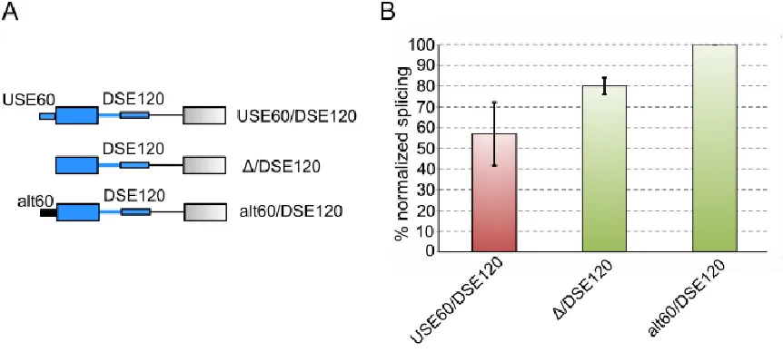
heterologous sequence. Figure 4.2 shows that the alt60/DSE also yields an increase in splicing
over the WT USE60/DSE120 construct, confirming the USE60 as a repressive sequence.
The roles of the USE60 as a splicing repressor and DSE120 as a splicing activator are
corroborated by in vivo minigene results. LEF1-E6 is more included when the USE60 is replaced
30
induced increase of binding of CELF2 is localized to the DSE120, the in vitro splicing results show
that the reason this increase in binding results in an increase in LEF1-E6 inclusion is because the
DSE120 is an activator of splicing. Since the USE60 is a splicing repressor, the effect of the USE60
dominates in unstimulated cells by the binding of CELF2 and upon stimulation this repression is
31
The differing roles that CELF2 plays upon binding to the USE60 versus the DSE120 is also
apparent when CELF2 is significantly knocked down in unstimulated and stimulated JSL1 cells.
Michael Mallory in the lab used a lenti-viral expression vector to cause very effective knockdown of
CELF2 in both US and S JSL1 cells. In unstimulated cells, knockdown of CELF2 caused an
increase in LEF1-E6 inclusion suggesting that CELF2 is functioning as a repressor in this condition.
In stimulated cells, knockdown of CELF2 caused a decrease of LEF1-E6 inclusion suggesting that
CELF2 serves as an activator in this condition. This is in line with the current model, wherein the
effect of CELF2 binding the repressor USE60 is the dominant effect in unstimulated cells. Upon
stimulation the effect of CELF2 binding the activator DSE120 is the dominant effect (Figure 4.4)
It is possible that the activating nature of the DSE120 is not solely due to the increased
binding of CELF2 but due to some other protein interacting with the DSE120. In order to confirm
that CELF2 binding downstream of LEF1-E6 leads to increased inclusion I created 4 minigenes
that replaced the DSE120 with increasing amounts the high affinity SELEX sequences used in the
32
to the 4xUGUU sequences. Therefore, these constructs allow for precise control over how much
CELF2 can bind downstream. LEF1-E6 inclusion can then be monitored under unstimulated and
stimulated states (Figure 4.5).
In in vivo minigene assays, if the amount of CELF2 binding downstream correlates with
LEF1-6 inclusion, then inclusion should increase from the 1x to the 4xUGUU minigenes. If only a
33
one of the UGUU minigenes and not the others. Finally, If CELF2 binding to the DSE120 is not the
sole cause for LEF1-E6 inclusion, then there should be no correlation between inclusion levels and
CELF2 binding. Figure 4.5 shows that increasing amounts of CELF2 binding downstream of
LEF1-E6 results in increasing amounts of LEF1-LEF1-E6 inclusion. Therefore, the signal induced enhancement
of LEF1-E6 is solely due to the increase in CELF2 binding to the DSE120 downstream of exon 6.A
striking result from Figure 4.5 however is that the 1x-4xUGUU sequences do not recapitulate the
signal induced enhancement of exon 6 inclusion. The lack of signal responsiveness is not due to a
lack of sensitivity caused by too much inclusion because the 1-3XUGUU minigenes are comfortably
within the range to observe an increase. There is therefore something unique about the DSE120
in the context of LEF1-E6 that is capable of regulating the amount of CELF2 that binds to it
unstimulated cells versus stimulated cells.
In order to probe whether there was a difference in the way CELF2 interacts with the
USE60 and DSE120 in the unstimulated versus the stimulated state, I isolated protein from each
condition. The first step towards isolating US and S-CELF2 was to stably express FLAG-tagged
CELF2 in JSL1 cells. In order to obtain protein from both the unstimulated and stimulated
conditions, I grew 30L of FLAG-CELF2 expressing JSL1 cells, and stimulated 15Lwith PMA. After
72 hours, both the unstimulated and stimulated cells were harvested. Since CELF2 is
predominantly a nuclear protein39, nuclear extract was separated from the harvest and subjected to a M2 FLAG affinity column to specifically pull out FLAG-tagged CELF2 (Figure 4.6).
To specifically probe how US and S-CELF2 interact with the USE60 and DSE120, I used
an Electro-Mobility Shift Assay (EMSA). For this assay, the USE60 and DSE120 were
radio-labelled and then incubated with increasing amounts of US-CELF2 or S-CELF2. The incubation
allows RNA-protein complexes to form and then these radiolabeled RNA-protein complexes, are
visualized in a non-denaturing acrylamide gel using autoradiography (Figure 4.6). Figure 4.6 shows
that for the both the USE60 and DSE120, US-CELF binds in three distinct modes even at the
highest CELF2 concentration. By sharp contrast, S-CELF2 binds very co-operatively to both the
35
The EMSAs were repeated 3 times and the results quantified showing the cooperativity present in
the binding of S-CELF2 to the RNA, that is absent from the US-CELF2 (Figure 4.6).
EMSAs were also repeated with US and S-CELF2 that was isolated in the presence of
DNase and RNase. The removal of nucleic acids provides for a cleaner pull-down and several
indirect associations that CELF2 made through RNA or DNA would be reduced (Figure 4.7). Figure
4.7 shows that the RNase treated US and S-CELF2 could not recapitulate the signal induced
change in interaction observed from the non-RNase treated protein. However, since the yield from
the pulldown was greatly improved, the EMSA was able to reach saturation and therefore apparent
Kds were calculated for these interactions. The binding of US and S-CELF2 to the USE60 and
DSE120 was compared to the high affinity CELF2 sequence 4xUGUU. Figure 4.7 shows that the
US-CELF2 and S-CELF2 in the presence of RNase are very potent binders of the USE60 and
DSE120 with a relative Kd of 15-20nM. This is a tighter interaction than the high affinity 4xUGUU
sequence which they bind with a relative Kd of 65nM (data not shown). The interaction of US and
S-CELF2 is non-cooperative and very similar to the binding of US-CELF2 in the absence of RNase
to these constructs.
Discussion:
CELF2 binding upstream of an alternative exon being repressive to inclusion and
downstream of an alternative exon being enhancing is a well-studied phenomenon in the
literature55,57–59,84. cTNT exon 555 and NMDAR1 exon 2158 have CELF2 regulatory elements downstream that contribute towards greater inclusion. NMDAR1 exon 558, CFTR exon 957, Tau
exon 285 and CELF2’s own exon 6 all have CELF2 regulatory elements upstream that contribute
towards exon exclusion.
The literature therefore supports the model where CELF2 binding upstream is repressive
and downstream is enhancing. The in vitro splicing results and the 1x-4xUGUU minigene results
confirm that the DSE120 activates exon inclusion while the USE60 represses exon inclusion.
Combining this with what was learnt about the pattern of CELF2 binding in unstimulated and
37
emerges. In unstimulated cells, the effects of CELF2 bound to the upstream repressor, USE60, are
dominant. Upon stimulation, this repression is relieved by a bolus of CELF2 binding downstream
to the activating DSE120. (Figure 4.3 panel C)
How is the binding of CELF2 so well regulated under these conditions? What is the
mechanism behind the preference for the USE60 in unstimulated cells, and DSE120 in stimulated
cells? One possibility is that there is an inherent difference in CELF2 itself between the two
conditions. Perhaps CELF2’s PTM landscape in unstimulated cells is inhibitory to binding the
DSE120. Upon stimulation this PTM landscape changes now promoting CELF2 binding to the
DSE120. A second possibility is that there are other splicing factors at play that actively keep
CELF2 from binding the DSE120 in unstimulated cells. Upon stimulation either a down-regulation
of this factor or the interference of another allows CELF2 to bind to the DSE120.
The EMSAs in the absence of RNase suggests that there is a difference in the way CELF2
pulldowns from US cells interact with the USE60 and DSE120 when compared to pulldowns from
stimulated cells. This difference in interaction can be attributed to either an inherent difference in
the CELF2 species between the two states or the presence of another splicing factor that was
pulled down with CELF2. The loss of differential binding upon Rnase addition suggests that there
was a factor present in the pulldown that lacked Rnase that was responsible for the change in
interaction. This doesn’t refute the necessity of a change in PTMs upon stimulation as it could be
how CELF2 regulates its binding with this other unknown regulatory protein. The next two chapters
of this thesis will discuss data that pertains to CELF2 PTMs in unstimulated and stimulated cells as