University of Pennsylvania
ScholarlyCommons
Publicly Accessible Penn Dissertations
1-1-2014
Mechanochemical Control of Stem Cell Biology in
Development and Disease: Experimental and
Theoretical Models
Polimyr Caesar Dave Pelisco Dingal
University of Pennsylvania, [email protected]
Follow this and additional works at:
http://repository.upenn.edu/edissertations
Part of the
Biomedical Commons,
Biophysics Commons, and the
Cell Biology Commons
This paper is posted at ScholarlyCommons.http://repository.upenn.edu/edissertations/1260
Recommended Citation
Dingal, Polimyr Caesar Dave Pelisco, "Mechanochemical Control of Stem Cell Biology in Development and Disease: Experimental and Theoretical Models" (2014).Publicly Accessible Penn Dissertations. 1260.
Mechanochemical Control of Stem Cell Biology in Development and
Disease: Experimental and Theoretical Models
Abstract
Whether a stem cell remains or egresses away from its physiological niche is a function of mechanical and soluble factors in a time-dependent manner, which implicates a `memory' of prior mechanochemical conditioning. Virtually every organ in the body contains resident stem or progenitor cells that contribute to organ homeostasis or repair. The wound healing process in higher vertebrate animals is spatiotemporally complex and usually leads to scarring. Limitations for the use of stem cells as regenerative therapy include the lack of expansion capabilitiesin vitroas well as materials issues that complicate traditional biochemical protocols. A minimal `scar in a dish' model is developed to clarify the kinetics of tension-sensitive proteins in mesenchymal stem cells (MSCs), which possess plasticity to mechanochemical changes of the
microenvironment that are typical of scars. The organization and expression of such proteins implicates transcription factors that ultimately steer cell fate. In contrast to classic mechano-transducers of matrix mechanics such as actin assembly-dependent serum response factor (SRF) signaling, a novel mechano-repressive role of NKX2.5 is implicated in maintaining intracellular tension in long-term stem cell cultures on stiff matrices via nucleo-cytoplasmic shuttling — ultimately setting up a 'mechanical memory'. Core gene circuits with known roles in stem cell mechanobiology are modeled based on the 'use it or lose it' concept: tension inhibits turnover of structural proteins such as extracellular collagens, cytoskeletal myosins and nucleoskeletal lamins. This theoretical approach is tested in a variety of processesin vitroandin vivothat involve forces including cardiac development, osteogenic commitment of MSCs, and fibrosis therapy. With the sophistication of the science and technology of biomaterials relevant to stem cell biology and medicine, matrix mechanics can thus be rigorously combined with biochemical instructions in order to maximize therapeutic utility of stem cells.
Degree Type
Dissertation
Degree Name
Doctor of Philosophy (PhD)
Graduate Group
Chemical and Biomolecular Engineering
First Advisor
Dennis E. Discher
Keywords
Biomaterials, Extracellular Matrix, Mechanobiology, Mechanorepressor, Stem Cell
Subject Categories
MECHANOCHEMICAL CONTROL OF STEM CELL BIOLOGY
IN DEVELOPMENT AND DISEASE: EXPERIMENTAL AND
THEORETICAL MODELS
Polimyr Caesar Dave P. Dingal
A DISSERTATION
in
Chemical and Biomolecular Engineering
Presented to the Faculties of the University of Pennsylvania
in
Partial Fulfillment of the Requirements for the
Degree of Doctor of Philosophy
2014
Supervisor of Dissertation
Dennis E. Discher, Professor of Chemical and Biomolecular Engineering
Graduate Group Chairperson
Raymond J. Gorte, Professor of Chemical and Biomolecular Engineering
Dissertation Committee
Scott L. Diamond, Professor of Chemical and Biomolecular Engineering
Rebecca G. Wells, Associate Professor of Medicine
Dedicated to
ACKNOWLEDGMENTS
These 5 years have been an adventure to the other side of the world. There are not enough
words to fully acknowledge all the persons that influenced my foray into academic enterprise.
To my advisor who never stops working and whom I never call by his first name (to his
face), I am grateful for the opportunities to pick your brain and to contribute to your academic
endeavors. The works that are presented here used to be riddled with his red ink, but are now
undoubtedly presentable.
To the Lamin Team (circa 2013-2014), I have enjoyed our sharing of personal stories and
scientific manuscripts as we share our meals.
To the University of Pennsylvania Glee Club (circa 2011-2013), thank you for filling my
nights and my head with music and for the unforgettable memories (Brothership Tour 2012!).
Most importantly, to my friends and family back home, thank you for always reminding me
ABSTRACT
MECHANOCHEMICAL CONTROL OF STEM CELL BIOLOGY
IN DEVELOPMENT AND DISEASE: EXPERIMENTAL AND
THEORETICAL MODELS
Polimyr Caesar Dave P. Dingal
Dennis E. Discher
Whether a stem cell remains or egresses away from its physiological niche is a function of
mechanical and soluble factors in a time-dependent manner, which implicates a ‘memory’
of prior mechanochemical conditioning. Virtually every organ in the body contains resident
stem or progenitor cells that contribute to organ homeostasis or repair. The wound healing
process in higher vertebrate animals is spatiotemporally complex and usually leads to scarring.
Limitations for the use of stem cells as regenerative therapy include the lack of expansion
capabilitiesin vitroas well as materials issues that complicate traditional biochemical protocols.
A minimal ‘scar in a dish’ model is developed to clarify the kinetics of tension-sensitive proteins
in mesenchymal stem cells (MSCs), which possess plasticity to mechanochemical changes
of the microenvironment that are typical of scars. The organization and expression of such
proteins implicates transcription factors that ultimately steer cell fate. In contrast to classic
mechano-transducers of matrix mechanics such as actin assembly-dependent serum response
factor (SRF) signaling, a novel mechano-repressive role of NKX2.5 is implicated in maintaining
intracellular tension in long-term stem cell cultures on stiff matrices via nucleo-cytoplasmic
shuttling – ultimately setting up a ’mechanical memory’. Core gene circuits with known roles
in stem cell mechanobiology are modeled based on the ’use it or lose it’ concept: tension
inhibits turnover of structural proteins such as extracellular collagens, cytoskeletal myosins and
nucleoskeletal lamins. This theoretical approach is tested in a variety of processesin vitroand
in vivothat involve forces including cardiac development, osteogenic commitment of MSCs, and
to stem cell biology and medicine, matrix mechanics can thus be rigorously combined with
Table of Contents
ACKNOWLEDGMENTS iii
ABSTRACT v
Table of Contents vi
List of Tables ix
List of Figures x
1 Matrix mechanics and soluble factors combine to steer stem cell fate 1
1.1 Introduction . . . 2
1.2 Lessons from the niche . . . 2
1.2.1 Hematopoietic stem cell niche . . . 3
1.2.2 Muscle stem cell niche . . . 4
1.2.3 Neurogenic niche . . . 5
1.2.4 Multifunctional mesenchymal stem cells . . . 6
1.3 Soluble factors with contrasting function . . . 7
1.4 Harnessing pluripotency . . . 10
2 A minimal ‘scar in a dish’ model reveals mechano-temporal effects of matrix stiffness on stem cell fate 18 Abstract . . . 19
2.1 Introduction . . . 19
2.2 Results and Discussion . . . 21
2.2.1 Hydrogels with scar-like collagen fiber heterogeneity and regional rigidity 21 2.2.2 ‘Scar in a dish’ impairs cardiomyocyte beating consistent with heart failure 23 2.2.3 MSCs durotax to the ‘scar in a dish’ . . . 24
2.2.4 Effective stiffness of ‘scar in a dish’ can be deduced from cell shape and cytoskeleton . . . 26
2.2.5 Stiffness of ‘scar in a dish’ increases lamin A and MSC osteo-commitment 27 2.2.6 Lamin-A & MMP9 in progenitor-derived macrophages also increase with matrix stiffness . . . 28
2.3 Conclusion . . . 29
2.4.1 Synthesis and functionalization of conventional and scar-like hydrogels
for cell culture . . . 31
2.4.2 Surface characterization and microrheology of hydrogels . . . 32
2.4.3 Cell culture, manipulation, and treatments . . . 33
2.4.4 Proteomics analyses of mouse tissue . . . 34
2.4.5 Bulk rheology . . . 35
2.4.6 3D reconstruction & photobleaching in confocal microscopy . . . 35
2.4.7 Decellularized Embryonic Heart Matrix. . . 36
2.4.8 Time-lapse Microscopy . . . 36
2.4.9 Histological analyses of osteogenic commitment . . . 37
2.4.10 Combined FluorescenceIn SituHybridization (FISH) and Immunofluorescence Microscopy . . . 38
2.4.11 Gelatin Zymography . . . 39
3 Role of a matrix stiffness-sensitive repressor, NKX2.5, in stem cell plasticity 58 Abstract . . . 59
3.1 Introduction . . . 59
3.2 Results and Discussion . . . 61
3.2.1 MSCs downregulate collagen-1 synthesis as they become more contractile 61 3.2.2 Matrix stiffness modulates NKX2.5 localization . . . 62
3.2.3 A long-term rigid-matrix phenotype: nuclear exit of NKX2.5 . . . 64
3.2.4 Crosstalk of NKX2.5 and SRF with TGF-βpathway decouples SMA & collagen-1 expression. . . 65
3.2.5 Other modifications of NKX2.5 . . . 66
3.3 Conclusion . . . 67
3.4 Materials and Methods . . . 68
3.4.1 Transcriptional profiling by DNA microarrays . . . 68
3.4.2 Promoter binding analyses . . . 68
3.4.3 Isolation of embryonic cardiomyocytes . . . 68
4 Systems Mechanobiology: Tension-inhibited protein turnover is sufficient to physically control gene circuits 90 4.1 Introduction . . . 91
4.2 Results and Discussion . . . 94
4.2.1 Tension-inhibited degradation of coiled-coil proteins. . . 94
4.2.2 Mechanical coupling of coiled-coil modules in series . . . 96
4.2.3 Logistic coupling between collagen and myosin in cardiac development . 99 4.3 Conclusion . . . 101
4.4 Methods . . . 102
5 Future challenges in characterization and mechanochemical control of stem cell fate 109 Abstract . . . 110
5.1 Introduction . . . 110
5.2 Soft matter control . . . 112
5.2.2 Advances in Soft Matter Research . . . 114
5.3 Hard matter approaches . . . 117
5.4 Conclusion . . . 118
A Supplementary Information for Chapter 2 119 A.1 Adhesion and Thin-film Corrections for scar-like0.3kP a gel . . . 119
A.2 Theoretical analysis of lateral pulling curves . . . 122
B Supplementary Information for Chapter 4 123 B.1 Mechanobiological gene circuits: systems of ordinary differential equations . . . 123
B.1.1 Single-module gene circuits: reaction-order stability. . . 124
B.1.2 Two-Module Gene Circuit . . . 127
B.1.3 Two-Module, Tissue-level Mechanobiological Gene Circuit . . . 128
B.2 Analytical Solution for Single-Module Gene Circuit . . . 129
B.3 Numerical Solution for Two-Module Gene Circuit . . . 130
B.4 Numerical Solution for Tissue-level Coupled Gene Circuit . . . 131
C Standard biological laboratory protocols and reagents used 132 C.1 Cell culture . . . 132
C.2 Quantitative Immunofluorescence Microscopy . . . 132
C.3 Cell Morphometrics . . . 133
C.4 Transfection protocol . . . 134
C.5 Western Blotting and Analysis . . . 134
C.6 Statistical Analyses . . . 135
List of Tables
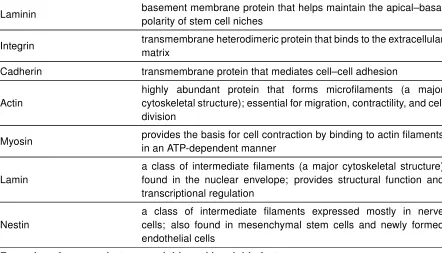
1.1 Glossary of factors and proteins . . . 13
3.1 Promoter analysis of transcription factor binding to ACTA2 and COL1A2. . . 70
C.1 Polyacrylamide gel precursor formulations . . . 136
List of Figures
1.1 The insoluble and soluble milieus in a stem cell niche. . . 15
1.2 Matrix and soluble factors converge to control lamin-A levels . . . 16
1.3 Stem cells have a time-dependent ’memory’ of prior conditioning . . . 17
2.1 Mechanics, transcriptomic, proteomic profiling of muscular dystrophy tissue. . . 40
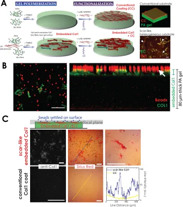
2.2 Synthesis and characterization of a ‘scar in a dish’ . . . 41
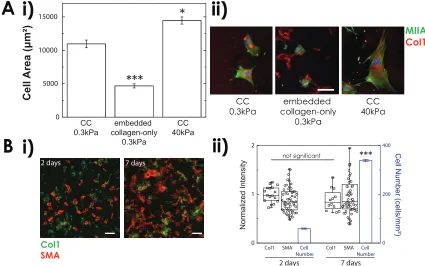
2.3 Embedded collagen is stable but does not induce cell spreading . . . 42
2.4 Bulk and surface rheology, and functionalization of PA gel . . . 43
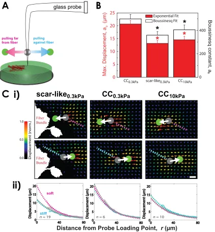
2.5 Lateral pulling microscopy . . . 44
2.6 Stem cell-derived cardiomyocytes beat less on scar-like gel . . . 45
2.7 Synthesis of a scar-like gradient gel . . . 46
2.8 MSCs durotax on a gradient of scar-like fibers . . . 47
2.9 Human MSCs home toward a ‘scar in a dish’ and crosslinked heart matrixex vivo 48 2.10 Matrix composition of decellularized embryonic chick heart . . . 49
2.11 Scar-like0.3kP agel exhibits matrix stiffness-dependent MSC response . . . 50
2.12 Local matrix stiffening couples lamin-A andα-smooth muscle actin upregulation 51 2.13 Matrix stiffness, but not retinoic acid, induces MSC osteogenesis . . . 52
2.14 Mechanochemical control of monocyte-derived macrophage differentiation . . . 53
2.15 Matrix stiffness, but not RA, induces lamin A in monocytes/macrophages . . . . 54
2.16 Matrix stiffness, but not RA, also induces MMP9 in monocytes/macrophages . . 55
2.17 Summary of transcription factor binding scores from ENCODE database . . . . 56
2.18 Retinoic acid, through RARα, reduced lamin A & MMP9 in monocyte/macrophage 57 3.1 Genes relevant to mechanobiology in development . . . 71
3.2 Transcriptomic and proteomic profiles of MSCs on gels . . . 72
3.3 Matrix stiffness downregulates collagen-I matrix synthesis in MSCs . . . 73
3.4 Stiff gels coated with collagen-2 reduced collagen-1 in MSCs . . . 74
3.5 TGF-β is sufficient but not necessary forα-smooth muscle actin expression . . 75
3.6 NKX family of transcription factors:ACTA2-promoter binding & gene abundance 76 3.7 Matrix stiffness modulates Nkx2.5 expression in embryonic cardiomyocytes. . . 77
3.8 MSCs express NKX2.5 with some modifications . . . 78
3.9 Matrix stiffness modulates NKX2.5 localization in MSCs . . . 79
3.10 NKX2.5 is a slow-acting mechanorepressor ofα-smooth muscle actin . . . 80
3.11 Subcellular localization of Nkx2.5 by mutating its nuclear localization signal . . . 81
3.13 Extranuclear localization of NKX2.5 . . . 83
3.14 Blebbistatin rescues NKX2.5 depletion from the nucleus . . . 84
3.15 NKX2.5 decreases with long-term stiff-substrate cultures of MSCs . . . 85
3.16 TGF-β inhibition recapitulates matrix stiffness-induced nuclear exit of NKX2.5 . 86 3.17 Crosstalk of NKX2.5 and Serum Response Factor with TGF-βsignaling . . . 87
3.18 Post-translational modifications of NKX2.5 in MSCs. . . 88
3.19 NKX2.5 is independent of retinoic acid/Lamin-A/SRF axis . . . 89
4.1 Systems-level view of structural molecules involved in mechanotransduction . . 103
4.2 Feedback-based gene circuit for lamin A exhibits polymer physics scaling if cell tension suppresses protein turnover . . . 104
4.3 Dynamic behavior of collagen-1 produced by mesenchymal stem cells . . . 105
4.4 Matrix stiffness-coupled lamin A and myosin turnover . . . 106
4.5 Sensitivity analysis of coupled structural modules . . . 107
Chapter 1
Matrix mechanics and soluble factors
combine to steer stem cell fate
1.1
Introduction
The broad promise of stem cells for regenerative medicine is founded on decades of clinical
success already achieved with bone marrow transplantation of hematopoietic stem cells.
Current capabilities to induce pluripotency with any person’s terminally differentiated cells could
also revolutionize therapies for repairing or replacing many other damaged tissues. Yet direct
implantation of such cells causes teratomas — that is, tumors with regions of disorganized
fat, bone, epithelium or other tissues. Expansion of stem cells with at least partial lineage
specificationin vitrothrough optimal control of the physicochemical milieu is therefore essential
to success in therapeutic applications. We highlight some key aspects of the interplay between
mechanical factors and soluble factors (Table 1.1) in both the physiological andin vitromilieu
of stem cells. We provide examples of how soluble-factor control of stem cell fate is modulated
by the physical microenvironment, and we discuss aspects of mechanobiological mechanisms
and potential research avenues requiring more attention and rigor.
1.2
Lessons from the niche
Stem and progenitor cells in adult organisms can maintain their multipotent state because of
‘signals’ that they receive in the protected, tissue-specific environment that is the stem cell niche
(Figure 1.1; Schofield, 1983). Signaling factors include soluble cytokines, cell–cell contacts,
and insoluble extracellular matrices with characteristic structure and physicochemical properties
(such as stiffness and softness) that define a cell orientation (O’Brien, 2013). Apical–basal
polarity is common, and seems to poise a stem cell for asymmetric division — that is, the
generation of one differentiated cell with the stem cell remaining in its niche. Otherwise, niches
vary with tissue type, as discussed below for soluble and insoluble microenvironments of four
1.2.1
Hematopoietic stem cell niche
Haematopoietic stem cells (HSCs) remain the best described somatic stem cells, but how they
are maintained in their adult niche(s) within the bone marrow or otherwise how they egress
into the circulation remains an active area of research. Embryonic niches for these cells are
distinct and precede establishment of bone. One niche for adult HSCs comprises nestin-positive
mesenchymal stem cells (MSCs) and endothelial cells that are spatially associated near the
perivascular microenvironment (Mendez-Ferrer et al., 2010); yet proximity to trabecular bone
and bone cells also seems important in another HSC niche (Morrison and Scadden, 2014).
All of these bone marrow cells, as well as factors present near the bone surface, could help
to promote HSC quiescence, with key niche cytokines including stem cell factor (SCF) and
stromal cell-derived factor 1 (SDF-1) (Morrison and Scadden,2014;Peled et al.,2000). MSCs
and other stromal cells play an important role in the homing of HSCs to the niche – especially
after myeloablative therapy, that is, the depletion of bone marrow cells by means of chemo- or
radiotherapy before transplantation — perhaps chemoctactically driven via SDF-1. For example,
SDF-1-induced expression of matrix metalloproteinase-9 (MMP9) in bone marrow cells can
enzymatically release matrix-bound SCF, which can then permit the recruitment of repopulating
cells and also promote the proliferation of normally quiescent HSCs (Heissig et al.,2002).
The ability of stem cells to divide asymmetrically is a characteristic niche process that
maintains stemness under homeostatic or inflammatory conditions to replenish blood cells. The
mobilization of HSCs through the induction of granulocyte colony-stimulating factor (G-CSF;
another soluble niche factor) was first demonstrated in the early 1990s, yet little is known about
HSC trafficking within the bone marrow before egress into the bloodstream, particularly towards
lineage specification and possibly to other sub-niches. Physiologically, HSC mobilization follows
circadian oscillations (Mendez-Ferrer et al.,2008), yet relations to local pressure, flow and other
circulating factors remain obscure; oxygen certainly regulates the behavior of stem cells (Keith
and Simon,2007).
(as suggested byLichtman and Kearneyin the 1970s, who anticipated key roles for
“contracting-relaxing [. . . ] macromolecules in motile cells”). In blood cells, a major impediment to migration is
the nucleus, and regulation of its deformability probably modulates hematopoietic cell trafficking
and lineage. Indeed, nucleoskeletal-lamin (Shin et al., 2013) and cytoskeletal-myosin (Shin
et al., 2014) isoforms differentially regulate egress and lineage maturation of HSCs. In
particular, myosin IIB promotes cell polarization to generate more blood progenitors, whereas
myosin IIA supports a more symmetric division. Interestingly, reversible inhibition of myosin
II, and soft matrices that mimic the compliance of marrow (and also tend to suppress myosin
II), both favor long-term HSC reconstitution (Shin et al., 2014). Tropoelastin, which is also
highly compliant, has an additive effect with serum-free cytokine cocktails (of IL-3, IL-6 and
SCF) in supporting repopulating HSCsin vitro; yet stiffening of tropoelastin by glutaraldehyde
crosslinking abrogated the effect (Holst et al., 2010). The myosin-II inhibition strategy also
enriches for megakaryocytes of high ploidy (Shin et al.,2013) and pro-platelet generation (Shin
et al., 2011), and thus seems to be key in directing at least some lineage programs. On the
whole, the tight regulation of HSC niche components plays a crucial role in stem cell quiescence,
with feedback and feed-forward mechanisms between cells and soluble and insoluble factors.
1.2.2
Muscle stem cell niche
Host muscle fibre, interstitial cells, basal lamina and a nearby blood circulation seem to be
the main components of the niche for muscle stem cells (also known as satellite cells) (Kuang
et al., 2007). In satellite-cell homing during development, canonical Notch signals that occur
asymmetrically at cell–cell contacts — which control multiple cell differentiation processes —
stimulate emerging satellite cells to assemble their own basal-lamina matrix (Brohl et al.,2012).
Integrin binding to laminin in this extracellular matrix localizes to the basal side of satellite
cells, whereas M-cadherin localizes to the apical side for attachment to the host muscle fibre
(Kuang et al., 2007). It has been hypothesized that the basal adhesion regulates stem cell
polarized distribution of adhesion molecules determines the asymmetric fate of daughter cells
after division (Kuang and Rudnicki, 2008). The one daughter cell that maintains contact with
the basal lamina maintains a self-renewal capacity, whereas the other is pushed into the host
myofibre and presumably differentiates (Kuang and Rudnicki,2008).
On injury, activated satellite cells remodel their niche through autologous expression
of fibronectin, feeding back to Wnt7a signaling (a pathway known to be implicated in
developmental processes), which in turn induces symmetric expansion of satellite cells
(Bentzinger et al., 2013). Collagen VI is also produced by satellite cells, with collagen-VI
knockout mice showing impaired muscle regeneration with decreased muscle stiffness (from
12 kPa to 7 kPa) (Urciuolo et al.,2013). Consistent with this,in vitrosatellite cell maintenance
and subsequent muscle engraftment is maximal when culturing on substrates that match normal
muscle tissue stiffness (∼12 kPa;Engler et al.,2004a;Gilbert et al.,2010). Nonetheless, such
cell cultures exhibited lower regenerative capacityin vivothan cells freshly isolated from muscle,
which suggests that some combination of biophysical and biochemical elements of the muscle
stem cell niche have yet to be identified for optimal cell culture.
1.2.3
Neurogenic niche
The relatively recent discovery of adult neural stem cells (NSCs) and of the signals regulating
adult neurogenesis illustrates differences with the early development of the brain. In embryonic
neurogenesis, all cells derive from neuroepithelial cells that divide symmetrically at first
(Chenn and McConnell, 1995). In adults, however, the NSC niche is restricted to the
ventricular–subventricular zone of the lateral ventricle and to the subgranular zone at the
interface of the hilus and dentate gyrus in the hippocampus (Alvarez-Buylla and Lim, 2004).
As with satellite cells, canonical Notch signaling is highly active and regulates maintenance of
both developing and adult brains (Imayoshi et al., 2010). More importantly, the neocortical
niche architecture is maintained by integrin-based adhesion of NSCs to laminin along the
ependymal cells at the apical surface maintains NSC quiescence, whereas SDF-1 secreted by
endothelial cells of the vascular basal region results in chemotaxis by both activated NSCs and
progenitors in an integrin-α6β1-dependent manner. Transition from progenitors to neuroblasts
is marked byβ1-integrin downregulation, enabling egress from the niche (Kokovay et al.,2010).
The process seems to prompt asymmetric cell division, with the NSC staying in its niche and
the resulting daughter cell moving into more differentiated basal regions of the niche. The
highly polarized NSC niche thus allows for compartmentalization of multiple signals derived
from cell–cell and/or cell–matrix adhesions as well as of soluble factor gradients that combine
to maintain NSC polarity and stemness (Fuentealba et al.,2012).
1.2.4
Multifunctional mesenchymal stem cells
MSCs can take on lineage characteristics of various soft- and hard-tissue cell types (such as
neurons, adipocytes, chondrocytes, osteoblasts, skeletal myoblasts and smooth muscle cells),
and thus they are an attractive choice for autologous transplantation (Pittenger et al., 1999).
Among the various types of adult stem cell, MSCs (from the bone marrow or other sources)
that have been isolated through strong adhesion to plastic seem to have the broadest lineage
potential (Colter et al.,2000); also, they expand for prolonged periods on tissue-culture plastic
while maintaining some degree of multipotency (Abdallah and Kassem,2009). Other materials
might do better, but traditional plastic has allowed MSCs to be used in additionalin vitrosystems
to identify soluble and insoluble cues that induce differentiation.
By mimicking the characteristic stiffnesses of different tissues (for instance, brain tissue is
always soft; bone tissue is rigid) with inert hydrogels functionalized with collagen I, it was shown
that matrix elasticity can direct MSC lineage specification from soft brain to stiff bone and that
myosin II has a key role in pulling on and feeling matrix elasticity (Engler et al.,2006). Microarray
analyses also indicated significant increases in some key factors of the transforming growth
factor beta (TGF-β) superfamily such as bone morphogenetic proteins (BMP) and myostatin
to such TGF-β-family growth factors (Wang et al., 2004), as expected of mesenchymal cells,
which means that growth factors could have key autocrine/paracrine signaling roles and that
these are triggered by matrix elasticity. For example, MSC injection into scars (which tend to
be stiff) such as those resulting from myocardial infarcts has sometimes led to bone formation
in the scar; in this case, matrix stiffness may trigger an increase in BMP, which is likely to be
the proximal cause for osteogenesis (Breitbach et al., 2007). Such potent growth factors may
thus contribute to lineage-inducing signals, even if differences in their expression or activation
imparted by physical features of the microenvironment such as matrix elasticity are small (say,
at the picomolar level). With respect to three-dimensional culture models (Khetan et al.,2013;
Huebsch et al.,2010), many questions arise that should be rigorously addressed, including how
cell-secreted growth factors and extracellular matrix binds or not to the surrounding scaffold or
gel, whether the various soluble serum or induction factors added to cultures permeate the gel
(including oxygen gradients studied decades ago in three-dimensional cultures;Colton,1995),
and also how far the cells reside above a rigid substrate that they might feel. Understanding
the various triggering mechanisms in two dimensions as well as three dimensions is likely to be
important in physiological conditions, yet it is also possible that adding a growth factor such as
BMP to cultures of cells in microenvironments with different stiffnesses will yield different cell
phenotypes.
1.3
Soluble factors with contrasting function
The main TGF-β reservoir in serum resides in platelets, and TGF-β is secreted on platelet
activation in a matrix-binding, latent complex form (Ahamed et al.,2008), from which it can be
released by high fluid shear. Evidence for TGF-β release resulting from cell tension was first
obtained with contractile myofibroblasts, the characteristic cell type in stiff, fibrotic tissue (Wipff
et al., 2007). Following release, TGF-β1 binds membrane receptors that activate ubiquitous
transcription factors (such as SMADs), which enter the nucleus to promote matrix synthesis
wound healing by myofibroblasts. Although intracellular processes are probably modulated
by the microenvironment, an interplay with TGF-β1 release mechanisms seems likely to occur
in vivo. For example, in patients with coronary artery disease an increase in active, but not
total, TGF-β1 in plasma correlates with the number of constricted vessels in the patient (Wang
et al.,1997); hence, constricted or stenotic vessels have higher shear stresses and thus higher
concentrations of active TGF-β(the release kinetics of active TGF-β with shear stress and time
characterizes the forced unfolding of a protein, and the unfolding seems to be roughly linear
with shear stress and lacks a threshold; Tenney and Discher,2009). Given its subnanomolar
concentrations in serum, however, TGF-β activation at low stress would take a long time. In
the marrow niche where flow is minimal, glial cells of the autonomic nervous system (known as
non-myelinating Schwann cells) produce TGF-β and activate it via integrinβ8 to help maintain
HSC dormancy (Yamazaki et al.,2011).
Unlike most protein-based growth factors, which cannot cross cell membranes, retinoic
acid (RA) is a much smaller molecule that readily permeates cell membranes and regulates
ubiquitous transcription factors that shuttle between the cytoplasm and the nucleus (oxygen is
another example). In adults, RA is converted in the liver from ingested vitamin A and circulates
in serum (∼10 nM). RA signaling seems somehow primed in adult HSCs in part by the niche
stroma (Ghiaur et al.,2013), and it is essential for the generation of early embryonic HSCs from
the haemogenic endothelium (Chanda et al.,2013). In HSCs, RA receptor-γ(RARγ) regulates
the balance of HSC self-renewal and differentiation (Purton et al.,2006), with RARαpromoting
granulocyte commitment. Indeed, RA suppresses erythropoiesis (Labbaye et al., 1994) and
enhances neutrophil differentiation (Shin et al., 2013). Although RARs regulate expression of
many genes (up or down), its downregulation of at least the nuclear structure protein lamin A
proved revealing: neutrophils have soft, floppy nuclei with low levels of lamin A, which allow
these cells to easily crawl through endothelial pores from marrow into blood, and also to enter
distal tissues. In contrast, erythroblasts normally maintain high levels of lamin A in their stiff,
condensed nuclei; these nuclei are easily ejected in final differentiation and retained in the
Lamin A is an intermediate filament protein related to keratins in nails, hair, beaks and
horns. In addition to controlling nuclear deformability, lamin A has a role in maintaining DNA
stability, and is also mechanoresponsive, as we have recently shown (Swift et al.,2013b). Its
levels scale with the stiffness of primary tissue (0.1–40 kPa), and in cultured MSCs the levels
increase with the stiffness of hydrogel substrates. Lamin-A levels in MSCs on stiff matrix are
also modulated by RA levels, with more RARγ entering the nucleus in cells on stiff matrices
than in cells on soft substrates (Figure 1.2). Brain is low in RARγ, but has other RARs (Swift
et al., 2013b); in serum-free NSC cultures adhering to a narrow range of flexible substrates
(10–1,000 Pa) and even under strong pro-neuronal conditions (1 µM RA), commitment to
specific lineages is stiffness-dependent (Saha et al., 2008). Similar observations were made
with NSCs encapsulated within alginate hydrogels of stiffness matching that of brain tissue
(Banerjee et al.,2009).
The regulation of the nuclear entry of transcription factors by matrix elasticity might actually
be general. Other transcription factors whose nuclear entry is also known to be regulated by
matrix stiffness include the YAP/TAZ co-activators (Swift et al., 2013b; Dupont et al., 2011;
Yang et al.,2014) and key lineage-specific factors for muscle (MyoD) and bone (core-binding
factor alpha 1; CBFA1) (Engler et al., 2006). Some of these participate in signaling pathways
(such as the Hippo pathway for YAP/TAZ) that are also regulated by soluble factors (such as
lipopolysaccharide). Nucleo-cytoplasmic shuttling of glucocorticoid receptors for natural and
synthetic glucocorticoids such as dexamethasone need to be studied thoroughly as a function
of matrix properties because glucocorticoids are frequently used in differentiation cocktails
(Grigoriadis et al.,1988). Target genes in these pathways that are differentially expressed as a
function of matrix properties and soluble-factor levels provide key evidence of functional nuclear
entry or exit of a transcription factor. Epigenetic memory is also key to stem cell responses. For
example, if MSCs are grown for one to three weeks on soft matrices (1 kPa) that tend to induce
expression of a neurogenic marker, and then an osteoinduction cocktail is added, one finds that
the neurogenic expression is largely reversed in the one-week cultures but not in the three-week
going downhill that does not require much steering to choose, but the further down a chosen
fork, the more difficult it is to reverse the fate/choice. Recent studies of the nuclear localization
of YAP/TAZ in MSCs cultured on stiffness-switchable hydrogels confirm the idea of a matrix
memory (Yang et al.,2014). How such transcriptional regulators are preferentially sequestered
in the cytoplasm rather than the nucleus is generally unknown, but immunoprecipitation of
the factors followed by mass spectrometry can help identify validatable binding partners in
the cytoplasm and nucleus, as recently shown for RARγ (Swift et al., 2013b). Far better
characterized in this respect is the actin-regulated serum response factor pathway (Miralles
et al.,2003; Swift et al., 2013b), which couples directly to the mechanosensitive assembly of
the actin–myosin cytoskeleton.
1.4
Harnessing pluripotency
Responses of embryonic stem cells (ESCs) to mechanical stress and stiffness are beginning
to be clarified. The softness of embryos (∼400 Pa) results from a relative lack of extracellular
matrix even when compared with the earliest beating heart (Majkut et al.,2013). The softness of
ESCs dictates the threshold for stress-driven, myosin-II dependent spreading and differentiation
of mouse ESCs, but not of ESC-differentiated cells that are ten times stiffer (Chowdhury et al.,
2010b). Similar to the matrix-modulated differentiation of MSCs, soft substrates that match
the intrinsic softness of ESCs (600 Pa) suppress cell–matrix tractions and maintain the ESCs’
self-renewal potential (Chowdhury et al.,2010a).
Inhibition of Rho-associated protein kinase (ROCK), an effector of myosin contractility,
dramatically enhances survival of dissociated human ESCs (hESCs), with no effects on ESC
differentiation (Watanabe et al., 2007). Decreased cell–cell contact has also been shown to
reduce survival of dissociated hESCs (Chen et al., 2010) because of irreparably disrupted
E-cadherin signaling (Xu et al.,2010). Enhancing cell attachment immediately after dissociation
greatly improves survival, and is suggested to be partly a consequence of the synergy between
on stiff substrates synergizes with soluble factors to inhibit the differentiation of pluripotent cells
and to enhance their self-renewal (Saha et al.,2006; the physiological relevance of the strain
needed, about 10%, is however unclear). Soft substrates might nonetheless be combined
with externally imposed strains and suitable biochemical signals to maximize pluripotent cell
numbers. For therapeutic applications, ESC expansion should be done without the usual
underlying feeder cells (commonly, fibroblasts), which introduce biological variation and sources
of immune rejection. The standard for ESC culture involves mouse tumor-derived Matrigel (∼1
kPa) to increase cell adhesion, and high concentrations of TGF-β and basic fibroblast growth
factor to antagonize differentiation (Rao and Zandstra, 2005). Current research is however
focused on synthetic surface-engineered surfaces for xeno-free human pluripotent stem cell
(hPSC) culture under chemically defined conditions, which should allow for scalability without
the risks of cellular or pathogenic contamination. Examples include ultraviolet-treated spatial
patterns (Saha et al.,2011) and peptide-decorated acrylate surfaces (Melkoumian et al.,2010).
A high-throughput study on biomaterials development also found acrylate surfaces to be optimal
for clonal growth of hPSCs (Mei et al., 2010). Materials that can accelerate hPSC expansion
(Celiz et al.,2014) are needed for regenerative-medicine applications.
The discovery and development of human induced pluripotent stem cells (hiPSCs;
Takahashi et al., 2007) was immediately followed by a flurry of research into therapeutic
applications, in particular neurological diseases. A recent kinome-wide study of iPSCs
highlighted the role of actomyosin polymerization through kinase-mediated phosphorylation
of cofilin (a protein that disassembles actin filaments) as one major barrier to efficient
reprogramming to iPSCs (Sakurai et al., 2014). It is then not surprising that NSC profiles are
more similar to ESCs than to NSC-derived differentiated cells (Ramalho-Santos et al., 2002),
not only because of default neurulation tendencies of ESCs (brain development is very early;
Tropepe et al., 2001) but also perhaps because of the similarity in softness of embryos and
the neural microenvironment relative to other tissues such as embryonic heart (Majkut et al.,
2013). Indeed, hPSCs integrate readily into the motor cortex and undergo cortical neurogenesis
hESCs into various other tissue sites produces heterogeneous teratomas (Prokhorova et al.,
2009). Reprogramming in vivo also produces teratomas as well as iPSCs with totipotency
features that are not possible in standardin vitroreprogramming (Abad et al.,2013).
Since the first bone marrow reconstitution after myeloablative therapy decades ago,
functional tissue regeneration with other types of stem cell has been the vision of many.
Enabling technologies include cell phenotyping by flow cytometry with antibodies against
lineage-distinctive arrays of cell-surface receptors. However, HSC transplantation is successful
because of the intrinsic ability of HSCs to ‘home’ to the bone marrow and find their niche(s). The
promise of iPSC technology to produce all diverse cell types in a given solid tissue is tempered
by a poor understanding of microenvironmental factors that direct stem cell fate, as well as of
lineage phenotyping and uncertainty over how to control the mobility and engraftment of cells
(Yu et al., 2013). Commitment to a lineage in vitro before implantation seems essential and
necessarily brings culture materials into a protocol, but also complicates the various efforts.
Yamanaka and Gurdon’s 2012 Nobel prize in Physiology or Medicine has largely fixed
the trajectory of most of the currently envisaged stem cell-based therapies into using iPSCs
generated from suitably matched donors. Yet high-efficiency reprogramming and tissue
specification remains a major challenge, as does delivery and survivalin vivo. Therapies with
any type of stem cell should eventually be compared to normal tissue, but technologies to
ascertain cell integration and contributions to diseased tissue require more rigor than is typical
with decades-old histological methods. For example, putting human cells in immunodeficient
mice is the current standard in the HSC field; however, for similar studies with solid tissues,
human versus mouse proteomics (Swift et al., 2013b) and RNA-sequencing can be used to
reveal the contributions of stem-cell-derived xenografts to obtain systems-level views of the
myriad of factors that come into play for fully functional transplants. With the wide range of
material and analytical technologies emerging to help the translation from in vitro to in vivo,
challenges in concepts and applications seem increasingly addressable with suitable rigor and
Table 1.1.Glossary of factors and proteins most relevant to stem cell mechanobiology
Growth Factors
SCF, stem cell factor
transmembrane or soluble cytokine produced by fibroblasts and endothelial cells that binds to the c-Kit receptor (CD117) to help HSC in homing to and maintenance of the niche
SDF-1, stromal cell-derived
factor soluble chemotactic cytokine for HSC homing to the marrow
G-CSF, granulocyte
colony-stimulating factor soluble glycoprotein that promotes stem cell granulopoiesis
TGF-β/BMP, Transforming growth factor-β/ Bone morphogenetic protein
soluble or matrix-bound growth factors that promote cell growth, differentiation and matrix synthesis
bFGF, basic Fibroblast growth factor
soluble or matrix-bound factor, used as supplement in culture medium for ESC maintenance
RA, Retinoic acid membrane-permeable vitamin-A metabolite with roles in neutrophil differentiation; implicated in nucleus deformability
IL, Interleukin
membrane-bound or matrix-bound cytokines produced mostly by the immune system to regulate production, differentiation and function of blood cells
Transcription factors
RAR, Retinoic acid receptor
on binding of RA, RAR heterodimerizes with retinoid X receptor (RXR) to bind to a retinoic acid response element (RARE) on a target gene, such asLMNA
SRF, Serum response factor
binds to serum response element (SRE) of genes responsible for controlling cytoskeletal structures involved in, for example, cell spreading, adhesion and contractility
SMAD, Sma and Mad related family
TGF-β/BMP family of ligands bind to their receptors, which in turn activate SMAD signaling and bind to SMAD-binding elements (SBE) in target genes, such asCOL1A1
YAP/TAZ, Yes-associated protein / Transcriptional co-activator of PDZ-binding motif
involved in the Hippo signaling pathway, responsible for cell-size control; recently shown to be crucial in transducing mechanical signals from ECM to nucleus, in an actomyosin and Rho-dependent manner and inRho-dependent of the Hippo pathway
Matrix, adhesion, and cyto-/nucleoskeletal proteins
Collagen the most abundant protein in animals; it confers the majority of tissue structure and stiffness
Table 1.1.(continued)
Matrix, adhesion, and cyto-/nucleoskeletal proteins
Laminin basement membrane protein that helps maintain the apical–basal polarity of stem cell niches
Integrin transmembrane heterodimeric protein that binds to the extracellular matrix
Cadherin transmembrane protein that mediates cell–cell adhesion
Actin
highly abundant protein that forms microfilaments (a major cytoskeletal structure); essential for migration, contractility, and cell division
Myosin provides the basis for cell contraction by binding to actin filaments in an ATP-dependent manner
Lamin
a class of intermediate filaments (a major cytoskeletal structure) found in the nuclear envelope; provides structural function and transcriptional regulation
Nestin
a class of intermediate filaments expressed mostly in nerve cells; also found in mesenchymal stem cells and newly formed endothelial cells
Examples of synergy between soluble and insoluble factors
Tropoelastin & serum-free cytokine cocktail (IL-3, IL-6 and SCF)
HSCs repopulate on soft tropoelastin but not on stiff, crosslinked tropoelastin even with the presence of cocktail
Matrix-bound SCF and SDF-1 induced MMP9 expression
MMP9 releases SCF and permit HSC repopulation and proliferation of quiescent HSCs
Matrix-bound TGF-β activated with shear flow or cell-derived force; implicated in
progressive fibrosis
Mechanosensitive lamin A and retinoic acid signaling
lamin A increases with tissue stiffness, and primes RARγ to enter nucleus and to promote RA signaling
Soft substrate and retinoic acid in NSC cultures
NSCs commit to neurons only on very soft substrates even under strong pro-neuronal conditions
A
C
B
Maintenance & Polarization Division & Differentiation Mobilization & Regeneration
LMNA
RARE
SBE
STIFF MATRIX
P P
LMNA
RARE
SOFT MATRIX
COL1A1
SBE
S O
FT
NUCLEU
S
COL1A1
Retinoic Acid
TGFβ
Integrin
TGFβ Receptors
Focal adhesion
G-Actin
MAL/SRF
Myosin
Lamin-A
Collagen
RARγ
SMAD2/3
SMAD4
0
Di
ffere
ntiat
ion
M
arker
Intensity
5
10
0
1
2
3
4
Plastic
Committed
time in culture (weeks)
Media Change: Osteo-Induction
neurogenic marker
osteogenic marker
Stem Cells on Soft, Brain-like Gel
Figure 1.3. Stem cells have a time-dependent ‘memory’ of prior conditioning. MSCs
Chapter 2
A minimal ‘scar in a dish’ model
reveals mechano-temporal effects of
matrix stiffness on stem cell fate
Portions of this work are under peer-review inNature Materials.
Abstract
Scarring in higher animals compromises tissue function and is highly complex, which motivates
the development of culture systems that clarify cellular mechanisms and that might also
help identify therapeutics. Co-polymerization of collagen-I with polyacrylamide produced
scar-like, crosslinked fiber bundles that segregated heterogenously atop the hydrogel and
directed the fate of multiple cell types towards scar-like phenotypes. As with scars, the gels
stain non-uniformly with Sirius Red dye and are regionally stiff. Cardiomyocytes derived
from pluripotent stem cells do not beat on scar-like gels, consistent with defective beating
of scarred hearts. Mesenchymal stem cells (MSCs), which traffic to injured sites, ‘durotax’
towards mimetic scars, undergo osteogenesis consistent with ossification of some scars,
and upregulate tension-sensitive proteins. Within hours, myosin IIB polarized rearward in
stress fibers and nucleoskeletal lamin-A levels increased as cells and nuclei spread, but
1-2 days were required to upregulate the ‘scar marker’ α-smooth muscle actin. Agonists to
one pathway involving retinoic acid receptors inhibited scar-induced differentiation, with similar
effects evident in the mechano-regulated fates of progenitor-derived macrophages that remodel
scars. Mechanobiological gene circuits are modeled mathematically, illustrating how key
features of scars in soft tissues can be usefully studied in culture.
2.1
Introduction
Scarring is an unavoidable consequence in mature higher vertebrates of survivable injuries that
range from external trauma to acute events such as myocardial infarction and various chronic
diseases (Gurtner et al.,2008). Fibrotic, heterogeneous matrix is the defining feature of a scar
and consists predominantly of excessive, crosslinked collagen-I bundles, which displace cells
and less oriented matrix and thereby limit tissue function (Martin,1997). Across striated muscle
diseases, for example, collagen-I expression increases up to ten-fold or more relative to normal
Tomita et al.,1999). An increase in tissue stiffness is predictable because collagen-I, being the
most abundant protein in animals, makes key structural networks (Munster et al., 2013), and
this is seen in most fibrotic tissues (Figure 2.1). However, many additional changes in scarred
tissue can be detected, including proportional increases in the nuclear structure protein lamin A
versus collagen-I (Figure 2.1B,C). For normal tissues, we have shown recently that lamin-A
levels scale with collagen-I and are mechanosensitivein vitro(Swift et al.,2013b), but given the
complexity of scarsin vivoin terms of diverse cell types, matrix, and soluble factors, it is unclear
whether lamin A and the transcription factors it regulates are responsive in any given cell type
to a heterogeneous scarring microenvironment.
Cell therapies for many types of scarred tissues are being pursued with a wide range of
cell types, and the list will no doubt grow with the emergence of various stem cells, including
pluripotent cells (Boiani and Scholer,2005) and adult mesenchymal stem cells (MSCs;Pittenger
et al., 1999). Whether stem cell-derived lineages are plastic and change phenotype when
engrafted into scars is a critical question for cell therapy, particularly for MSCs that are
multipotent as well as immunomodulatory. A mechanochemical gradient is also likely wherever
normal tissue transitions to scar (Tomita et al., 1999; Pfeffer and Braunwald, 1990), and an
impact on cell trafficking into scars will likely affect cell and tissue fate (Orlic et al.,2001).
Matrix ligand type and density provide signals complementary to matrix mechanics (Engler
et al., 2004b) in influencing cell behavior such as MSC differentiation (Engler et al., 2006;
Huebsch et al.,2010;Trappmann et al.,2012;Khetan et al.,2013), but recent studies of matrix
tethering (Trappmann et al., 2012), topography (McNamara et al., 2010), crosslinking, and in
three-dimensional (3D) models (Huebsch et al.,2010;Khetan et al.,2013;Liu et al.,2012;Ulrich
et al., 2009; Levental et al., 2009) have stirred debate on the influence of matrix mechanical
properties in cell-fate decisions. Stiffness of scars (e.g. in skin) measured in the macroscopic
(in MPa; Corr et al., 2009) versus microscopic (in kPa; Achterberg et al., 2014) scales yield
vastly different numbers. A clear relationship to specific tissue parameters is an important goal
of new cell-culture material systems (MacQueen et al.,2013). Here we focused on microscale
scar-like culture platforms here, we make a range of comparisons to scarred tissue (Figure 2.1) and
ultimately show that the effective stiffness of a scar is key to cell fate signals.
2.2
Results and Discussion
2.2.1
Hydrogels with scar-like collagen fiber heterogeneity and
regional rigidity
Starting with an allylsilane-modified ‘bottom’ coverslip that anchors polyacrylamide (PA) chains,
we discovered that mixing collagen-I (Col1) heterotrimers into the PA precursor solution
promotes Col1 fibril formation with segregation to within ∼10 microns of the gel interface
during free-radical polymerization (Figure 2.2A,B). Importantly, the embedded Col1 gels stain
positively but heterogeneously for the histochemical dye, Sirius Red (Figure 2.2C), which is
widely used to assess tissue fibrosis (Turgeman et al.,2008;Mann et al.,2011;Georges et al.,
2007). Sirius Red staining was not observed, in contrast, with Col1 coated (CC) conventionally
on the PA gel surface (Figure 2.2C) via a standard sulfo-SANPAH-mediated covalent reaction
widely used for culture substrates (Dembo and Wang,1999). The embedded Col1 was varied to
approximate the Sirius Red area fraction visible in typical sections of scarred tissue (20–30%,
Figure 2.1). Surprisingly, initial studies of cells cultured on the embedded Col1 gels showed
that ligand density on fiber bundles was below the threshold needed for cell adhesion, and so
the conventional ‘CC’ modification was used to obtain cell-adhesive scar-like0.3kP a gels with
bundles of collagen that were stable for at least 7 days in cell culture (Figure 2.3). This minimal
matrix model of a scar provides separate control over matrix elasticity and ligand density,
which is not possible in pure collagen gels where ligand density changes with collagen density.
Furthermore, collagen gels exhibit non-linear elasticity (Storm et al., 2005) and stiffen under
strain induced by incorporating cells (Vader et al.,2009), which complicate matrix-mechanical
effects on cell responses.
w/v) showed no significant effect on the gel’s bulk modulus (E ≈ 2.9G0 = 0.3 kPa; 3%
acrylamide, 0.07% bis-acrylamide w/v), and for all gels, the elastic modulus dominated the
viscous modulus (Figure 2.4A). Unlike PA gels, collagen fiber networks typically show
non-linear strain stiffening independent of crosslinking (Munster et al., 2013), which becomes
insignificant in the embedded system here that is predominantly elastomeric. However, because
a surface-adherent cell feels only a few microns deep into a gel (Buxboim et al., 2010), the
surface microelasticityEsurf seemed critical to measure. We started with conventional
micro-indentation by atomic force microscopy (AFM;Engler et al.,2007), with results for pristine or CC
gels identical to bulk rheology (Figure 2.4Bi). Microelasticity maps of scar-like0.3kP agel surfaces
proved to be far more heterogeneous than those of CC gels, consistent with Sirius Red imaging,
and the scar-like0.3kP a gels also appeared 2–3-fold stiffer than the same gels without collagen
fibers (Figure 2.4Bii). However, such a nominal shift for a stratified material is indicative of a
stiffer material on top of a softer bulk. We calculated therefore a suitable thin-film correction and
also measured gel tackiness for a second estimation ofEsurf (see section A.1, Appendix A).
Both measurements suggestedEsurf ∼10 kPa, which is far stiffer than the bulk gel. A recent
report showed that extracellular matrix tethering, by varying sulfo-SANPAH concentration, could
influence stem cell fate regardless of hydrogel stiffness (Trappmann et al., 2012). However,
we found that reducing Sulfo-SANPAH reduced Col1 coating density (Figure 2.4C), which may
complicate the reported matrix-tethering effects as they may also be attributed to cell responses
to ligand-density variation (Engler et al.,2004a).
Cells apply tractions predominantly along the matrix surface (Dembo and Wang, 1999),
whereas the AFM probes primarily normal to the substrate. Lateral pulling of the gel surface
with a glass microprobe at constant displacement was therefore used to apply a constant
force on scar-like0.3kP a gels (Figure 2.5). Displacements of microbeads embedded in the
gels exhibited the expected Boussinesq-like profiles (Figure 2.5B,C; also see section A.2,
Appendix A). Displacement profiles far from a fiber bundle were strikingly similar to those
for homogeneous 0.3-kPa gels, whereas profiles closer to fiber bundles appeared similar to
to 10 kPa for scar-like0.3kP a gels, which highlights the regional rigidity near the fiber bundles.
The imaging and physical measurements therefore suggest that the scar-like matrix is a stiff
fibrous meshwork within a background of otherwise soft, tissue-like matrix. Whether cells would
respond in a scar-like manner, despite matrix heterogeneity, was the next critical question.
2.2.2
‘Scar in a dish’ impairs cardiomyocyte beating consistent
with heart failure
Following a heart attack, regions of necrotic myocardium that result from acute ischemic injury
are replaced by scars that are too rigid for the heart to contract, causing large decreases in
cardiac output and eventual heart failure. This is a major clinical problem today, and there is
great interest in using cardiomyocytes differentiated from pluripotent stem cells to help replace
damaged myocardium (Laflamme et al., 2007). However, how such cells respond to a
scar-like matrix when engrafting is crucial to understand. Stem cell-derived cardiomyocytes were
therefore seeded onto CC3kP a or scar-like3kP agels, with 3 kPa chosen as the nominal stiffness
because embryonic heart is mostly collagenous matrix and is ∼1–6 kPa in stiffness (Majkut
et al., 2013; Engler et al., 2008). Collagen-coated tissue culture polystyrene (CC-TCP) was
used as a standard control. After 10 days of culture, the number of beating cells was minimal
on scar-like3kP a gels compared to CC3kP a gels (28% vs 100%) with large differences likewise
in beating frequencies (0.24±0.03 Hz vs. 0.61±0.06 Hz) (Figure 2.6A). Results for CC-TCP
were the same as scar-like3kP agels, consistent with expectations that heart cells simply cannot
contract rigid matrices and will eventually stop beating.
The handful of cardiomyocytes that beat at high rates on the stiff scar-like3kP a and
CC-TCP substrates exhibited informative beating patterns. CC-TCP is too rigid to contract, but local
regions of some cells did appear to twitch at the normal rate. Likewise, on scar-like3kP a gels,
any cardiomyocyte that was half-attached to a scar-like fiber bundle showed no measurable
contraction on the fibers, while the other half of the same cell beat normally on the CC part
nuclear beating (Figure 2.6Biv) suggested that nuclear phenotypes could also emerge in
scar-like cultures. Ultimately, individual cardiomyocytes could not beat when the matrix was too rigid.
These initial findings with cells confirm our physical measurements showing scar-like gels are
regionally soft or rigid, but they also suggest that the overall cell response is dominated by the
rigid regions.
2.2.3
MSCs durotax to the ‘scar in a dish’
MSCs show some therapeutic utility in heart repair (Berry et al.,2006;Hare et al.,2009), and
although mechanisms remain unclear, there is also evidence of homing to the injury site (Orlic
et al.,2001). MSC migration towards a site of injury within tissue will typically involve a large
gradient in stiffness, such as in an infarct border zone (∼9 kPa/mm;Berry et al.,2006;Tse and
Engler,2011). In several other homogeneous gel systems, various cell types including MSCs
have been reported to ‘durotax’ from soft to stiff matrix (Tse and Engler,2011;Isenberg et al.,
2009;Raab et al.,2012;Lo et al., 2004). A gradient of increasing scar-like fiber bundles was
therefore made here to resemble a transition from homogeneous soft tissue to a heterogeneous
scar (Figure 2.7A). A PA gel precursor solution was mixed with or without Col1, and drops of
each solution were deposited next to each other; Sirius Red staining confirmed such a gradient
of scar-like fibers over a length scale ( 2–4 mm) that ultimately saturates to a 20–30% area
fraction (Figure 2.7B), similar to that seen in diseased tissue (Pfeffer and Braunwald, 1990;
Turgeman et al.,2008;Mann et al.,2011).
Live cell imaging allowed us to track cells on either side of the transition zone (Figure 2.8A).
Net displacements of cell centroids every 15 min towards either the CC half or scar-like half
were used to calculate a Durotaxis Index (Raab et al., 2012), by counting the net number of
steps a cell takes to move towards the scar-like region. Strikingly, only MSCs migrating in the
transition region exhibited strongly positive net displacements (Figure 2.8B) and Durotaxis Index
(Figure 2.8C) towards the scar-like0.3kP a region. Far from the gradient and in both CC0.3kP a
in Durotaxis Index with the underlying elasticity contrast (∆E) was previously documented for
vascular smooth muscle cells (Isenberg et al., 2009), and a very similar relationship for MSCs
implies that scar-like0.3kP a gels have an effective Esurf ∼ 10–14 kPa (Figure 2.8Ciii). This
Esurf estimated from cell motility agrees with the maximumEsurf obtained from our physical
measurements (Figure 2.5). Heterogeneity of our matrices did not obscure durotaxis probably
because the turnover and hysteresis of cytoskeletal structures (e.g. myosin IIB) takes hours
(Raab et al., 2012), suggesting a short-term memory (rather than long-term memory; Yang
et al., 2014) in the crawling cells as they average over matrix heterogeneities and ‘remember’
only the stiffer fiber bundles. Moreover, the minimum area fraction of ‘scar’ for imparting a
memory seems low since MSCs durotax into an increasingly heterogeneous scar-like region
with migration results that fit well to findings for homogeneous gels.
Knockdown of myosin IIB (MIIB) in MSCs eliminated durotaxis (Figure 2.8Ci,ii), consistent
with our recent demonstration that MIIB polarizes rearward only on stiff matrices and is
needed for durotaxis where persistent migration increases from soft to stiff (Raab et al.,2012).
Immunostaining and imaging of MSCs on both ends of the soft-to-scar gradient confirmed
the expected MIIB polarization differences as well as increasing expression of α-smooth
muscle actin expression (SMA; Figure 2.9A), which is a myogenic marker for intracellular
tension that is commonly elevated in scarred tissue (Hinz, 2007). However, results for
day-1 versus day-2 showed that MIIB polarization preceded SMA upregulation on the scar-like
matrix (Figure 2.9Aii). Thus, the ‘scar marker’ SMA is not required for early stiffness sensing,
consistent with studies of chemically induced liver fibrosis (Georges et al., 2007). Rearward
nucleus polarization during cell migration on stiffer matrices (Figure 2.9Ai) is also suggestive of
higher contractility in the rear, consistent with MIIB polarization seen recently (Raab et al.,2012).
Among myosin-II isoforms (A, B and C), MIIB accounts for only 5–10% of total myosin-II in MSCs
(Raab et al.,2012), and cell traction forces do not change drastically with MIIB knockdown (Cai
et al.,2006). Interestingly, a cell size-dependent ‘attraction’ to the underlying scar-like fibers was
evident (Figure 2.9B), consistent with a local, myosin-dependent durotaxis in which well-spread
MSCs did not spread on scar-like fibers without the additional collagen coating (Figure 2.3A).
To confirm and extend these findings, an additional model for scarred heart matrix was
made. Ventricular myocardium was decellularized and half of the collagen-I-dominated matrix
(Majkut et al., 2013; Figure 2.10) was stiffened by chemical crosslinking (Figure 2.9C). The
apparent E of this 3D matrix determined by AFM (Figure 2.9Cii) approximates the results
above from AFM for CC0.3kP a and scar-like0.3kP a gels (0.3–1 kPa; Figure 2.4Bii). Seeding
of MSCs (transduced with GFP for visualization) showed a uniform distribution along the entire
heart matrix, as verified 4 hrs post-seeding. After 1–2 days, cell numbers were depleted in the
gradient region as a wave of MSCs accumulated in the crosslinked, stiff region (Figure 2.9Ciii).
The findings are consistent with the net effect of durotaxis-based accumulation as illustrated
with the simpler ‘scar in a dish’.
2.2.4
Effective stiffness of ‘scar in a dish’ can be deduced from
cell shape and cytoskeleton
Systematic analyses of cell morphologies and key markers of cytoskeleton uniformly indicate
the scar-like matrix is ∼10–14 kPa with respect to the cell. Most cells spread more on stiffer
substrates (Engler et al., 2006; Rehfeldt et al., 2012), and by fitting a characteristic hyperbola
in response to matrixE (Rehfeldt et al., 2012;Zemel et al., 2010), the scar-like0.3kP a gel has
an effective elasticity (Eef f) of∼12 kPa (R2 = 0.98), which is similar to the estimate obtained
from analyses of cell aspect ratio (Figure 2.11Ai,ii). The latter also proved sensitive to myosin-II
inhibition, even on the scar-like matrix. Nuclear shapes also respond similarly to matrix
rigidity-induced contraction (Figure 2.11Aii). Localization of MIIB to the rear of the cell (Raab et al.,
2012) and the expression of SMA are both hyperbolic with E (R2 ≥ 0.97), and once again
indicate that the scar-like0.3kP a gel has anEef f ∼12 kPa (Figure 2.11B,C). SMA incorporation
into stress fibers as well as its increased expression is sustained up to 7 days on stiff matrices
2.2.5
Stiffness of ‘scar in a dish’ increases lamin A and MSC
osteo-commitment
Our analyses of striated muscle diseases prone to scarring revealed an increase in lamin A
(Figure 2.1), which is a nucleoskeletal protein implicated in various differentiation and
maturation processes (Swift et al.,2013b;Majkut et al.,2013;Shin et al.,2013). Measurements
of lamin A here likewise showed an increase on stiffer matrices (Figure 2.12A) as early
as 24 hours (Figure 2.12B), which is also consistent with our recent report that lamin A is
mechanosensitive to matrix stiffness (Swift et al., 2013b), likely through cytoskeletal stress
exerted on the nucleus (Swift et al.,2013b;Liu et al.,2014). Knockdown of lamin A followed by
in situ hybridization shows that lamin-A protein levels increase withLMNAtranscript numbers
(Figure 2.12C). Lamin A is highly expressed in bone (Swift et al.,2013b), and MSCs injected into
infarcted hearts have sometimes been found to form ossified structures (Breitbach et al.,2007)
that would be detrimental for the beating heart. Lamin A regulates nuclear entry of a retinoic acid
(RA) receptor (RARγ) transcription factor (Swift et al., 2013b) that is known to promote bone
formation (Shimono et al.,2011), while SMA is a product of the serum response factor (SRF)
pathway that also contributes to osteogenesis (Chen et al.,2012). Studies of osteogenesis on
the scar-like matrix were thus strongly motivated. A mechanobiological gene circuit (MGC) is
proposed to formalize the various pathways in Chapter4 (Figure 2.12D), starting with matrix
stiffness that induces cytoskeletal tension (e.g. myosin II or SMA) that is, in turn, applied to the
nucleus, specifically regulating the stability of lamin A. RARγ-dependent lamin-A expression is
upstream of SMA via SRF (Swift et al.,2013b), while positive mechanical feedback by SMA-rich
stress fibers on lamin A leads to fold changes of both proteins in response to matrix E –– as
modeled by the MGC (Figure 2.12A).
Compared to MSCs on CC0.3kP a gel, culturing MSCs for a week on both scar-like0.3kP aand
CC40kP a gels with osteogenic induction media (OIM) increased Alkaline Phosphatase (ALP)
activity, which is an early marker of osteogenic commitment (Figure 2.13A). Calcification was
of significant Alizarin-Red staining on scar-like0.3kP a gels conforms to the observations that its
Eef f is well below what is needed for MSC osteogenesis eitherin vitro(Engler et al.,2006) or
in vivo(Berry et al.,2006;Breitbach et al.,2007). In all cases, RA treatment inhibited both ALP
activity and Alizarin-Red staining that is likely due to reduction of stress-stabilized lamin A (Swift
et al.,2013b). The results indicate that the scar-like matrix tends to be osteogenic, and that even
though the vitamin-A derivative RA is normally nM in serum, higher doses of RA agonists show
therapeutic promise for inhibiting calcification of scar tissue.
2.2.6
Lamin-A & MMP9 in progenitor-derived macrophages also
increase with matrix stiffness
Monocyte-derived macrophages are essential in solid tissue remodeling, as exemplified by
regeneration of salamander limbs (Godwin et al., 2013), liver (Ramachandran et al., 2012)
and, to a more limited extent, skeletal muscle (Tidball and Wehling-Henricks, 2007). Distinct
macrophage subtypes and plasticity complicate our understanding of repair (Ramachandran
et al., 2012; Tidball and Wehling-Henricks, 2007; Nahrendorf et al., 2007), and specialized
resident macrophages in various tissue compartments exhibit a spectrum of activation states
(Mosser and Edwards, 2008) that seem, in part, mechanically regulated (Yang et al., 2000;
McWhorter et al., 2013). We hypothesized therefore a differentiation circuit for macrophages
that depends on matrix stiffness and RA (Figure 2.14A) and affects not only lamin-A levels but
also proteases known for roles in liver repair (Ramachandran et al.,2012).
Monocytes differentiate to surface-adherent macrophages by addition of phorbol esters
(PMA) that induce expression of lamin A (Olins et al., 2001) and matrix metalloproteinases
(MMP; Worley et al., 2003), which RA inhibits (Olins et al., 2001; Worley et al., 2003;
Figure 2.14B). As a function of increasing matrix stiffness, PMA increased protein levels of
lamin A and MMP9 (but not of lamin B or MMP2), and subsequent RA treatment generally
suppressed expression (Figs. 2.15 and 2.16). Chromatin immunoprecipitation followed by