Tuberculosis - Human Immunodeficiency Virus Coinfection: Bidirectional effect
Andargachew Mulu1*, Afework Kassu1, Kahsay Huruy2, Gobena Ameni3
1Department of Microbiology and Parasitology, College of Medicine and Health Sciences,
University of Gondar,
2Department of Medical Laboratory Technology, College of Medicine and Health Sciences,
University of Gondar,
3Aklilu Lemma Institute of Pathobiology, Addis Ababa University
Summary
Tuberculosis (TB) remained a major public health problem all over the world with high incidence particularly in developing countries. There has been a global resurgence of TB in the last decades and the majority of these cases occur in the impoverished countries of Africa, Asia and South America. This is largely due to the pandemic of the Human Immunodeficiency Virus (HIV); the direct effect being immunodeficiency caused by HIV, which leads to an increased risk of developing TB in those with latent infection, increased risk of re-infection and a higher probability of new infection, with a rapid progression of active TB. On the other hand, TB shortens the survival of patients affected with HIV infection and accelerates the progression of HIV infection to Acquired Immunodeficiency Syndrome (AIDS). The objective of this manuscript was to review the existing literature on TB/HIV co-infection, compile and avail for the readers. Furthermore, the pathological and immunological mechanisms in TB/HIV co-infection were discussed. Due emphasis was given on the effect of one on the other. Lastly, relevant recommendations were forwarded.
Key words: M. tuberculosis, HIV, AIDS, Active tuberculosis, Latent tuberculosis
*Correspondence: Andargachew Mulu
Department of Microbiology and Parasitology, College of Medicine and Health Sciences, University of Gondar P. O Box 196, Gondar, Ethiopia
1. Introduction
M. tuberculosis, the agent that cause tuberculosis (TB), is estimated to infect about 1/3 of the world
population and although only about 5-10 % develop active disease during the first few years following exposure (Mario et al., 2001); which results in a massive case load with eight million
cases each year and 3 million deaths. Moreover, the percentage that progress to disease is increasing. The progress of infection towards disease depends on the cell-mediated immunity. T cells plays a pivotal role in combating mycobacterial infection; antigen presenting cells (APC) process and present mycobacteria both to CD4 and CD8 T cells, which then release cytokines such as the gamma interferon (γ-IFN). The latter leads to activation macrophages and thereby control of the infection (Chan et al., 1994).
Worldwide, TB is the most frequent occurring co infection in subjects with HIV type 1 infection and TB is one of the first secondary infections to be activated in HIV positive individuals. HIV-1 infection remains the most common risk factor for the development of active TB (Dye C et al.,
1999). Both reactivation of latent M. tuberculosis infection and progressive primary TB are
substantially more common in HIV-1 infected subjects (Mario et al., 2001). The resurgence of TB
has been attributed, in part, to HIV-1 epidemic in developing and developed countries. In developing countries 60-70% of TB cases occur in HIV-1 infected individuals (Elliott et al., 1993;
WHO, 2001). There is a mutual interaction between HIV-1 and M. tuberculosis infection i.e. HIV-1
infection predisposes to the development of active TB while the course of HIV-related immunodeficiency is worsened by active TB infection. Indeed, the immunosuppression induced by HIV modifies the clinical presentation of TB and its management, while immune restoration induced by highly active anti-retroviral therapy (HAART) may be associated with paradoxical manifestation related to immune reconstitution. And, of course, TB influences the prognosis of HIV infection, and anti-TB drugs interfere with anti-retroviral drugs, including protease inhibitors (PIs) and non-nucleoside reverse transcriptase inhibitors (NNTRIs) (Rook et al., 2001; Toossi, 2003).
2. Biology of M. tuberculosis and HIV
2.1. Biology of M. tuberculosis
Mycobacteria belong to the family Mycobacteriacea, and the pathogenic species belonging to the M.
tuberculosis complex. M. tuberculosis is a rod-shaped, non-spore-forming, thin aerobic bacterium
measuring 0-5 µm by 3µm. It is often neutral in Gram’s staining. However, once stained, the bacilli cannot be decolorized by acid alcohol, a characteristic justifying their classification as acid-fast bacilli. Acid fastness is due to mainly to the organisms’ high content of mycolic acid, long chain cross-linked fatty acid and other cell walls lipid (Jawetz et al., 2004). The cell wall lipids (e.g.
mycolic acids) are linked to under lying arabinogalactan and peptidoglycan. This structure confers very low permeability of the cell wall, thus reducing effectiveness of most antibiotics. Another molecule in the mycobacterial cell wall, lipoarabinomammnan, is involved in the pathogen-host interaction and facilitates the survival of M. tuberculosis with macrophage. A large proportion of
genes are devoted to the production of enzymes involved in cell wall metabolism (Jawetz, 2004; Mario et al., 2001).
2.2. Biology of HIV
HIV is an RNA virus belonging to the lentivirus, subfamily of retroviruses. It has two types named HIV-1 and HIV-2. HIV-1 is the main cause of AIDS all over the world. HIV-2 also causes AIDS but is mainly present in West African countries (Jawetz, 2004). Both HIV-1 and HIV-2 are enveloped viruses with positive-sense; single stranded ribonucleic acid (RNA) genome. The viron contains two copies of genomic RNA and a number of proteins. The viral genes env encode for the
glycoprotein (gp) 160, gp 120 and gp41, gag for p24, p17, p7, p9 and pol for p32, p66, p51, p11
(Schupbach et al., 1999). HIV is known for its rapid mutation and genetic recombination. This has
resulted in the evolution of different groups and subtypes of HIV. HIV-1 has groups M, O and N. Group M is subdivided into 10 subtypes (A to J). Six subtypes, A to F, have been defined in HIV-2. M group viruses are responsible for the AIDS pandemic. During replication, single stranded RNA of the HIV virion is reverse transcribed to DNA and it is integrated into the host cell genome, which is then called the provirus (Schupbach et al., 1999; Wiliam, 2004). HIV measures 100 to 150 nm in
The major structural and core proteins of HIV are synthesized from gag and as a large, myristoylated precursor protein (pr 55), which is subsequently cleared by the viral protease to yield the matrix (MA) (p 17), capsid (CA) (p24), and nucleocapsid (NC) (p7) proteins. The matrix protein is primarily a peripheral membrane protein located along the inner leaflet of the viral lipid envelope, where it directs the incorporation of the envelope glycoproteins (ENV) into the forming viron. Some p 17 is also found in the viron core, where it participates in the transport of the viral pre-intergaration complex to the nucleus. The capsid protein assembles to form the conical core of the viron. The nucleocapsid protein (p7) is an RNA binding protein required for packaging of the genomic RNA into the viron (John, 2004; Anthony et al., 2001).
3. Pathogenesis and Immunity
3.1. Pathogenesis of M. tuberculosis
M. tuberculosis is most commonly transmitted from a patient with infectious pulmonary TB to other
persons by droplet nuclei, which are aerosolized by coughing, sneezing or speaking. The probability of contact with a case, intimacy and duration of the contact, degree of infectiousness of the case, and the environment of the contact are all-important determinant of transmission (Mario et al.,
2001). TB patients whose sputum contains acid-fast bacilli (AFB) visible by microscopy play the greatest role in the spread of infection. These patients often have cavitary pulmonary TB of the respiratory tract and produce sputa containing as many as 105 AFB/ml (WHO, 2003). Patients with sputum smear-negative/culture positive TB are less infectious, and those with culture negative pulmonary disease and extra pulmonary TB are essentially non infectious (WHO, 2003). Generally, the risk of acquiring M. tuberculosis infection is determined by exogenous factors. But, the risk of
developing disease after being infected depends largely on endogenous factors, such as the individual’s innate susceptibility to disease and level of function of cell mediated immunity. This risk is, however, greatly increased among HIV infected individuals (WHO, 2003).
The interaction of M. tuberculosis with the human host begins when the droplet nuclei containing
Several genes thought to confer virulence to M. tuberculosis have been identified (Jayasankear et
al., 1999): Kat G encodes for catalase -an enzyme protective against oxidative stress, rPOV – is the
main sigma factor initiating transcription of several gene and erp gene encoding a protein required for multiplication also contributes to virulence. Defect of the first 2-genes result in loss of virulence.
About 2 to 4 weeks after infection, two additional host responses to M. tuberculosis develop: a
tissue-damaging response and a macrophage activating response. The tissue damaging response is the result of a delayed type hypersensitivity (DTH) reaction to various bacillary antigens; it destroys non-activated macrophages that contain multiplying bacilli. The macrophage activating response is a cell-mediated phenomenon resulting in the activation of macrophages that are capable of killing and digesting tubercle bacilli. Although both of these responses can inhibit mycobacterial growth, it is the balance between the two that determines the form of TB that will develop subsequently (Schlesinger et al., 1996). With the development of specific immunity and the accumulation of large
numbers of activated macrophage at the site of the primary lesion, granulomtous lesion (tubercles) are formed which consist of lymphocyte & activated macrophage, such as epitheliod cell and giant cells (Mario et al., 2001).
3.2. Immunity of M. tuberculosis
After the bacilli are processed and presented to T cells, the T cells produce IFN-γ, interleukin –2 (IL-2), tumor necrosis factor alpha (TNF-α) and macrophage colony-stimulating factor, which activate macrophages and cytotoxic cells to inhibit the intracellular growth of the bacilli (Selvaraj et
al., 1998). TB appears when the immune response inducing granuloma is insufficient to limit the
growth of mycobacteria. IFN-γ plays a pivotal role at this stage and individuals harboring genetic defects that result in reduced production of either IFN-γ or its cellular receptors develop severe and fatal TB (Ottenhoff et al., 1998). Alveolar macrophages secret a number of cytokines including IL-1
(induces fever), IL-6 (causes hyperglobulinemia), and TNF-α (kills mycobacteria, contribute to the formation of granuloma, and a number of systemic effects such as fever and weight loss) (Selvaraj
et al., 1998). Macrophage also process and present antigens to T lymphocyte and result in
proliferation of CD4+ lymphocyte, which is crucial to the host defense against
Reactive CD4+ lymphocyte produce cytokines of the TH 1 pattern and participate in MHC class II– restricted killing of cells infected with M. tuberculosis. IFN-γ may induce release of nitric oxide and
TNF-α also seems to be important (Selvaraj et al., 1998; Vishwanath et al., 1998).
3.3. Pathogenesis of HIV
HIV is transmitted by homosexual and heterosexual contact, blood and blood products, and infected mothers to infants either intrapartum, perinatally, or via breast milk. Nevertheless, there is no evidence that HIV is transmitted by casual contact or that the virus can be spread by insects such as mosquito bite (UNAIDS/WHO, 2003). The hallmark of HIV disease is a profound immunodeficiency resulting primarily from a progressive quantitative and qualitative deficiency of CD4 T cells. CD4 molecule serves the primary cellular receptor for HIV. A number of mechanisms responsible for cytopathicity and immune dysfunction of CD4+ T cells have been demonstrated in
vitro, it remains unclear as to which mechanisms or combination of mechanisms are primarily
responsible for the in vivo progressive depletion and functional impairment. When the number of
CD4+ T cells declines below a certain level, the patient is at high risk of developing a variety of opportunistic disease, particularly the infections and neoplasms that are AIDS defining illness (John, 2004; Anthony et al., 2001).
The combination of viral pathogenic and immunogenic events that occur during the course of HIV disease from the moment of initial (primary) infection through the development of advanced-stage disease is complex and varied. So it is important to appreciate that the pathogenic mechanisms of HIV diseases are multifacoral and multiphabic and are different at different stages of the disease. Therefore, it is essential to consider the typical course of untreated HIV-infected individuals in order to more fully appreciate these pathogenic events (John, 2004; Anthony et al., 2001;
Schlesinger et al., 1996).
3.4. Immunity of HIV
Activation of B cells leads to hypergammaglobulinemia while lymphocytes proliferate and cause activation of monocytes. In the early phase of the disease, lymph nodes become hyperplasic. Secretions of proinflammatory cytokines is increased, and the levels of neopterin, Beta 2-microgobulin, acid-labile interferon and soluble IL-2 receptors are elevated. On top of these, autoimmune phenomenon develops (John, 2004; Anthony et al., 2001). In addition to endogenous
factors such as cytokines, a number of exogenous factors such as other microbes those are associated with lightened cellular activation can enhance HIV replication and thus may have important effects on HIV pathogenesis. Co-infection or simultaneous cotransfection of cells with HIV and other viruses or viral genes can up regulate HIV expression. Cytokines that are important components of this immunoregulatory network have been demonstrated to play a major role in the regulation of HIV expression. In vitro cytokines which induce HIV expression include IL-1, IL-2,
IL-3, IL-6, IL-12, TNF -α, TNF-β, Macrophage colony stimulating factor (M-CSF), Granulocyte monocyte colony stimulating factor GM-CSF. Among these cytokines, the most consistent and potent inducers of HIV expression are the proinflammatory cytokines, which consists of TNF-α, IL-5 & IL-6. IFN-α and IFN-β suppress HIV replication, whereas IL-4, IL-10 & IFN-γ can either induce or suppress HIV expression, depending on the system involved (John, 2004; Anthony et al.,
2001; Feng, 2000). Figure 1 shows the network of cytokines during HIV infections.
4. Interaction between TB and HIV in co-infection
TB is the most common serious opportunistic infection in HIV positive patients and is a manifestation of AIDS in more than 50% of cases in developing countries. TB shorten the survival of patients affected with HIV infection and may accelerate the progression of HIV infection to AIDS and, hence is a cause of death in one third of people with AIDS worldwide (UNAIDS/WHO, 2003). Higher mortality observed in co-infected individuals is due to progression into AIDS rather TB due to the fact that M. tuberculosis increases viral replication. On the other hand, the HIV
epidemic has the potential to worsen the TB situation as has happened in certain African countries. HIV is the most potent risk factor for the progression of TB infection to active TB. Individuals infected with M. tuberculosis have an approximately 10% life time risk of developing active TB,
compared to 60% or more in persons dually infected with HIV/TB (Anthony et al., 2001). Most
Thus, active TB can accelerate the progression of HIV to AIDS and as such HIV infection facilitates the progression of M. tuberculosis infection to active TB.
During the course of HIV infection, IFN-γ production is decreased dramatically in parallel with the reduction of CD4+ T-lymphocytes, which in turn leads to increased risk of developing reactivation or reinfection by M. tuberculosis (Havlir et al., 1999). Conversely, TB may also influence HIV
evolution in that proinflammatory cytokine production by TB granulomas (in particular TNF-α) has been associated with increased HIV viraemia, which may accelerate the course towards severe immunosuppresion. The risk of death in HIV infected patients with TB is twice that of patients infected in HIV without TB (Garrait et al., 1997).
A number of studies have indicted that the development of TB is associated with increased HIV-1 replication. Both HIV-1 load and heterogeneity appear to be affected by M. tuberculosis infection.
For example, Goletti et al. (1996) showed increased viral load in serum sample from HIV-1 infected
patients at the time of diagnosis of TB, compared to serum samples obtained before diagnosis. Similarly in a survey of purified protein derivatives (PPD) skin test positive HIV-1 infected subjects who were evaluated for preventive chemotherapy, HIV activity was shown to be enhanced at the time of diagnosis of TB (Toossi et al., 2001; Whalen et al., 1996).
Limited studies conducted in developed countries (Dean et al., 2002) have shown significant
reductions in HIV-1 plasma viral load in patients after successful treatment of TB. Those conducted in a few sub-Saharan African countries, however, did not find significant decrease in HIV-1 viral load months after anti-TB therapy (Kalou et al., 2005; Wolday et al., 2005). In Ethiopia, a
significant decline in the HIV plasma viral load of 5 patients after deworming and anti-TB treatment was observed (unpublished data). Similarly, Wolday et al. (2002) demonstrated a significant decline
in viral load following de-worming of the patients.
During M. tuberculosis infection, excess proinflammatory cytokines, such as TNF-α may be critical
to the expansion of virus burden. The interaction of M. tuberculosis with mononuclear phagocyte
induces the expression of TNF-α before and at the phagocytosis of the bacilli due to exposure to M.
tuberculosis protein. In patients co-infected with HIV-1/TB, the level of circulating TNF-α activity
correlate with HIV activity (Aung et al., 1996). TB/HIV co-infection can also result in severe
Variation in the concentration of essential trace elements was reported among patients co-infected with TB and HIV in Gondar, Ethiopia (Kassu et al., 2006). The result of this study showed that TB
patients with HIV co infection had significantly lower serum Z and Se concentration and significantly higher Cu/Zn ratio compared TB patients without HIV co infection.
5. Epidemiology of TB-HIV co-infection
Almost half of the world’s population is infected with M. tuberculosis with 20 million new
infections occurring annually worldwide, 90% of them are from developing countries and hence at risk of developing active disease. About 8.4 million people develop active TB every year, and 3 million die of the disease each year (WHO, 2003). In Africa, the incidence of TB in the year 2000 was 290/100,000 population.
The burden of TB in Ethiopia is one of the highest in the world. The prevalence of TB in Ethiopia is not well known, but it is estimated to range from 0.36%- 0. 62% per year in the general population (MOH, 2002). According to the 2002 National TB and Leprosy control Program report, 94,957 cases of TB from the Directly Observed Treatment (DOTs) implementing areas of the country have been registered, among which 33, 028 were new smear-positive pulmonary TB cases (36 % of the total new cases)(MOH, 2002).
T o t a l: 4 0 . 3 ( 3 6 . 7 – 4 5 .3 ) m ill io n
W e s te rn & C e n tra l E u ro p e
7 2 0 0 0 0 7 2 0 0 0 0 [5 7 0 0 0 0 [5 7 0 0 0 0 ––8 9 0 0 0 0 ]8 9 0 0 0 0 ]
N o rth A fric a & M id d le E a s t
5 1 0 0 0 0 5 1 0 0 0 0 [2 3 0 0 0 0
[2 3 0 0 0 0 ––1 .4 m illio n ]1 .4 m illio n ]
S u b -S a h a ra n A fric a
2 5 .8 m illio n 2 5 .8 m illio n [2 3 .8
[2 3 .8 ––2 8 .9 m illio n ]2 8 .9 m illio n ]
E a s te rn E u ro p e & C e n tra l A s ia
1 .6 m illio n 1 .6 m illio n [9 9 0 0 0 0
[9 9 0 0 0 0 ––2 .3 m illio n ]2 .3 m illio n ]
S o u th & S o u th -E a s t A s ia
7 .4 m illio n 7 .4 m illio n [4 .5
[4 .5 ––1 1 .0 m illio n ]1 1 .0 m illio n ]
O c e a n ia
7 4 0 0 0 7 4 0 0 0 [4 5 0 0 0 [4 5 0 0 0 ––1 2 0 0 0 0 ]1 2 0 0 0 0 ]
N o rth A m e ric a
1 .2 m illio n 1 .2 m illio n [6 5 0 0 0 0
[6 5 0 0 0 0 ––1 .8 m illio n ]1 .8 m illio n ]
C a rib b e a n
3 0 0 0 0 0 3 0 0 0 0 0 [2 0 0 0 0 0 [2 0 0 0 0 0 ––5 1 0 0 0 0 ]5 1 0 0 0 0 ]
L a tin A m e ric a
1 .8 m illio n 1 .8 m illio n [1 .4
[1 .4 ––2 .4 m illio n ]2 .4 m illio n ]
E a s t A s ia
8 7 0 0 0 0 8 7 0 0 0 0 [4 4 0 0 0 0
[4 4 0 0 0 0 ––1 .4 m illio n ]1 .4 m illio n ]
Figure 2: Distribution of HIV in the world as of end 2005 (UNAIDS/WHO, 2004)
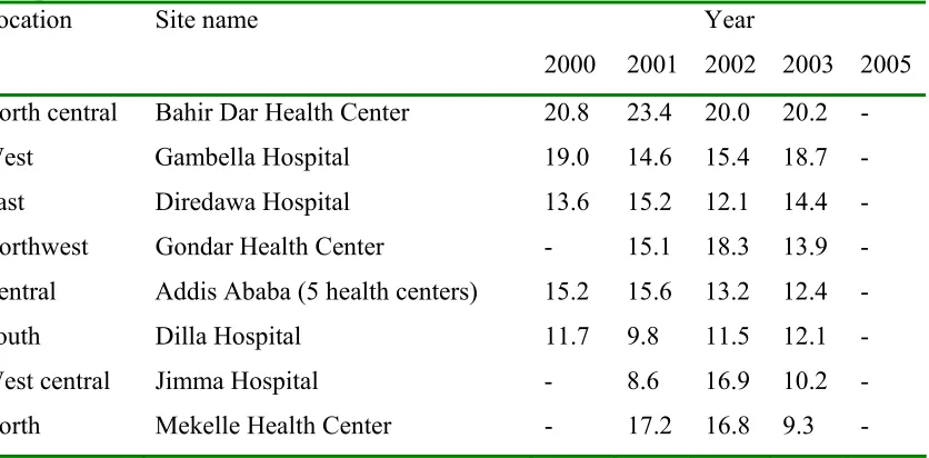
The HIV prevalence in Ethiopia among adults at the end of 2004 was 4.4% with 1.5 million people living with HIV/AIDS, new infections of 197,000, new AIDS cases of 98,000 and AIDS deaths of 90,000 (UNAIDS/WHO, 2004). The trends in HIV-1 prevalence at urban antenatal care sites in various parts of Ethiopia are summarized in Table 1.
Table 1: Trend in HIV-1 prevalence (%) at urban antenatal care (ANC) sites in various parts of Ethiopia (MOH, 2004)
Year Location Site name
2000 2001 2002 2003 2005
North central Bahir Dar Health Center 20.8 23.4 20.0 20.2 -
West Gambella Hospital 19.0 14.6 15.4 18.7 -
East Diredawa Hospital 13.6 15.2 12.1 14.4 -
Northwest Gondar Health Center - 15.1 18.3 13.9 -
Central Addis Ababa (5 health centers) 15.2 15.6 13.2 12.4 -
South Dilla Hospital 11.7 9.8 11.5 12.1 -
West central Jimma Hospital - 8.6 16.9 10.2 -
There has been a global resurgence of TB in the last decades and the majority of these cases occur in the impoverished countries of Africa, Asia and South America. This is largely due to the pandemic of the HIV, which has worst hit on Sub-Saharan African countries (UNAIDS/WHO, 2004). The occurrence and distribution of TB/HIV co-infection is depicted in Figure 3. In New Delhi, a study of 555 patients with TB demonstrated an HIV seropositivity of 9.4% vs an overall
seropositivity of 0.4% during 1994-1999. Prevalence rates among TB patients of 30% in Mumbai11, and 40% in Northern Thailand have been noted (Preetish et al., 2003).
Studies from Uganda and Zambia have recorded HIV rates of 50-70% among TB patients (Jai et al.,
2004). In Tanzania, the survey conducted during 1994-1998 on 10,612 new smears positive TB patients reveled 40% HIV prevalence (Range et al., 2001). In general, the proportion of TB
infections due to HIV varies between countries and has reached level above 80% in some African countries. In Ethiopia, the prevalence of HIV among TB patients is variable from area to area. Study conducted in 2002 showed 57% prevalence of TB/HIV co-infection (Bruchfeld, 2002). Another study conducted in the southern Ethiopia showed prevalence of 19% and 26% TB/HIV co-infection among smear positive and smear negative PTB cases, respectively (Mohammed et al., 2004). More
recently a prevalence of 47% TB/HIV coinfection has been reported from the Northwest part of Ethiopia (Kassu et al., 2006).
6. Laboratory diagnosis of TB-HIV infection
6.1. Diagnosis of M. tuberculosis
The clinical diagnosis of TB differs with the degree of immunity. The classic picture of pulmonary TB is seen mainly in non-severely immunocompromised patients (CD4> 200), and is secondary to a recent infection. Pulmonary involvement is associated with cough, sputum, and more rarely haemoptysis, thoracic pain and dyspnoea. A typical feature consist of lower lobe involvement with a trend towards diffuse infection rather than cavitations, are seen frequently. Cavitary lesions are encountered rarely in patients with a CD4 T-lymphocyte count < 200. TB can be diagnosed by AFB microscopy, Mycobacterial culture, nucleic acid amplification, radiographic examinations, PPD skin test and cytokine release assay (Mario C et al., 2001).
6.2. Diagnosis of HIV infection
HIV infection can be detected by serologic assays for detecting antibodies to HIV and by direct detection assays. The latter involves p24 antigen detection, nucleic acid detection and quantification (detection of proviral DNA and HIV-1 RNA) and culture of HIV (Demeter et al., 2000).
6.3. Diagnostic problems in TB/HIV co-infection
The diagnosis of TB in HIV-positive patients is difficult for three main reasons: a) the sensitivity of the direct sputum smear examination is reduced in positive patients. Compared to HIV-negative patients with pulmonary TB, a lesser proportion of HIV-positive patients with pulmonary TB will have positive sputum smears; b) X-ray abnormalities, which are not specific for TB in HIV-negative patients, are even more non-specific in the HIV-infected, with only minor abnormalities seen on chest X-ray or with abnormalities which do not look like classical TB, and c) Patients infected with HIV have frequent illnesses with pulmonary involvement caused by agents other than
M. tuberculosis ( Jai et al., 2004; Anthony et al., 2001).
6.4. Monitoring of patients with HIV infection
And, thus determination of CD4 T cells count and, measurement of the levels of HIV RNA in serum or plasma provide a powerful set of tools for determining prognosis and monitoring of response to therapy. CD4 T cells count provides information on the current immunological status of the patients and the HIV RNA level predicts what will happen to the CD4 T cells count in the near future, and hence provides an important piece of prognostic information (John, 2004; Anthony et al., 2001).
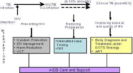
7. Combating TB/HIV co-infection
Figure 4 shows the schematic presentation of TB/HIV combating. In most cases, TB infection comes first and HIV is contracted subsequently when the person achieves adolescence or adulthood. Once co-infected, the progression to active TB occurs quite rapidly, which could be prevented through the use of TB prevention therapy. Those who progress to active TB could be managed with DOTS and through provision of care and support, including the use of antiretroviral therapy. Therefore, at each point in the scheme, interventions can be planned and implemented to try and interrupt TB and HIV infection from progressing to active TB and/or AIDS. Management of HIV- associated TB through DOTS can prevent community transmission of TB (Jai et al., 2004).
Figure 4: Combating TB/HIV: The conceptual framework and intervention points (Jai et al., 2004).
Keys: ART, antiretroviral therapy; TPT, TB preventive therapy; STI, sexually transmitted
7.1. Preventing HIV infection
Action for prevention of HIV transmission must include: a) information and education aimed at all men and women, particularly those at high risk of infection, including sex workers and injecting drug users; b) health and social services especially for the purpose of providing condoms, clean needles and syringes to reduce harm, and the early diagnosis and treatment of sexually transmitted infections using syndromic approach; and c) creating an enabling environment, in the absence of stigma and discrimination directed against people living with HIV/AIDS or those at risk (Jai et al.,
2004).
7.2. Reducing progression to active TB
Preventing the occurrence of clinical TB among co-infected persons requires the people with HIV to be diagnosed in the first place. The primary intervention required is availing the Voluntary Counseling and Testing (VCT) services to those with HIV infection. Those found infected both with TB and HIV can use TB preventive therapy with isoniazid (INH) to prevent progression to TB (Jai
et al., 2004 ;). The efficacy of INH in preventing TB in HIV-positive people has been proven.
However, it must be administered to the patient for a long period, for at least six months. A two-month course of rifampicin and pyrazinamide daily can also be used instead of INH alone (Jai et al.,
2004; Mario et al., 2001).
Short-course chemotherapy under the directly observed treatment (DOTS) strategy is as effective among HIV positive TB patients as in HIV negative patients in curing patients of TB. Thus, besides lowering individual suffering, implementing DOTS through effective TB control programme can reduce the transmission of TB infection, even in the context of increased HIV prevalence (Jai et al.,
2004; Mario et al., 2001).
8. Conclusions and recommendations
As evidenced from the reviewed literatures, TB/HIV co-infection is an important emerging crisis all over the world particularly in Africa, including Ethiopia. Both re-activation of latent M.
tuberculosis infection and primary infection of M. tuberculosis are common in HIV infected
individuals. A mutual relationship has been observed between HIV and M. tuberculosis infections
in that HIV infection leads to the progression of M. tuberculosis infection to active TB; while on the
Thus it can be suggested that the resurgence of TB is attributed to HIV epidemic in developing as 60-70% of TB cases occur in HIV-1 infected individuals in developing countries.
On the basis of these remarks, the following recommendations were forwarded:
Establishing functional collaboration between National HIV/AIDS and TB programme, Preventing HIV through behavioral change in the context of sexual practices, injecting drug
uses and harmful traditional practice,
Preventing the progression of TB infection to clinical TB through TB preventive therapy, Effective case management of patients with HIV associated TB or those with AIDS,
Partnership- building for surveillance, advocacy and program management for the control of TB and HIV.
9. References
1. Ahmed Yassin M, Takele L, Gebresenabet S, et al. HIV and Tuberculosis coinfection in Southern Region of Ethiopia: A prospective Epidemiological study. Scand J Infect Dis 2004; 36: 670-673
2. Anthony S, Fauci H. Human immunodeficiency viruses disease: AIDS and related Disorders. In. Braunwald E., Faucl A., Hauser S., Longo D., Jameson L. Harrison’s Principles of Internal medicine (editors), 15th Ed. McGraw-Hill, New work USA 2001; 1076-1139
3. Aung H, Toossi Z, Wisnieski JJ, et al. Induction of monocyte expression of tumor necrosis factor alpha by the 30 KD alpha antigen of M. tuberculosis and synergism with fibronectin. J
Clin Invest 1996; 64: 399-405
4. Bruchfeld J, Aderaye G, Palme IB, et al. Evaluation of outpatient with suspected pulmonary tuberculosis in a high HIV prevalence setting in Ethiopia: clinical, diagnostic, and epidemiological characteristics. Scand J Infect Dis 2002; 34: 331-7
5. Chan J, Kaufmann SHE. Immune mechanism of protection. In: Tuberculosis pathogenesis, protection and control (ed. BR Bloom) Amercan Society for Microbiology, Washington, DC, 1994, 389-415
6. Dean GL, Edwards SG, Ives NJ, Matthews G, et al. Treatment of TB in HIV infected persons in the era of highly active antiretroviral therapy. AIDS 2002; 16: 75-83
7. Demeter L.M. and Reicman, R.C., 2000. Detection of human immunodeficiency virus infection. In: Mandell, G.L., Bennett, J.E. and Dolin, R., Editors, 2000. Mandel, Douglas
and Bennett's Principles and Practice of Infectious Diseases (5 ed.), Churchill Livingstone,
Philadelphia, pp. 1369–1374
9. Elliott AM, Halwiindi B, Hayes RJ, et al. The impact of Human immunodeficiency Viruses on presentation of and diagnosis of tuberculosis in chort study in Zambia. J Trop Med Hyg 1993; 96: 1-11
10.Failla ML. Trace elements and host defense: recent advance and continuing challenges, J Nutr 2003; 1443S-1443S
11.Feng CG, Bean AGD, Hooi H, Briscoe H, britton WJ. Increase in gamma interferon secreating CD8 +, as well as CD4+ t cells in lung following aerosol infection with m. tuberculosis. Infect Immun 1999; 67: 3242-7
12.Garrait V, Cadranel J, Esvant H et al. TB generates a microenvironment enhancing the productive infection of local lymphocytes by HIV. J Immunol 1997; 169: 2824-2830
13.Goletti D, Weissman D, Jackson RW, et al. Effect of M.tuberculosis on HIV repilication: role of immune activation. J Immunol 1996; 157: 1271-8
14.Havlir DV, Barnes PF. Current concept. Tuberculosis in patients with HIV infection. N Engl J Med 1999; 340: 367-373
15.Jai P, Ying- Ru LO. Epidemiology of HIV-TB in Asia. Indian J Med Res 2004; 120: 277-289
16.Jayasanar K, Ramanathn VD. Biological abd histochemical changes relating to fibrosis following infection with M. tuberculosis in the guineapig. Indian J Med Res 1999; 110: 91-7 17.Jawetz, Melnick, Adelbergis. Medical Microbiology. 19ed. Prentice-Hall International Inc 18.John C, Guatelli, Robert F. Siliciano, Daniel R. Kuritzkes, Douglas D. Richard. William A.
Blattner. Human immunodeficiency viruses. In: Gouglas D, Richard J. Whitley, Frederick G.
19.Hayden (editors), Clinical Virology. 2nd ed. Amercican Society for Microbiology, 2004 : 685-730
20.Kalou M, Sassan-Morokro M, Abouya L, Bile C et al. Changes in HIV RNA viral load,
CD4 T cells counts, and levels of immune activation markers associated with anti tuberculosis therapy and cotrimoxazole prophylaxis among HIV infected Tuberculosis patients in Abidjan. J Med Virol 2005; 75: 202-8
21.Kassu A, Yabutani T, Mahumud ZH, et al. Alteration in trace elements in tuberculosis and
HIV infections. Euro J Clin Nutri 2006; 60: 580-86
22.Mario C, Raviglione, Richard J, O’ Brien. Tuberculosis. In. Braunwald E., Faucl A., Hauser S., Longo D., Jameson L. Harrison’s Principles of Internal medicine, 15th Ed. McGraw-Hill, New work USA 2001; 1024-1034
23.Ministry of Health. AIDS in Ethiopia. 5th ed. Disease Prevention and Control Department, Ministry of Health, Addis Ababa, Ethiopia, 2004
24.Ottenhoff THm, Kumararatne D, Casanova JL. Novel HIV reveals the essentail role of cytoknes in immunity to intracelllar bacteria. Immunol Today 1998; 19: 491-94
25.Preetish S, Vaidyanathan, Sanjay S. TB-HIV coinfection in India; NTI Bull 2003; 39: 11-18 26.Range N, Louge YA, Obrien Jr, Egeaga SM, et al. Trend in HIV prevalence among
Tuberculosis patients in Tanzania. AIDS 2001; 5: 401-407
27.Rook GAW, Seah G, Ustianowski A. M. tuberculosis: Immunology and Vaccionolgy. Eur
Respir J 2001; 17: 537-557
28.Schlesinger LS. Role of mononuclear pahgocytes in M. tuberculosis. J Invest Med 1996; 44:
312-23
29.Schupbach J., Human immunodeficiency viruses. In: Murray, P.R., Baron, E.J., Pfaller, M.A., Tenover, F.C. and Yolken, R.H., Editors, 1999. Manual of Clinical Microbiology (7th
30.Selvaraj P, Venkataprasad N, Vijayan VK, Prabhaha R, Narayanan PR. Alveolar macrophage in patients with Pulumonay Tuberculosis. Lung India 1988; 6: 71-74
31.Toossi Z, Mayanija-Kizza H, Hirsch CS, et al. Impact of Tuberculosis on HIV-1 activity in dually infected patients. Cli Exp Immunol 2001; 123: 233-8
32.Toossi Z. Virological and Immunological Impact of Tuberculosis on HIV-1 Disease. The J Infect. Diseases 2003; 188: 1146-55
33.UNAIDS/WHO. Joint United Nation Program on HIV-1/AIDS. UNAIDS/WHO. AIDS epidemic update. 2004. World Health Organization, Geneva, 2004
34.Vishwanath V, Narayanan S, Narayanan PR. The fate of M. tuberculosis in activated
macropahges. Curr Sci 1998; 75: 942-946
35.Whalen CC, Johnson JL, Okwera A, et al. A trial of three regimens to prevent tuberculosis in Uganda adults infected with HIV. N Engl J Med 1997; 337: 801-8
36.Wolday D, Mayaan S, Mariam ZG, Berhe N, Seboxa T, Britton S et al. Treatment of intestinal worms is associated with decreased HIV plasma viral load. J Acquir Immune Defic Syndr 2002; 31: 56-62
37.Wolday D, Tegbaru B, Kassu A, Messele T, Coutinho R, Van Baarle D et al. Expression of chemokine receptors CCRS and CXCR4 on CD4 T cells and plasma chemokine levels during treatment of active tuberculosis in HIV-1 coinfected patients. J Acquir Immune Defic Synde 2005; 39: 265-271
38.World Health organization (WHO). Report on Global Tuberculosis Control. 2002. WHO, Geneva, Switzerland