Research Article
CODEN: IJPNL6
EFFECTS OF THE PROPERTIES OF CREAMS ON SKIN PENETRATION
Yutaka Inoue
1,*, Arisa Shimura
1, Misa Horage
1, Rikimaru Maeda
1, Isamu Murata
1, Masahiro
Sugino
2, Kazuhiko Juni
2, Ikuo Kanamoto
11
Laboratory of Drug Safety Management, Faculty of Pharmaceutical Sciences, Josai University,
Japan
2
Laboratory of Pharmaceutics, Faculty of Pharmaceutical Sciences, Josai University, Japan
*Corresponding author e-mail:
[email protected]
ABSTRACTThe current study conducted human sensory testing and temperature-dependent measurement of the creams to examine their viscosity and viscoelasticity. A retention on filter paper test and skin penetration test involving Yucatan micropigs (YMPs) were conducted to measure penetration of the skin and transdermal transfer. The relationship between testing results and physical properties of the creams was then examined. When viscosity was measured, NDFX-A and NDFX-B displayed similar behavior as gauged by viscosity and shear stress. NDFX-C produced a large flow curve, with a larger area under the flow curve than NDFX-A or NDFX-B. Measurement of viscoelasticity indicated that the storage modulus G’ and the loss modulus G” for NDFX-A and NDFX -B increased with a change in temperature (60˚C to 10˚C) while G’ and G’’ decreased for NDFX-C. The loss tangent tan δ was determined for each cream. Prior to and after a rise in temperature to 60˚C, NDFX-A had a tan δ of +0.48 at 20˚C, NDFX-B had a tan δ of −0.34, and NDFX-C had a tan δ of ±0. In a retention on filter paper test, NDFX-B had the highest level of drug retention, followed by NDFX-C and then NDFX-A. Water content presumably plays a role in this phenomenon. Results of a skin penetration test indicated that NDFX-B and NDFX-A had approximately the same amount of skin penetration and the same amount of transdermal transfer after 24 hrs while NDFX-C had less skin penetration and less transdermal transfer. This is because crystals were noted in NDFX-C. Microscopy revealed oil droplets in that cream. Thus, these aspects presumably affected its skin penetration and transdermal transfer. Skin penetration amount is directly related to efficacy, so physical properties of creams may be an important aspect to consider.
Keywords: Creams, Skin Penetration, viscosity and viscoelasticity
INTRODUCTION
Over the past few years, generic drugs have been developed as oral agents and topical agents1). Differences in the additives and vehicles in brand-name and generic topical agents are reported to result
in different physical properties, such as
viscoelasticity and yield values2). Many additives are used in topical agents to increase the usefulness in those drugs. In fact, topical preparations mostly consist of additives, and contain only small amounts of active ingredients. Generic preparations must contain the same active ingredients in the same content as brand-name preparations, but they need not contain the same types or amounts of additives. Brand-name and generic preparations are reported to
differ in terms of characteristics such as physical properties and the rate of drug release3). Even if preparations contain the same additives, they may
have different properties than brand-name
preparations whatever the method of manufacture or manufacturing process differs. Additives in brand-name and generic tulobuterol patches are reported to differ, and these differences are reported to affect on drug release4).
An in vitro skin penetration study has been widely used to develop preparations that applied to the skin5). Use of human tissue and organs is difficult because of ethical issues. However, animal skin is considered to be an acceptable way to estimate a drug’s penetration of human skin when developing transdermal preparations. Skin from numerous
International Journal of Pharmacy
animals, including mice, rats, and pigs, has been used as an alternative method to human skin penetration studies6). Pigs are known to have large bodies and a skin morphology likes that of humans, and similar drug penetrate profiles are observed with the skin of pigs and humans, so pigs have been used in numerous experiments7). Yucatan micropigs (YMPs) have little body and their skin is similar to human
skin in aspects, such as structural and
immunohistochemical properties and the rate of drug penetration. YMPs reported to be a useful model to predict a drug penetration of human skin8). Franz type diffusion cells are used to assess skin permeation; these devices facilitate the measurement of the amount of a drug that permeates through the skin. Use of Franz cells allows sampling over time starting with the receiver phase, and a study has reported using Franz cells to assess a drug penetration of the skin9). According to the Fick’s 1st law of diffusion, the rate of skin penetration is proportional to the concentration (of a drug) applied. Thus, the drug concentration in a cream is a key factor that determines the amount of its penetration into the skin.
Previously, we examined the physical properties of brand-name and generic versions of NDFX creams. Microscopy revealed crystals in NDFX-C, and water content in different creams differed4). The current study conducted human sensory testing and temperature-dependent measurement of the creams to examine their viscosity and viscoelasticity. In addition, this study examined the retention on filter paper test and the skin penetration of those preparations resulting from differences in additives they contained. Testing results should provide useful information for future development of creams and allow better selection of creams in accordance with their intended use.
MATERIALS AND METHODS
Reagents: Three NDFX creams were used. The brand-name NDFX cream Acuatim® (from Otsuka Pharmaceutical Co.), the generic NDFX cream Nadiflo® (from POLA Pharma Inc.), and the generic
NDFX cream Nadiroxisan® (from Towa
Pharmaceutical) were respectively designated
NDFX-A, NDFX-B, and NDFX-C. Purchased from Sigma Aldrich, NDFX powder was used as. Other reagents used were of special commercial grade (Wako Pure Chemical Industries).
Human sensory testing: Test subjects were 24 healthy adults volunteer (9 men, 15 women) with a mean age of 23.3 years (range: 20–38 years). A
single-blinded human sensory test was conducted, and feel was assessed using an assessment form (Dia. 1). Each cream was randomly designated A, B, or C. Prior to the test, subjects washed their hands with tap water and then wiped them with a paper towel before allowing their hands to dry for 5 min. Afterwards, each subject selected either cream A, B, or C. Creams had been portioned into dollops of 0.1 g beforehand, and subjects used their index finger to rub the cream into their arm 10 times in a circular motion. After 5 min, subjects assessed the aspects of the cream listed on the assessment form. They then rinsed the area where the cream had been applied with tap water. Four aspects were assessed: adherence, spreadability, tightness, and feel. Subjects followed the same steps of applying and assessing subsequent creams. The human sensory testing in this study was approved by the Ethics Committee of the Life Science Research Center of Josai University. Testing was fully explained to subjects and their written consent to participate in the test was obtained.
Measurement of viscosity: Viscosity was measured using a HAAKE MARS Rheometer (Thermo Scientific Co.). Measurement conditions were a 35 mm cone rotor, 1° angle, a gap of 0.05 mm, and a sample amount of 0.051 mL. For each sample, shear stress (Tau (Pa)) was measured in 1-sec intervals at 20˚C and 35˚C. The shear rate was 0 s-1
- 1000 s-1(1 min)→1000 s-1- 0 s-1(1 min).
Measurement of viscoelasticity: Viscoelasticity was measured using a dynamic viscoelasticity measuring
device with a vertically vibrating super
magnetostrictor (MX-1000 MG Rheoanalyzer, Sons Corporation). Measurement was performed with an amplitude of 35 μm and frequency of 5 Hz. The measurement temperature was raised from 10ºC to 60ºC at a rate of 1ºC / min. Once the temperature reached 60ºC, viscoelasticity at 60ºC was measured for 10 min. The measurement temperature was lowered from 60ºC to 10ºC at a rate of 1ºC / min and viscoelasticity was similarly measured.
The storage modulus G’ (Pa), which represents the elastic component of a sample, and the dynamic loss modulus G” (Pa), which represents that viscous component of that sample, were measured. In addition, the loss tangent tan δ was determined. A ratio of G’ and G” and an indicator of viscoelasticity, tan δ was calculated using the formula tan δ = G”/ G’.
tan δ= G”/ G’ ; tan δ: loss tangent G”: loss modulus (Pa)
Diagram 1: Scores for NDFX creams.
Cohesiveness
Extensibility
Tension feeling
Usability
1
2
3
4
5
No
Yes
1
2
3
4
5
1
2
3
4
5
1
2
3
4
5
Retention on filter paper test
About 0.1 g of each cream was applied to filter paper (Tokyo Roshi Kaisha) and then left to stand for 10 min and 30 min at room temperature. Afterwards, the filter paper was placed in a centrifuge tube containing 50 mL of physiological saline. The tube was shaken at 100 rpm for 5 min at 25ºC or at 32ºC. After shaking, the filter paper was removed in order to extract NDFX in physiological saline. A solution of chloroform/NaOH (100 mmol/ L)=1/ 1 was added and the mixture was agitated for 10 min. The mixed solution was centrifuged at 4000 rpm for 30 min and the supernatant was then filtered with a 0.45-μm membrane filter. Afterwards, the solution was assayed using HPLC. The assay was performed with a high-performance liquid chromatograph (HPLC: Waters E2695).
Skin penetration test
Skin Yucatan micropigs (YMPs, females) purchased from that had been cryopreserved at −80ºC was thawed at about 4ºC for 12 hrs before use. After thawing, subcutaneous fat was removed from the
skin. After fat was removed, the skin was cut into pieces of about 2.5 cm ×2.5 cm. The stratum corneum was removed by tape stripping (30 times)
10). Once the stratum corneum had been removed
from YMP skin, the skin was placed with the epidermis up on a paper towel moistened with physiological saline. The skin was stored at 4ºC for 12 hrs before use.
Franz type diffusion cells (PermeGear) (effective penetration area: about 0.95 cm2) were used for skin penetration and transdermal transfer study. The receiver solution (about 9.5 mL) used was 5% albumin dissolved in physiological saline. The receiver chamber of the Franz diffusion cell was oriented toward the dermis of the YMP skin. The receiver solution was kept at 32ºC. Receiver solution was agitated and then used in the penetration test. About 0.5 g of each cream was applied to the epidermis of the YMP skin and the penetration test began. Once the cream was applied, 400 μL of the receiver solution was collected over time at 1, 3, 6, 9, and 24 hrs. The same amount of receiver solution was replenished. After sampling at 24 hrs, the
Diagram 1
effective area of YMP skin penetration was excised. One mL of sodium hydroxide was added to deproteinization. The skin was homogenized, and chloroform/NaOH (100 mmol/L)= 2 mL/2 mL was added. The mixture was centrifuged (25ºC, 4000 rpm, 30 min) and assayed using a high-performance liquid chromatograph (HPLC: Waters E2695). Protein was removed from the collected receiver solution using acetonitrile/sodium hydroxide=200 μL/200 μL, and the solution was then centrifuged (4ºC, 4000 rpm, 10 min). YMP skin was assayed using a high-performance liquid chromatograph in a similar manner to the assay technique used in the retention on filter paper test.
Assay: An assay was performed using a high-performance liquid chromatograph (HPLC: Waters E2695). Assay conditions were an Inertsil ODS-3 column (φ5 μm, 4.6×150 mm, GL Sciences Inc.), a column temperature of 35ºC, a mobile phase of water/acetonitrile/acetic acid=130/70/1, a detection wavelength of 280 nm, and an injection volume of 20 μL. Conditions were tailored for NDFX to produce a peak at 15 min.
Statistical Analysis: Results are presented as mean ± standard deviation, and statistical significance was evaluated using the Tukey Test.
RESULTS
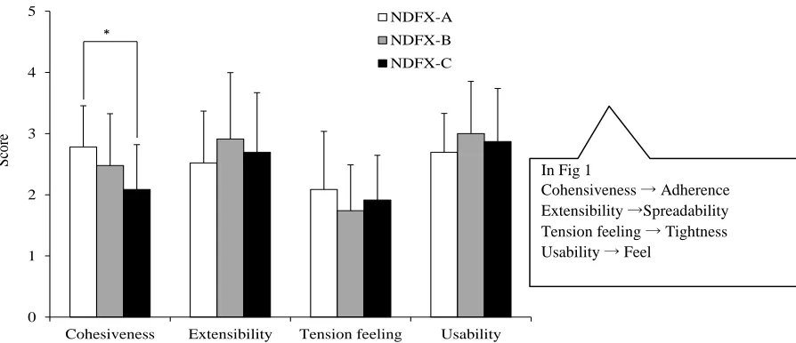
Human sensory testing: Sensory testing was conducted with regard to 4 aspects (“adherence,” “spreadability,” “tightness,” and “feel”) in order to examine the feel of each cream (Fig. 1). NDFX-A was rated as having the best adherence, followed by NDFX-B and then NDFX-C. There were significant differences in the adherence of A and NDFX-C (p<0.01). Significant differences in the spreadability, tightness, and feel of each cream were not noted. However, NDFX-B had a high level of spreadability while NDFX-A tended to have a low level of spreadability (B> C> NDFX-A). NDFX-A had a high level of tightness while NDFX-B tended to have a low level of tightness (NDFX-A> NDFX-C> NDFX-B). NDFX-B was rated as having a better feel while NDFX-A tended to have a worse feel (NDFX-B> NDFX-C> NDFX-A).
0 1 2 3 4 5
Cohesiveness Extensibility Tension feeling Usability NDFX-A NDFX-B NDFX-C *
Figure 1: Human sensory testing of each NDFX creams. *p<0.01, Tukey test (mean±S.D. n=24)
S
cor
e
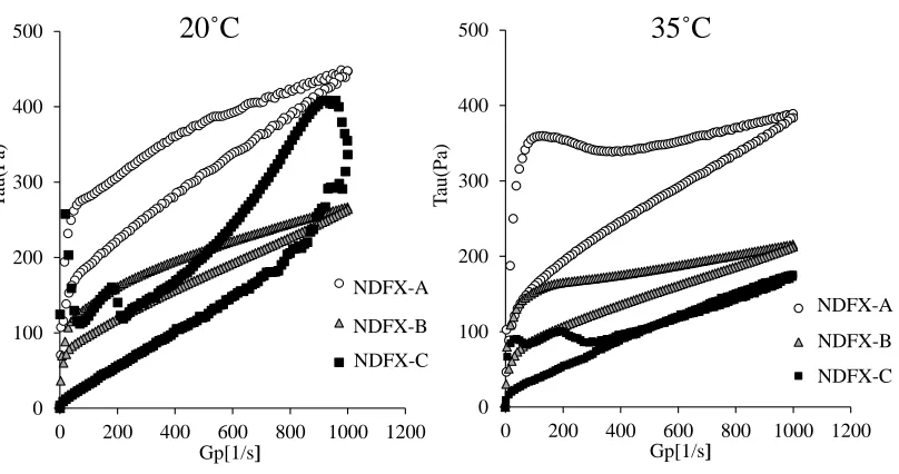
Measurement of viscosity: There were significant differences in adherence of the creams in sensory testing, so viscosity was measured and the relationship between adherence and viscosity was examined (Fig. 2). The storage temperature for a typical drug is 20ºC, so a measurement temperature of 20ºC was used. At that temperature, NDFX-A and NDFX-B displayed similar behavior, i.e. they had similar flow curves. NDFX-C had a large flow curve, with a larger area under the flow curve than
NDFX-A or NDFX-B. Assuming that the temperature of human skin would be 35ºC when a cream was rubbed in, a measurement temperature of 35ºC was used. At that temperature, NDFX-A and NDFX-B displayed almost the same behavior, i.e. flow curves, as they did at 20˚C. However, stress decreased for NDFX-C, with a smaller area under the flow curve. NDFX-A had the largest flow curve, followed by NDFX-B and then NDFX-C. This result was the same as for the aspect of adherence in sensory testing.
In Fig 1
0 100 200 300 400 500
0 200 400 600 800 1000 1200 Gp[1/s]
0 100 200 300 400 500
0 200 400 600 800 1000 1200 Gp[1/s]
NDFX-A NDFX-B NDFX-C
20˚C
35˚C
NDFX-A NDFX-B NDFX-C
Figure 2: Shear stress Tau(Pa) versus shear rate Gp[1/s] curves for NDFX creams.
T
au(
Pa)
T
au(
P
a)
Viscoelasticity of each creams: Viscoelasticity was measured with respect to temperature, and the storage modulus G’ and dynamic loss G’’ were determined for each cream. The adherence of the creams was examined based on their loss tangent tan δ=G”/ G’. When the temperature was lowered from 60ºC to 10ºC, G’ and G’’ increased for A and NDFX-B. In contrast, G’ and G’’ decreased for NDFX-C when the temperature was lowered from 60ºC to 10ºC (Fig. 3). When the temperature was raised to 20ºC, NDFX-A had a loss tangent tan δ of 0.38, NDFX-B had a loss tangent tan δ of 1.07, and NDFX-C had a loss tangent tan δ of 0.32. When the
temperature was raised to 32ºC, NDFX-A had a loss tangent tan δ of 0.32, NDFX-B had a loss tangent tan δ of 1.07, and NDFX-C had a loss tangent tan δ of 0.53 (Fig. 4). When the temperature was raised to 60ºC and lowered to 32ºC, NDFX-A had a tan δ of 0.97, NDFX-B had a tan δ of 0.71, and NDFX-C had a tan δ of 0.48. When the temperature was then further lowered to 20ºC, NDFX-A had a tan δ of 0.86, NDFX-B had a tan δ of 0.73, and NDFX-C had a tan δ of 0.32. Prior to and after a rise in temperature to 60˚C, NDFX-A had a tan δ of +0.48 at 20˚C, NDFX-B had a tan δ of −0.34, and NDFX-C had a tan δ of ±0.
0 2 4 6 8 10 12 14
0 10 20 30 40 50 60
Temperature(˚C)
G'(kPa) G"(kPa)
0 2 4 6 8 10 12 14
0 10 20 30 40 50 60
Temperature(˚C)
G'(kPa) G"(kPa)
0 2 4 6 8 10 12 14
0 10 20 30 40 50 60
Temperature(˚C)
G'(kPa) G"(kPa)
Figure 3: Changes in viscoelasticity G(kPa) of NDFX creams with respect to temperature. a) NDFX-A, b) NDFX-B, c) NDFX-C
a) b) c)
V
iscoe
lasticit
y
;
G(
kpa
)
0 1 2 3 4 5
0 20 40 60
Temperature(˚C)
0 1 2 3 4 5
0 20 40 60
Temperature(˚C)
0 1 2 3 4 5
0 20 40 60
Temperature(˚C)
Figure 4: Changes in tan δ values with respect to temperature for NDFX creams. a) NDFX-A, b) NDFX-B, c) NDFX-C)
a) b) c)
tanδ tanδ tanδ
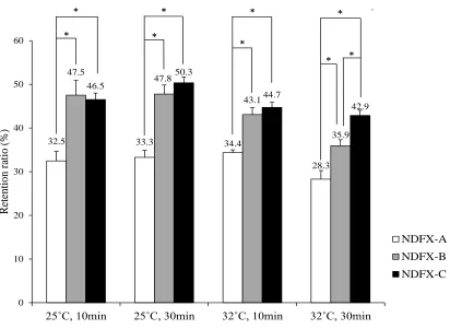
Retention on filter paper: The ease with which creams were removed was examined based on the retention on filter paper test. NDFX-A had the least retention on filter paper at 25ºC for 10 min, followed by NDFX-C and then NDFX-B. Significant differences between NDFX-B and NDFX-A and between NDFX-C and NDFX-A in terms of retention
were noted. At 25ºC for 30 min and at 32ºC for 10 min, retention results were similar (Fig. 5). At 32ºC for 30 min, NDFX-B still had the greatest retention,
followed by NDFX-C and then NDFX-A.
Significant differences in the retention of the creams were noted.
*
32.5 33.3 34.4
28.3 47.5
47.8
43.1
35.9 46.5
50.3
44.7
42.9
0 10 20 30 40 50 60
25˚C, 10min 25˚C, 30min 32˚C, 10min 32˚C, 30min
R
etention
ra
ti
o (%
)
NDFX-A NDFX-B NDFX-C *
*
* *
* *
* *
Skin penetration and transdermal transfer:
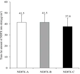
Skin penetration was measured for 24 hrs (Fig. 6) and transdermal transfer was measured after 24 hrs (Fig. 7). Whether or not there were differences in the penetration and transdermal transfer of each cream was examined. In terms of the amount of skin penetration (μg/cm2) after 3 hrs, NDFX-A had skin penetration of 16.33 μg/cm2, NDFX-B had skin
penetration of 14.76 μg/cm2, and NDFX-C had skin
penetration of 1.14 μg/cm2. There were significant
differences between NDFX-C and NDFX-A and between NDFX-C and NDFX-B in terms of skin penetration (p< 0.05). In terms of the amount of skin penetration 24 hrs after application, NDFX-A had skin penetration of 46.41 μg/cm2, NDFX-B had skin penetration of 45.70 μg/cm2, and NDFX-C had skin
penetration of 36.65 μg/cm2. NDFX-A and NDFX-B tended to have approximately the same amount of skin penetration while NDFX-C had less skin penetration. However, significant differences in skin penetration were not noted.
In terms of the amount of transdermal transfer amount of NDFX after 24 hrs, NDFX-A had transdermal transfer of 41.5 μg/cm2, NDFX-B had transdermal transfer of 41.5 μg/cm2
, and NDFX-C had transdermal transfer of 37.6 μg/cm2. NDFX-A
and NDFX-B tended to have approximately the same amount of transdermal transfer while NDFX-C had less transdermal transfer. However, significant differences in transdermal transfer were not noted.
0
10
20
30
40
50
60
0
4
8
12
16
20
24
Cu
m
u
lat
iv
e
am
o
u
n
t
o
f
N
D
FX
p
erm
eat
ed
(µ
g
/cm
2
)
Time (hr)
NDFX-A
NDFX-B
NDFX-C
Figure 6: Skin penetration-time profile of NDFX,
*p<0.05 vs. NDFX-C, Tukey test (mean
±
S.D. n=3)
*
41.5 41.5
37.6
0 10 20 30 40 50 60
NFX-A
NFX-B
NFX-C
T
ra
ns fa
r
amount
of N
DF
X
int
o
skin(
μg
/cm
2
)
NDFX-B NDFX-C NDFX-A
Figure 7: Amount of transdermal NDFX transfer.
No significant differences, Tukey test (mean ±
S.D. n=3)
DISCUSSION
Sensory testing was conducted with 3 kinds of topical NDFX creams. NDFX-A was rated as having the best adherence, followed by NDFX-B and then NDFX-C. Significant differences between NDFX-A and NDFX-C in terms of adherence were noted. Differences in the physical properties of creams may be evident in the assessment of adherence in human sensory testing. A viscosity test, a viscoelasticity test, a retention on filter paper test, a skin penetration test, and a transdermal transfer test were also conducted. Test results indicated differences in the viscoelasticity of the creams.
Present study measured viscosity at 20˚C, which is the storage temperature for a typical drug, and at 35˚C, which is the temperature of the surface of human skin when a cream is rubbed in. In the measurement of viscosity, shear stress and the shear rate are known to be indicators of a substance’s viscosity, resistance to force, and the strength of its internal structure, i.e. its stability11). A larger area under the flow curve means that a substance has a strong internal structure that is less susceptible to
disruption4). NDFX-A and NDFX-B behaved
similarly, i.e. they had similar flow curves, presumably indicating that these 2 creams had an internal structure with similar small strength.
The effect of heating on physical properties was examined via temperature-dependent measurement of viscoelasticity using the storage modulus G’, which represents the elastic component, and the loss modulus G”, which represents the viscous elastic component. When heated and cooled, only NGFX-C had a G’ and G’’, i.e. viscosity and viscoelasticity, that decreased. This is because heating of NDFX-C resulted in the dispersal of its ingredients while cooling resulted in those ingredients clumping together. The less viscous ingredients in NDFX-C clumped together, which is presumably why its elasticity and viscosity both decreased.
Based on the results in Fig. 4, the viscosity of NDFX-A increased when the temperature of the creams was raised to 60˚C and then lowered. The elasticity of NDFX-B increased. However, changes in NDFX-C prior to and after the rise in temperature were not noted. This relates to the constituents in the cream when its temperature is raised. A cream’s elasticity presumably increases if its structure is further disrupted. Presumably, this is because water content led to structural differences in the creams. We previously reported that NDFX-B has the highest water content, followed by NDFX-C, and then NDFX-A2). A cream’s internal structure is disrupted if it has a higher water content. Its elasticity presumably decreases when its temperature is raised to 60˚C.
The retention on filter paper test presumably indicated that relatively small amount of NDFX-A was retained on filter paper. A previous study on the characteristics of NDFX-A in comparison to those of NDFX-B and NDFX-C reported that NDFX-A has a low water content and a high viscosity2). These findings presumably indicate that a cream separates from physiological saline more readily if it has a low water content, i.e. it is oilier. Presumably, such a cream is less apt to remain and is easier to remove. NDFX-C had less skin penetration and transdermal transfer than NDFX-A and NDFX-B. One characteristic of NDFX-C is that it had gaps and needle-shaped crystals 2). In the current sensory testing, NDFX-A and NDFX-B were rated as having better adherence than NDFX-C. NDFX-C contains the same amount of principal ingredient, but the NDFX crystals in the cream precipitate. This results in less dissolution of the principal ingredient into the preparation in comparison to the other 2 creams. As a result, application of an adequate concentration
cannot be ensured. This is presumably why its penetration of the skin decreased. The same holds true for its transdermal transfer. The principal ingredient is not taken up by the skin, which is presumably why its transdermal transfer also decreased. In contrast, NDFX-A contains a high concentration of the principal ingredient. The ingredient quickly begins to penetrate the skin, and this is presumably why the amount of its penetration also increases.
CONCLUSION
In sensory testing, NDFX-C was rated as having the worst adherence in comparison to NDFX-A and NDFX-B (NDFX-A>NDFX-B>NDFX-C). With this in mind, a viscosity test and a viscoelasticity test were conducted. Test results coincided with the assessment of adherence in sensory testing (NDFX-A>NDFX-B>NDFX-C). The association between adherence to and penetration of the skin was examined. A cream that was less adherent was found to be less apt to penetrate the skin. These findings are presumably due to the fact that there are NDFX crystals in NDFX-C. NDFX is not fully dispersed in the cream and NDFX is also dispersed unevenly. This is presumably why the viscosity and viscoelasticity of the cream decreased. In addition, the presence of crystals means that there is less dispersal of NDFX in the cream. This is presumably why NDFX-C had a lower rate of skin penetration and less skin transfer than NDFX-A and NDFX-B. Thus, this study has indicated that the efficacy of a cream may be closely related to its physical properties.
ACKNOWLEDGEMENTS
The authors wish to thank the subjects who participated in the sensory test. The authors also wish to thank Dr. Masahiro Sekine of the Saitama Industrial Technology Center for advice on measuring viscoelasticity and Daiichi Sankyo Healthcare for advice on measuring viscosity.
CONFLICTS OF INTEREST
REFERENCES:
1. Nakazono S, Ogura S. Expert Rev Pharmacoecon Outcomes Res, 2014; 14(2): 181-94.
2. Inoue Y, Matsumoto M, Kimura M, Tanaka T, Kanamoto I. Int J Pharm, 2011; 47(11): 616-22. 3. Inoue Y, Furuya K, Maeda R, Murata I, Kanamoto I. Int J Pharm, 2013; 447(1-2): 158-64. 4. Yoshihara S, Fukuda H, Abe T, Arisaka O. Biol Pharm Bull, 2010; 33(10): 1763-65.
5. Takeuchi H, Terasaka S, Sakurai T, Furuya A, Urano H, Sugibayashi K. Biol Pharm Bull, 2011; 34(4): 555-61. 6. Wang X, Shi L, Tu Q, Wang H, Zhang H, Wang P, Zhang L, Huang Z, Zhao F, Luan H, Wang X. Int J
Nanomedicine, 2015; 10: 347-55.
7. Summerfielda A, Meurensb F, Ricklin ME. Mol Immunol, 2014; 66(1): 1-8.
8. Uchino T, Matsumoto Y, Murata A, Oka T, Miyazaki Y, Kagawa Y. Int J Pharm, 2014; 464(1-2): 75-84. 9. Franz TJ. J Invest Dermatol, 1975; 64(3): 190-95.