www.pharmascholars.com
159
Original Article
CODEN: IJPNL6
COMPARATIVE EVALUATION OF CYCLODEXTRIN COMPLEXATION IN
IMPROVING DISSOLUTION OF RITONAVIR
Bharani S. Sogali
a*, Sushant M. S
a, Ramana Murthy K. V
ba
Department of Pharmaceutics, Krupanidhi College of Pharmacy, ChikkaBellandur,
Bangalore-560035, India.
b
Department of Pharmaceutics, Andhra University, Visakhapatnam-530003, India.
*Corresponding author e-mail: [email protected]
Received on: 18-02-2016; Revised on: 29-03-2016; Accepted on: 01-04-2016
ABSTRACT
The present study was focused on comparing different methods of preparations and the effect of hydroxy propyl beta cyclodextrin (HPβCD) and randomly methylated beta cyclodextrin (RMβCD) in enhancing dissolution and oral bioavailability of poorly soluble, antiretroviral protease inhibitor, ritonavir. Complexes of ritonavir-CD complexes were prepared at 1:1 ratio using physical mixing, co evaporation, spray drying and freeze drying methods. The prepared complexes were characterized using FTIR, differential scanning calorimetry (DSC), x-ray diffraction studies (XRD), nuclear magnetic resonance spectroscopy (NMR) and scanning electron microscopy. In vitro
dissolution studies were performed in 0.1N HCl and the dissolution data were subjected to one way ANOVA. Phase solubility studies proved complexation of ritonavir and were of AL type. All complexes showed enhanced
dissolution rate compared to ritonavir but the complex prepared with RMβCD using freeze drying had shown complete and fast drug release. On the basis of above results, it was concluded that complex prepared with RMβCD by freeze drying was successful in improving solubility, dissolution rate and oral bioavailability of ritonavir.
Key words: Randomly methylated βcyclodextrin (RMβCD), Freeze drying, Ritonavir, Complex, Bioavailability.
INTRODUCTION
Human immunodeficiency virus (HIV) infection, which leads to acquired immunodeficiency syndrome (AIDS), remains a serious worldwide health problem. HIV continues to be a major global public health issue, claiming more than 25 million lives over the past three decades. There were approximately 34 [31.4–35.9] million people living with HIV in 2011. In 2012, more than 9.7 million people living with HIV were receiving antiretroviral therapy (ART) in low and middle-income countries [1]. The discovery of HIV protease inhibitors introduced new and effective first-line therapies for HIV/AIDS. Helping to combat HIV-related diseases and prolong survival, protease inhibitors are commonly administered with reverse transcriptase inhibitors. However, poor patient compliance, noxious side effects, and viral
resistance have led to a recommendation to treat with different kinds of protease inhibitors.
Ritonavir, a widely prescribed antiretroviral protease inhibitor drug belongs to Class II under BCS and exhibits low and variable oral bioavailability due to its poor aqueous solubility. Its oral absorption is dissolution rate limited and it requires enhancement in solubility and dissolution rate for increasing its oral bioavailability. Ritonavir, a CYP3A and P-gp inhibitor, helps to increase saquinavir oral bioavailability [2-4]. However, because HIV/ AIDS patients must take other drugs known to be metabolized by CYP3A or they are P-gp substrates, ritonavir has been shown to cause additional toxicity and safety issues [5]. Therefore, safe pharmaceutical formulations that enhance the bioavailability of ritonavir are needed.
International Journal of Pharmacy
www.pharmascholars.com
160
Several approaches could be investigated to improve oral bioavailability of poorly soluble drugs, among them, complexation with cyclodextrins has drawn considerable attention, because of their low toxicity, low cost, biocompatibility, biodegradability and abundant availability. Drug-CD complexation and improvement in solubility and dissolution rate are influenced by both nature of the cyclodextrin (native or chemically modified, crystalline or amorphous) and the method of complexation, viz co-grinding, kneading, solid dispersion, solvent evaporation, co-precipitation, spray drying, or freeze drying. The effectiveness of a method depends on nature of the drug and CD [6-8]. In many cases, spray drying and freeze drying were found to be more effective for drug complexation [9-13].
Therefore, in the present work, a series of binary systems of ritonavir with amorphous and highly soluble derivatives of cyclodextrins, HPβCD and RMβCD were prepared by using four different methods. The main objective of the study was to carry out comparative studies on effectiveness of the method of preparation and better cyclodextrin derivative in enhancing solubility and dissolution rate of ritonavir so that the drug dosing can be reduced for minimizing side effects and also the utilization of large amounts of cyclodextrins can be lowered.
MATERIALS AND METHODS
Materials: Ritonavir was kindly gifted from Hetero Chemicals, Hyderabad, India. HPβCD and RMβCD were gift samples received from RoquettePharma, Italy. All other chemicals used were of analytical grade.
Methods
Phase solubility studies: Phase solubility studies were performed as mentioned by (Higuchi and Connors, 1965) [14] in 0.1 N HCl to determine suitable cyclodextrin derivative in solubility enhancement. Solubility studies were carried out by adding excess amounts of drug separately in quantities exceeding its aqueous solubility to 50 mL of 0.1 N HCl, containing increasing concentrations of cyclodextrins (2-12 mM). The resulting suspensions were shaken at room temperature for a period of 72 hrs, until equilibrium was established. The samples were filtered through a 0.45 µm membrane filter (Millex-HA filter units, Millipore) and suitably diluted with 0.1 N HCl before analysis. All studies were performed in triplicate.
Apparent 1:1 stability constants were calculated from the straight-line portion of the phase solubility diagrams, according to the Equation 1.
-Eq.1
where So is the solubility of the drug in examined
buffer solution in the absence of ligand.
Preparation of drug-CD complexes
In the present work, drug-CD complexes were prepared using physical mixing, co evaporation, spray drying and freeze drying.
Physical mixture [15,16]: Drug and cyclodextrins at equimolar ratio (1:1) were weighed and mixed in a mortar by geometric dilution method for sufficient time (~5 min) to obtain a homogenous powder blend, passed through sieve no. 80 and stored in a sealed glass vials and kept in desiccator over fused calcium chloride until further use.
Co evaporation [16-18]: Required quantities of drug and cyclodextrin were dissolved in sufficient quantity of water-ethanol solvent mixture (1:1) and evaporated on a water bath at 50°C with stirring. Each solid product was sieved through #80 and stored in desiccator.
Spray drying: Spray drying was performed using Labultima LU22 spray dryer for the preparation of inclusion complexes of both drugs and cyclodextrins. Equimolar ratio of drug and cyclodextrins were dissolvedin sufficient quantity of ethanol and water mixture (1:1). The solutions were atomized at a flow rate 2 mL/min using fixed values for compressed air (500 L/h) and aspirator 40-50 m3/hr with T
in value of
70°C, corresponding to a Tout of 48°C. After spray
drying, each resulting powder was collected by cyclone separation and transferred to glass vials. After spray drying, all complexes were kept in an oven at 40°C for 24 hrs to remove traces of moisture present in the complexes.
Freeze drying/lyophilization[19,20]: Required quantity of cyclodextrin was dissolved in sufficient quantity of water and required quantity of drug was added and dispersed. The dispersion was kept under magnetic stirring at 200 rpm for 72 hrs in closed vials for complex formation which resulted in the formation of clear solution. The resulting solution was fast frozen at -20°C using liquid nitrogen and dried at -50°C and 0.0070 mbar pressure in a freeze dryer (Model MODUL YOD 230, Thermo Electron Corporation, India) for 48 hrs. The obtained freeze dried product in the glass vial was stored in desiccator.
www.pharmascholars.com
161
equivalent to 100 mg of drug was used. 5 mL sample was withdrawn at intervals of 5, 15, 30, 45, 60 min using a syringe fitted with prefilter (0.45 µm). An
equal amount of fresh dissolution medium
maintained at the same temperature was replaced immediately after withdrawal of the test sample. Test samples were suitably diluted wherever necessary and the absorbance was measured as per the analytical procedures described. The mean percent of drug dissolved and the standard deviations were calculated.
Drug-excipient interaction studies
Drug excipient interaction studies were conducted for ritonavir, pure CDs, physical mixtures and freeze dried complexes to gain insight into interactions between drug and respective CDs in the complexed form.
Fourier transform infrared spectroscopy (FTIR):
Fourier transform infrared spectra (FTIR) were used to identify the formation of complex. Samples were analyzed by potassium bromide pellet method in an IR spectrophotometer (Shimadzu, FTIR 1300) in the region between 400-4000 cm-1. Complex formation
was evaluated by comparing the IR spectra of the solid complex with drug. For comparison purposes, FTIR spectra were obtained for ritonavir, each βCD being studied as well as physical mixtures of both drugs with each cyclodextrin.
Differential scanning calorimetry (DSC): The thermal behavior of ritonavir and cyclodextrin inclusion complexes was studied using Differential scanning calorimetry in order to confirm the formation of solid complex. When guest molecules are incorporated in the cyclodextrin cavity or in the crystal lattice, their melting, boiling and sublimation points are usually shifted to a different temperature or disappear within the temperature range. The samples were heated from 0 to 350°C at a heating rate of 10°C/min under a nitrogen flow, flowing at a rate of 40 mL/min through the DSC cell.
Powder X-Ray diffraction study (XRD): X-Ray diffraction of inclusion complexes and the pure drug were performed to identify the interaction of the drug with cyclodextrins using a PW 1720 X-ray generator and a PW 1710 diffractometer control (Philips Electronic Instrument). The scanning range (2θ) was from 5° to 90°, and the scan step and scan speed were 0.04° and 0.02°/s, respectively.
Nuclear magnetic resonance (NMR) spectroscopy:
1H-NMR studies were conducted to determine the
electronic interactions between drug and
cyclodextrins were obtained from a Bruker AM-400 spectrophotometer. Samples were prepared by dissolution of the drug in methanol as solvent for RTV. 1H-NMR spectra of inclusion complexes and
pure components were recorded. Chemical shifts were reported in ppm (δ) downfield from tetra methyl silane (TMS) (internal reference).
Scanning electron microscopy (SEM): The morphological properties of the ritonavir, physical mixture, freeze dried powders of ritonavir and RMβCD complex were characterized by scanning electronic microscopy (Cambridge instrument: Stereoscan 360).
RESULTS AND DISCUSSION
Phase solubility studies: Ritonavir is practically insoluble in water. The phase solubility diagrams obtained were linear as shown in (Figure 1). Profiles obtained were according to Higuchi and Connors are of AL type. Because these profiles were characterized
by a slope of less than one, it was assumed that the solubility increase was due to the formation of a complex in 1:1 molar ratio. Stability constant values obtained for ritonavir cyclodextrin complexes were 119.01 M-1 for HPβCD and 192.96 M-1 for RMβCD
(Table 1). 1.6 fold higher stability constant was obtained with RMβCD compared to HPβCD proved that RMβCD is having higher complexation efficiency.
Complexation is generally due to the hydrophobic interaction between poorly water soluble guest moleculeritonavir and the apolar cavity of cyclodextrin molecule. The hydrophobicity and geometry of the guest molecule as well as the cavity size and the derivative groups of the cyclodextrin are important for complex formation. In the present study, the enhancement of ritonavir solubility was highly dependent on the type of cyclodextrin used. RMβCD is more effective in solubilising ritonavir in 0.1N HCl than with HPβCD. The different complexation constants found for different cyclodextrin molecules indicated that the derivative groups on cyclodextrins appear to play an important role in the incorporation of ritonavir into cyclodextrin cavity, since much higher complexation constants were obtained with RMβCD for ritonavir.
Dissolution studies of RTV-CD complexes: The dissolution parameters of pure ritonavir and complexes prepared with HPβCD and RMβCD by physical mixing, co evaporation, spray drying and freeze drying are represented in Table 2. Dissolution profiles of RTV complexes prepared with both cyclodextrins by freeze drying are represented in
www.pharmascholars.com
162
64.45-72% respectively. Complete drug release was obtained with freeze dried complex of RTV-RMβCD within 10 min and RTV- HPβCD complex released 98.87% RTV within 20 min. A marked enhancement in dissolution was observed in complexes prepared by freeze drying technique. The order of drug release based on method of preparation was as follows, FD>SD>COE>PM. RMβCD complexes of ritonavir gave higher dissolution compared to HPβCD. This may be due to RMβCD reduced the interfacial tension between the solid particles of RTV and the dissolution medium, leading to a greater rate of dissolution.
The higher drug release observed with inclusion complexes prepared by freeze drying method may be due to better interaction of drugs and cyclodextrins. Similar way, the dissolution efficiencies were 9 fold higher with RMβCD and 8.16 fold higher with HPβCD in DE10 compared to RTV. 8.2 fold with
HPβCD and 1.96 fold with RMβCD higher values in %DE10 offreeze dried products corresponding
physical mixture were observed. High dissolution efficiency and low T50 values were obtained with
freeze dried complexes of RMβCD. Both RTV-CD complexes prepared by co evaporation showed higher DE60, DE10, DP30 and low T50values compared
to spray dried products. Spray dried products showed very slow release but co evaporated products showed initial quick release followed by slowing down of release of drug. This may be due to long heating time in co evaporation could help in complex formation. Higher DE60 and DE10 and lower T50 were observed
with freeze dried complexes of RTV-RMβCD and HPβCD complexes.
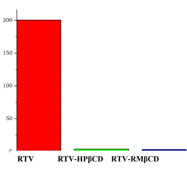
Bar diagrams of MDT values for RTV-CD complexes prepared by freeze drying are shown in
Fig 3. A drastic decrease in MDT values was observed with freeze dried complexes of ritonavir with both HPβCD and RMβCD. Ritonavir had shown higher MDT value of 200.6. The reduction in MDT to 3.0 was found with RTV-RMβCD complex indicated that effective complexation of RTV with RMβCD has been taken place. Freeze drying method proved in enhancing dissolution rate.
The enhancement with freeze dried complexes may be due to increased interaction and conversion of drug into highly amorphous state in freeze drying method. Increase in dissolution parameters of RTV complex with RMβCD has been attributed to the formation of an inclusion complex in the solid state and formation of readily soluble complexes in the dissolution medium with these cyclodextrins. One way ANOVA was performed for freeze dried
complexes with two CDS using Graph Pad Prism 5.03 (Graph Pad Software, Inc.CA, USA) software (trial version) at a level of significance of p<0.05 and the results are shown in Table 3 and 4.
Null hypothesis was rejected in the one way ANOVA test as p value is less than 0.05. Therefore it indicated statistically a significant difference in the dissolution profiles of RTV complexes. The results showed that there is significant difference among RTV-CD complexes prepared with four methods compared to ritonavir alone. But, no significant difference was observed between methods physical mixing and co evaporation. T50 values showed no significant
difference as all complexes showed quick release compared to pure RTV [24].
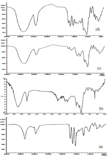
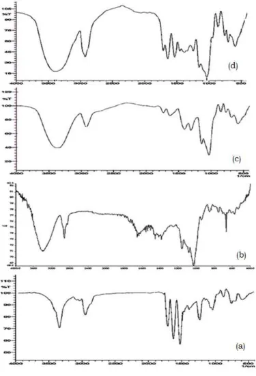
FTIR studies: FTIR patterns of ritonavir complexes with HPβCD and RMβCD are represented in Figure 4 and 5 respectively. When FTIR was performed, the drug spectrum indicated characteristic peaks at 3344.88 cm−1 (N–H stretching amide group), 2951 cm−1 (hydrogen-bonded acid within the molecule), 1714 cm−1 (ester linkage), 1627.97 and 1525.74 cm−1 (–C=C– stretching aromatic carbons).
The FTIR spectra of ritonavir cyclodextrin complexes showed considerable difference compared to pure ritonavir and cyclodextrin spectra and showed decrease in frequency of a specific peak which is generally seen in complexation. Physical mixtures showed broadening of peak and shifting from 3344.68 cm-1 to 3338.89 cm-1 (NH stretching), 2952
cm-1 to 2922 cm-1 hydrogen bonded acid peak, 1734
cm-1 (ester linkage) and 1639 cm-1 C=C group with
HPβCD. RMβCD physical mixture showed peaks with less intensity and broadening at 3452.39 cm-1,
2939 cm-1, 1716 and at 1631 cm-1 regions. Freeze
dried complexes showed similar pattern with widening. Freeze dried complexes with RMβCD showed one small additional peak at 2407.24 cm-1.
Intermolecular hydrogen bonding was observed in ritonavir cyclodextrin complexes prepared by freeze drying, due to broad absorption at 3389.04 cm-1. The
broadening and widening of peak at 1730 cm-1 region
may be due to the interaction between cyclodextrins at the aldehydic link. The results confirmed the formation of stable hydrogen bonds and interaction at the aldehydic group was responsible in complex formation, giving increase in solubility of the drug.
DSC studies: The thermograms of ritonavir, cyclodextrins and the complexes are presented in
www.pharmascholars.com
163
HPβCD showed a broad endothermic peak from 60-80°C. In case of RTV and HPβCD complex prepared by physical mixture showed and endothermic peak showed at 118.99°C and freeze dried complex showed broad endothermic peak at 107.19°C that
strongly indicates strong interaction between
ritonavir and HPβCD and hence confirms
complexation.
Thermogram of RMβCD showed a broad endotherm in the range of 80-100°C which can be attributed to desolvation. Physical mixture of RTV and RMβCD showed broadening of endothermic peak in the range of 100.41°C and freeze dried complex showed a broad endothermic peak at 100.63°C due to formation of inclusion complex.
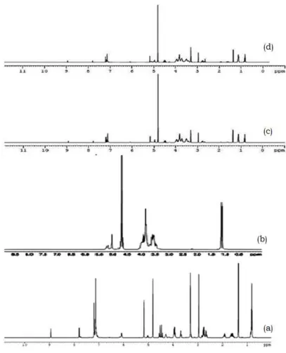
Nuclear magnetic resonance spectroscopy (NMR):
1H-NMRspectra of ritonavir, cyclodextrins, physical
mixtures and its freeze dried complexes were carried out and are presented in Figure 8 and 9. All the proton signals in the 1H-NMR spectrum of the pure
ritonavir are essentially located in a narrow range 1.0 to 5.2 ppm including the aliphatic amine peak at 3.2 ppm and other prominent peaks at 7.0 and 7.3 ppm.
1H-NMR signals reported for HPβCD was found at
1.12, 3.8, 4.7 and 5.19 ppm. 1H-NMR spectrum of
RMβCD showed proton signals at 3.48, 3.78, and 4.65 ppm.
In NMR spectra of ritonavir, NH and OH groups must have undergone hydrogen bonding and other molecular forces of attraction which can be shown by NMR data by disappearance of many OH, NH signals between 3.6-5.2 ppm and disappearance of few signals near 7.0-7.3 ppm that indicates strong interaction between ritonavir and cyclodextrins. Around 3.0 ppm, N-H in ritonavir has intense peak where as in freeze drying of ritonavir with HPβCD and RMβCD complex, peak was disappeared indicating complexation between ritonavir and cyclodextrins.
X-ray diffraction studies: XRDof ritonavir, cyclodextrins, physical mixture and freeze dried complexes were performed and are shown in Figure 10 and 11.In the X-Ray diffractogram of ritonavir, peaks at a diffraction angle (2θ) of 17°, 18° and 22° were present but the peak intensity was less in pure ritonavir which suggested that the drug is not completely crystalline but it is a mixture of both crystalline and amorphous forms. In the physical
mixture complexes, prepared with HPβCD and RMβCD, the presence of drug was revealed by peaks of low intensity at 21°. Peaks were not observed in freeze dried complexes of ritonavir with HPβCD and RMβCD indicating that the drug is completely converted into amorphous form. A similar behavior was previously reported for ketoprofen and ibuprofen [25].
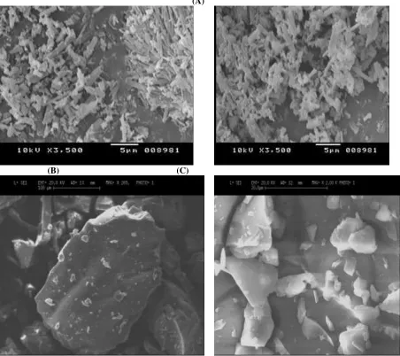
Scanning Electron Microscopy (SEM): The SEM images of RTV, RTV complexes prepared with HPβCD and RMβCD by freeze drying method are shown in Figure12. Ritonavir has appeared as needle shaped crystals. Freeze dried samples appeared as elongated particles. The change of the particle’s shape and disappearance of needle structure of pure RTV in freeze dried complexes confirmed single phase existence and loss of needle structure indicates loss of crystallinity, thus supporting with XRD studies. The present study clearly indicated the superiority of RMβCD in improving the solubility and dissolution of poorly soluble drug RTV. Among the methods of preparation freeze drying technique was found to be more useful compared to other methods.
CONCLUSION
For the first time, comparative studies with different CDs like HPβCD and RMβCD for improving bioavailability of BCS class II drug, ritonavir were carried out. Simultaneously, the effect of method of preparation and influence of CD on enhancement of solubility and dissolution were studied. With the help of phase solubility studies, quantities of CDs were minimized in the preparation of drug-CD complexes. Formation of inclusion complexes was established with the help of FTIR, DSC, 1H-NMR, XRD and
SEM studies. Freeze drying method was found to be useful in improving the solubility and dissolution of ritonavir and the in vivo studies confirmed the improvement in oral bioavailability of these drugs. Further studies on these complexes may lead to reduction of the dose of the drug due to improved bioavailability.
ACKNOWLEDGEMENTS
www.pharmascholars.com
164
Table 1: Stability constants of RTV with two CDsCyclodextrin K1:1 (M-1) Phase diagram
HPβCD 119.01 AL Type
RMβCD 192.96 AL Type
Table 2: Dissolution parameters of RTV inclusion complexes
Type of complex
Method of preparation
DE60 DP30 DE10 T50(min) MDT
Pure Drug --- 17.97±0.95 16.80±0.55 8.02±0.35 --- 200.46±4.5
RTV-HPβCD
PM 46.76±0.58 52.13±0.56 8.40±0.30 27.5±0.15
COE 55.83±0.78 58.73±0.69 36.13±0.55 7.50±0.25
SD 50.81±0.75 42.96±0.45 27.97±0.56 35.0±0.25
FD 93.80±0.65 84.69±0.52 69.08±0.45 2.50±0.15 3.67±5.2
RTV-RMβCD
PM 41.05±0.86 59.29±0.55 37.50±0.91 8.50±0.12
COE 56.86±0.75 59.25±0.45 36.69±0.95 8.01±0.32
SD 58.81±0.42 61.54±0.60 31.44±0.85 25.00±0.15
FD 94.76±0.35 92.86±0.42 72.40±0.75 2.00±0.15 3.00±4.9
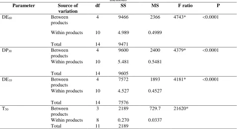
Table 3: ANOVA for dissolution parameters of RTV- HPβCD complexes
Null hypothesis: Ho= there is no significant difference in dissolution parameters between products prepared by various methods
Parameter Source of
variation
df SS MS F ratio P
DE60 Between
products
4 9466 2366 4743* <0.0001
Within products 10 4.989 0.4989
Total 14 9471
DP30 Between
products
4 9600 2400 4379* <0.0001
Within products 10 5.481 0.5481
Total 14 9605
DE10 Between
products
4 7572 1893 4181* <0.0001
Within products 10 4.527 0.4527
Total 14 7576
T50 Between
products
3 2189 729.7 21620*
Within products 8 0.270 0.0337
Total 11 2189
www.pharmascholars.com
165
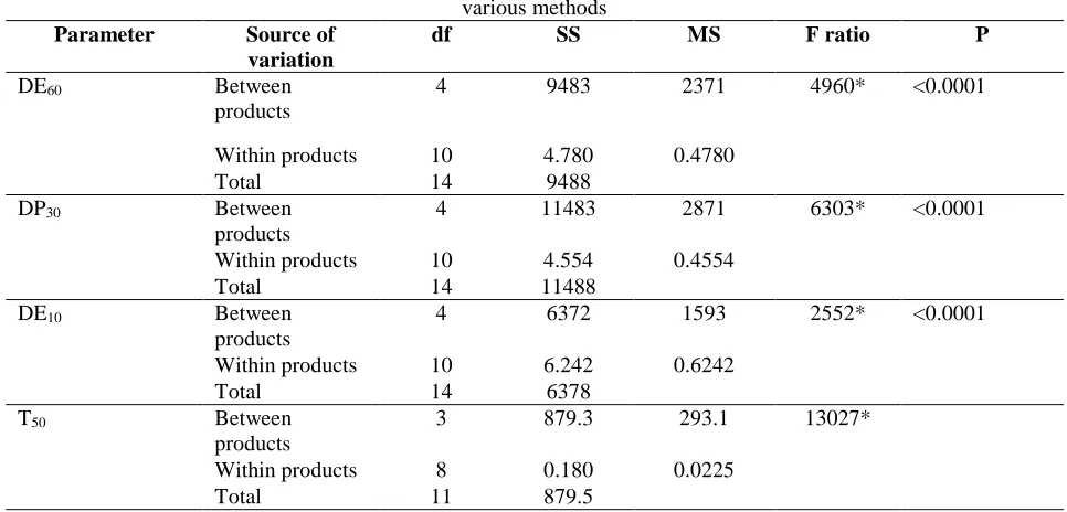
Table 4: ANOVA for dissolution parameters of RTV- RMβCD complexesNull hypothesis: Ho= there is no significant difference in dissolution parameters between products prepared by various methods
Parameter Source of variation
df SS MS F ratio P
DE60 Between
products
4 9483 2371 4960* <0.0001
Within products 10 4.780 0.4780
Total 14 9488
DP30 Between
products
4 11483 2871 6303* <0.0001
Within products 10 4.554 0.4554
Total 14 11488
DE10 Between
products
4 6372 1593 2552* <0.0001
Within products 10 6.242 0.6242
Total 14 6378
T50 Between
products
3 879.3 293.1 13027*
Within products 8 0.180 0.0225
Total 11 879.5
df: Degrees of freedom, SS: Sum of square, MS: Mean square p: Significance level, *Significant difference (Null hypothesis rejected)
www.pharmascholars.com
166
Figure 2: Comparative dissolution profiles of RTV inclusion complexesprepared by freeze drying
0 50 100 150 200
Figure 3: Bar diagram of MDT values for RTV-CD complexes prepared by freeze drying
www.pharmascholars.com
167
Figure 4: FTIR spectra of a) RTV b) HPβCD c) RTV-HPβCD physical mixturewww.pharmascholars.com
168
Figure 5: FTIR spectra of a) RTV b) RMβCD c) RTV-RMβCD physical mixturewww.pharmascholars.com
169
Figure6: DSC spectra of a) RTV b) HPβCD c) RTV- HPβCD physical mixture d) RTV- HPβCD complexwww.pharmascholars.com
170
Figure 7: DSC spectra of a) RTV b) RMβCD c) RTV- RMβCD physical mixture d) RTV- RMβCD complexwww.pharmascholars.com
171
Figure 8: 1H NMR spectra of a) RTV b) HPβCD c) RTV- HPβCD physical mixture d) RTV- HPβCD complexwww.pharmascholars.com
172
Figure 9: 1H NMR spectra of a) RTV b) RMβCD c) RTV- RMβCD physical mixture d) RTV- RMβCDwww.pharmascholars.com
173
Figure 10: XRD patterns of a) RTV b) HPβCD c) RTV- HPβCD physical mixture d) RTV-HPβCD complexwww.pharmascholars.com
174
Figure11: XRD patterns of a) RTV b) RMβCD c) RTV- RMβCD physical mixture d) RTV- RMβCD complexwww.pharmascholars.com
175
(A)(B) (C)
Figure 12: SEM images of A) RTV B) RTV- HPβCD complex C) RTV-RMβCD complex by freeze drying method
REFERENCES
1. Data Base on HIV/AIDS. Compilation prepared by World Health Organization (WHO)
2. Huisman MT, Smit JW, Wiltshire HR, Hoetelmans RMW, Beijnen JH, Schinkel AH. Mol Pharmacol, 2001; 59: 806–813.
3. Gao W, Kishida T, Kageyama M, Kimura K, Yoshikawa Y, Shibata N, Takada K. Antiviral Chem Chemother, 2002; 13(1): 17–26.
4. Farrar GAM, Mitchel H, Hooper FS, Malcolm SL. Br J Clin Pharmacol, 1994; 38: 162.
5. Pathak M, Musmade P, Dengle S, Karthik A, Bhat K, Udupa N. Eur J Pharm Sci, 2010; 41(3-4): 440-51. 6. Chowdary KPR, Nalluri BN. Drug Dev Ind Pharm, 2000; 26: 1217-1220.
7. Palmeiri GF, Angeli DG, Giovannnucci G, Martelli S. Drug Dev Ind Pharm, 1997; 23: 27-37. 8. Palmieri GF, Wehrle P, Stamm A. Drug Dev Ind Pharm,1993;19: 875–85.
9. Nagase Y, Hirata M, Wada K. Int J Pharm, 2001; 229: 163–72.
www.pharmascholars.com
176
11. Castillo JA, Canales JP, Garcia JJ, Lastres JL, Bolas F, Torrado JJ. Drug Dev Ind Pharm,1999;25: 1241– 48.
12. Diaz D, Escobar Lianos CM, Bernad MJB. Drug Dev Ind Pharm, 1999;25: 107–110. 13. Stella VJ, Rajewski R. United States Patent 5, 1992.
14. Higuchi T, Connors KA. Adv Anal Chem Instr, 1965; 4: 117-212.
15. Hirayama F, Uekama K. Methods of investigating and preparing inclusion compounds. In: Duchene D. (ed.) Cyclodextrins and their industrial uses. Paris, Editions de Sante :1987, pp. 131-172.
16. Hegdes AR. Chem Rev, 1998;98: 2035-2044.
17. Rajendrakumar K, Madhusudan S, Prahlad T. Eur J Pharm Biopharm, 2005; 60(1): 39-46. 18. Nagase Y, Hirata M, Wada K. Int J Pharm, 2001; 229: 163–72.
19. Miller LA, Carrier RL, Ahmed I. J Pharm Sci, 2007; 96: 1691-1707.
20. Salustio PJ, Feio G, Figueirinhas JL, Pinto JF, Cabral Marques HM. Eur J Pharm Biopharm, 2009 ;71(2): 377-86
21. Orienti I, Cerchiara T. Int J Pharm, 1999;190: 139-53.
22. Thoresteinn M, Brewster ME. J Pharm Sci, 1996; 85(10): 1017-26.
23. Shilpi S, Mushir A, Sanjula B, Alka A, Anil Kumar, Javed A. AAPS Pharm Sci Tech, 2010; 2(2): 518-527. 24. Bolton S and Bon C. Nonparametric methods. Pharmaceutical statistics: Practical and clinical applications.
5th ed, New York; Informa Healthcare: 2010, pp.390-424.