R E S E A R C H
Open Access
Functional analysis of the impact of
ORMDL3
expression on inflammation and activation of the
unfolded protein response in human airway
epithelial cells
Karolynn J Hsu and Stuart E Turvey
*Abstract
Background:The geneORMDL3was shown to be associated with early-onset asthma susceptibility in multiple independent genome-wide and candidate-gene association studies. Asthmatic patients have elevated expression levels of this gene.ORMDL3encodes a transmembrane protein localized in the endoplasmic reticulum (ER) that may be involved in ER stress and inflammation. It is essential to validate the genetic associations linkingORMDL3 with asthma through functional studies that confirm the biological relevance of this gene in disease. We investigated the effects of manipulatingORMDL3expression levelsin vitroin airway cells on innate immune inflammatory responses, ER stress and activation of the unfolded protein response (UPR).
Methods:ORMDL3expression levels were manipulated in airway cells using an overexpression plasmid and siRNA technologies. Successful modulation of ORMDL3 was confirmed at both the gene and protein level. The functional impact of modulation ofORMDL3expression levels on inflammatory responses and activation of the UPR were quantified using complementary cellular and molecular immunology techniques.
Results:Cells with alteredORMDL3levels responded equally well to innate immune stimuli and produced similar levels of pro-inflammatory cytokines compared to wild-type cells. Treatment with ER stress inducers, thapsigargin and tunicamycin, resulted in activation of the unfolded protein response (UPR). However, we observed no difference in UPR activation in cells withORMDL3knockdown compared to cells with normalORMDL3levels.
Conclusions:Our results suggest thatORMDL3variation in the airway epithelium is unlikely to play a significant role in modulating innate immune responses and the UPR in the lung.
Keywords:Immune response, Epithelium, Cytokines, Chemokines, Host defense
Introduction
Asthma and allergic diseases are rapidly becoming the most common chronic diseases in the developed world. Current asthma therapy treats symptoms of the disease, however it is ineffective in up to 25% of patients [1]. Asthma and allergic diseases are complex disorders caused by the interaction of various genetic and environ-mental factors [2-4].
Genome-wide association studies (GWAS) have been used to identify genes that may be involved in asthma pathogenesis [5]. Moffatt and colleagues first reported that multiple single nucleotide polymorphisms (SNPs)
on chromosome 17q21 linked ORMDL3 (orosomucoid
1-like 3) to the risk of developing childhood asthma [6]. This association has since been reproduced in multiple independent studies [7-14]. However, little work has been done to elucidate the biological and functional rele-vance of this gene in asthma. The disadvantage of these association studies is that they cannot differentiate be-tween true causal SNPs and non-causal variants simply in linkage disequilibrium with disease-causing genes. It * Correspondence:[email protected]
Division of Infectious and Immunological Diseases, Department of Pediatrics, BC Children’s Hospital and Child & Family Research Institute, University of British Columbia, Vancouver, BC, Canada
is therefore imperative to validate GWAS data through functional studies that confirm the biological relevance of a gene in disease.
SNP variants have also linked ORMDL3 to
inflamma-tory bowel disease (IBD) and Type I diabetes, suggesting that ORMDL3 may be involved in dysregulation of the
immune system [15,16]. Association ofORMDL3in both
asthma and IBD is of interest because the lung and gut are composed of similar mucosal surface cells and these tissues are exposed to many potentially harmful antigens and allergens requiring tight regulation of the mucosal immune system [17]. This unique system is responsible for maintaining a delicate equilibrium between antigen re-sponsiveness and tolerance and is therefore responsible for preventing hyper-reactivity [17]. Inappropriate immune responses to foreign components or commensal bacteria can lead to inflammation characteristic of asthma and IBD. Furthermore, the polymorphisms may be involved in regu-lation of mRNA expression of 17q21 locus genes, including ORMDL3 [6]. The expression of ORMDL3 was recently associated with elevated levels of IL-17 secretion [18] and ORMDL3was expressed at higher levels in the peripheral blood of patients with recurrent wheeze compared to con-trols [19]. This correlation further supports the hypothesis thatORMDL3is involved in immunity.
TheORMDL3gene is a member of a family of conserved endoplasmic reticulum (ER)-localized transmembrane pro-teins [20]. The functions of theORMDLproteins are cur-rently unknown, but a recent study suggested that
ORMDL3 is involved in ER-mediated Ca2+ homeostasis
and activation of the unfolded protein response (UPR) –
ORMDL3 may inhibit sarco/endoplasmic reticulum Ca2+
ATPase (SERCA) activity [21,22]. Disruptions to ER Ca2+ concentrations can cause protein misfolding, and accumu-lation of these unfolded proteins can lead to ER stress [23,24]. UPR signaling cascades are initiated in response to this stress and have been shown to activate the JNK-AP-1 and NF-κB-IKK pathways [25-27]. The ER stress response
and UPR, caused by changes in ORMDL3 expression,
can initiate inflammation through induction of cytokine production. This mechanism may explain the role of ORMDL3 in asthma pathogenesis. Indeed, Milleret al.
have shown that in mice ORMDL3 is an allergen and
cytokine (IL-4 or IL-13) inducible ER gene expressed predominantly in airway epithelial cells, and that it acti-vates the ATF6 pathway of the ER localized UPR regu-lating expression of metalloprotease, chemokine, and oligoadenylate synthetase genes [28].
Although the symptoms of asthma are largely driven by dysregulated T helper type 2 (TH2) responses, innate
im-mune responses are also involved in asthma pathogenesis [29,30]. Airway epithelia are central to host defense and immune regulation. These cells are among the first to en-counter environmental insults and play an important role
in shaping downstream immune responses. Any dysregu-lation of the innate immune response can result in hyper-sensitivity to environmental factors, leading to asthma symptoms.
Given the multiple lines of evidence suggesting that ORMDL3is involved in immunity, we investigated the role of the gene in innate immune responses of airway cells.
We hypothesized that elevated ORMDL3 levels result in
heightened inflammatory responses that are associated with the asthmatic phenotype. Increased levels of ORMDL3 pro-tein may in turn disrupt ER homeostasis, leading to ER overload and activation of the UPR, initiating
inflamma-tory responses. Using an in vitro model, we manipulated
ORMDL3expression in airway cells to determine whether a difference in basalORMDL3expression affected inflam-matory responses or activation of the UPR before and after stimulation.
Materials and methods Cell culture
1HAEo¯ (1HAE) cells (SV40-transformed normal human
airway epithelial cells) were cultured in DMEM-high glu-cose medium with 10% fetal calf serum (FCS), 2 mM L-glutamine, and 1 mM sodium pyruvate (HyClone). A549 cells (adenocarcinomic human alveolar basal epithelial cells) were cultured in F-12K medium supplemented with 10% fetal calf serum (FCS), 2 mM L-glutamine, and 1 mM so-dium pyruvate (HyClone). Cells were incubated in a 37°C,
5% CO2 incubator. All cells were cultured under
non-polarizing conditions.
Cloning ORMDL3 cDNA into pEGFP-N1 vector
TheORMDL3gene was amplified from cDNA using
for-ward primer 50-CTAAGAATTCATGAATGTGGGCAC
AGCGCAC-30 and reverse primer 50-TACTGGTACCCC
GTACTTATTGATTCCAAAAATCCGGACT-30,
introdu-cing EcoRI and KpnI restriction endonuclease sites,
re-spectively. TheORMDL3PCR product was then inserted
into a pEGFP-N1 eukaryotic expression vector (Clontech). ORMDL3 and eGFP are in frame and produce a fusion protein with eGFP expressed at the C-terminus of ORMDL3. The construct was verified by sequencing and is denoted as pEGFP-ORMDL3. Protein is denoted as ORMDL3-eGFP.
Cell transfection
A549 and 1HAE cell lines were transfected with
pEGFP-ORMDL3, scramble (non-specific) or ORMDL3-specific
siRNA (pre-designed by Qiagen) using AmaxaW Cell
Line NucleofectorW Kit T (Lonza). Two ORMDL3
density of 2x10⁵cells/well for A549 cells or 1x10⁵cells/ well for 1HAE cells.
Cell stimulation and immune response quantification Twenty-four hours post-transfection, cells were
stimu-lated with TNF-α (200 ng/ml) (eBioscience), E. coli
K12 LPS (100 μg/ml) (InvivoGen), S. typhimurium
fla-gellin (10-200 ng/ml) (InvivoGen), or IL-1β(200 ng/ml) (eBioscience). Stimulants and their concentrations were chosen based on published literature or past experiments [31-34]. Cells were stimulated for 24 hours. Supernatants were collected and analyzed for cytokine secretion. Pro-inflammatory cytokines, IL-6 and IL-8, were detected and
quantified using Human IL-6 and IL-8 Ready-Set-Go!W
ELISA kits (eBioscience). Experiments were repeated three times (n = 3).
ER stress induction and UPR activation
Cells were stimulated with tunicamycin (200μg/mL) (Cal-biochem) or thapsigargin (10μM) (Sigma) for 2 or 4 hours
to activate the UPR. For ORMDL3 knockdown cells,
stimulation was performed 24 hours post-transfection.
RNA was extracted and expression of genes XBP-1u,
XBP-1s, and CHOP were then quantified as markers of
UPR activation. For measurement of p-eIF2α levels by
Western blot, lysates from unstimulated cells with ORMDL3knockdown were collected 24 hours post-trans-fection. Experiments were repeated three times (n = 3).
RNA isolation and reverse transcription
RNA was extracted from lysates using E.Z.N.A.W Total
RNA Kit (Omega Bio-Tek) according to the manufac-turer’s protocol. Extracted RNA was reverse transcribed
into cDNA using the SuperScriptW VILO™ cDNA
Syn-thesis Kit (Invitrogen). Complement DNA was diluted to 200 ng/μl prior to quantification of gene expression by qPCR. This method was followed for all samples, unless otherwise stated (see PCR Array).
Quantification ofORMDL3mRNA expression
Gene expression was calculated relative to GAPDH or
PPIA (encoding cyclophilin A) and was quantified by
SYBR Green chemistry (PerfeCTa™ qPCR SuperMix,
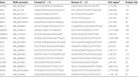
Quanta Biosciences) using a 7300 Real Time PCR Sys-tem (Applied BiosysSys-tems). Reactions were performed in triplicate using the following cycling conditions: 50°C for 2 mins, 95°C for 10 mins, [95°C for 15 s, 60°C for 1 min] x 40. The relative expression of the measured gene was calcu-lated by the Pfaffl method [35]. The primers used are listed in Table 1.
Western blot analysis
Cells were lysed in 50μl RIPA Buffer + 1x HALT™ prote-ase inhibitor (Thermo Scientific). Cell debris were removed by centrifugation: 18,000 x g for 10 min at 4°C. Proteins were analyzed by standard Western blotting protocols where they were transferred onto ImmobilonW-FL transfer membrane (Millipore). Antibodies used for Western blot
Table 1 Quantitative PCR primer sequences
Gene NCBI accession Forward (50→30) Reverse (50→30) CDS region† Product size
GAPDH NM_002046.4 GCACCGTCAAGGCTGAGAACGG CGACGTACTCAGCGCCAGCATC c.173-286 114
PPIA NM_021130 TAAAGCATACGGGTCCTGGCATCT ATCCAACCACTCAGTCTTGGCAGT c.269-369 101
ACTB NM_001101.3 GTTGCGTTACACCCTTTCTT ACCTTCACCGTTCCAGTTT c.*16-*162 147
DDIT3 NM_001195053.1 GAAATGAAGAGGAAGAATCA TTCTCCTTCATGCGCT c.197-437 241
XBP-1s NM_001079539.1 ATGGATGCCCTGGTTGCTGAAGA TGCACCTGCTGCGGACTCA c.415-504 90
XBP-1u NM_005080.3 AGCACTCAGACTACGTGCACCTCT CCAGAATGCCCAACAGGATATCAG c.495-624 130
ORMDL1 NM_016467.4 AATGGCTGGTCCTTCAAGTGCT ACCCTCACTGTGATGCCCTTTA c.*121-*269 149
ORMDL2 NM_014182.4 ACACACTGGGAGCAAATGGACT AGTGCGCAGCATCATACTTGGT c.250-370 121
ORMDL3 NM_139280.2 TCAGGCAGCCAAAGCACTTTAACC ACCCATCCCACACTTGCTTCCATA c.*358-*496 139
BCL6 NM_001706.4 ACAATCCCAGAAGAGGCACGAAGT GCTCGAAATGCAGGGCAATCTCAT c.790-952 163
CCL2 NM_002982.3 TCGCTCAGCCAGATGCAATCAATG TGGAATCCTGAACCCACTTCTGCT c.65-259 195
CCL5 NM_002985.2 TGCCTGTTTCTGCTTGCTCTTGTC TGTGGTAGAATCTGGGCCCTTCAA c.*36-*127 92
CSF2 NM_000758.3 AAATGTTTGACCTCCAGGAGCCGA GGTGATAATCTGGGTTGCACAGGA c.185-357 173
IL12A NM_000882.3 ATGATGGCCCTGTGCCTTAGTAGT AGGGCCTGCATCAGCTCATCAATA c.457-611 155
IL13RA1 NM_001560.2 GTCCCAGTGTAGCACCAATGA CAGTCACAGCAGACTCAGGAT c.297-391 95
ADRB2 NM_000024.5 TCATCATGGGCACTTTCACCCTCT AGCTCCTGGAAGGCAATCCTGAAA c.830-1016 187
VEGFA NM_001025366.2 TTCAGGACATTGCTGTGCTTTGGG TGGGCTGCTTCTTCCAACAATGTG c.*778-*969 192
IL23A NM_016584.2 TCGGTGAACAACTGAGGGAACCAA TGGAATCTCTGCCCACTTCCACTT c.-140- -54 87
analysis were: monoclonal anti-GFP antibody 1:10,000 (Clontech), anti-ACTB antibody 1:6,000 (Cell Signaling),
anti-p-eIF2α 1:500 (Cell Signaling) and IRDyeW 680 or
800 secondary antibodies 1:8000 (Li-cor). Western blots were visualized using an Odyssey Infrared Imaging Sys-tem (Li-cor).
PCR array
1HAE cells co-transfected with pEGFP-ORMDL3 and ORMDL3 siRNA (low ORMDL3 expression) were com-pared to cells co-transfected with pEGFP-ORMDL3 and
scramble siRNA (highORMDL3expression) at two
time-points (2 and 24 hours) after TNF-αstimulation. Extracted RNA was reverse transcribed into first strand cDNA using the RT2First Strand Kit (SABiosciences, Qiagen). Protocol as described by the manufacturer was followed.
Two RT2Profiler PCR arrays (SABiosciences, Qiagen),
profiling expression of 84 genes each, were used: Human Cytokines & Chemokines and Allergy & Asthma (see Additional file 1 for complete list). Complementary DNA
template was mixed with RT2SYBRW Green qPCR
Mas-termix (SABiosciences, Qiagen) as follows: 1350μL SYBR
Green Master Mix, 1248μL nuclease-free H2O, and
102μL cDNA (~200 ng/μL). Note: these volumes were
used as recommended by the manufacturer for use with a 7300 Real Time PCR System (Applied Biosystems). Tem-plate was then aliquoted into PCR Tem-plates containing pre-dispensed primers. Cycler program as provided by the manufacturer was used. Results were analyzed using the PCR Array Data Analysis Web Portal.
Statistical analysis
Data are shown as mean ± SEM of three separate experi-ments. Results were analyzed using one-way ANOVA with Bonferroni post-test. Statistical analysis was performed using GraphPad Prism5 (GraphPad Software, Inc.). Differ-ences with p < 0.05 were considered significant.
Results
ORMDL3modulation in airway cells
In order to determine functional impact of ORMDL3
modulation, knockdown of the gene was performed using siRNAs. A549 and 1HAE cells were transfected with
scramble (non-specific) or ORMDL3-specific siRNA.
Modeling variation expected to occur in the human
popu-lation, we achieved 40-70% knockdown ofORMDL3gene
expression using siRNA concentrations of 50 nM-500 nM.
We also confirmed thatORMDL3-specific siRNA did not
affect expression of genes in the same family,ORMDL1or ORMDL2 (Additional file 2). Sequences of primers used for qPCR are listed in Table 1.
ORMDL3 has 84% and 80% protein sequence homology to ORMDL1 and ORMDL2, respectively. This presented a challenge for confirming knockdown of ORMDL3 protein
because commercially available antibodies detect all three proteins. Therefore, we were unable to show ORMDL3 protein knockdown in cells transfected with siRNA alone. To overcome this limitation, we constructed a fusion protein, where ORMDL3 is tagged with an eGFP protein. Co-transfection of this overexpression
plas-mid withORMDL3-specific siRNA enabled us to
knock-down ORMDL3 and detect changes at the protein level using an anti-GFP antibody.
Airway cells were co-transfected with both
pEGFP-ORMDL3 and siRNA (scramble orORMDL3). Protein and
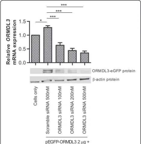
mRNA were analyzed for gene knockdown. At the tran-script level, we observed (Figure 1) a small but significant increase (p < 0.05) in ORMDL3 expression in the cells transfected with pEGFP-ORMDL3 and scramble siRNA compared to cells alone, confirming successful
overexpres-sion of the ORMDL3 gene. Transfection with
pEGFP-ORMDL3 and increasing concentrations of ORMDL3
siRNA resulted in a titration effect of increasingORMDL3 knockdown. One advantage to constructing a fusion pro-tein, ORMDL3-eGFP, is that eGFP is only expressed with expression of ORMDL3. Therefore, when cells are co-transfected with pEGFP-ORMDL3 and siRNA, knockdown of ORMDL3 protein could subsequently be detected by immunoblot analysis using the anti-GFP antibody. Expres-sion of the ORMDL3-eGFP protein was confirmed by
Western blot of whole cell lysate collected from the cells in each condition. Figure 1 shows knockdown of
ORMDL3-eGFP protein, confirming thatORMDL3siRNA
affects protein expression. The double band may be explained by either variation in mRNA splicing or post-translational modifications to the fusion protein, such as acetylation, methylation, myristylation, phosphorylation, or glycosylation.
ORMDL3knockdown does not affect IL-6 or IL-8 production following innate immune activation
1HAE cells transfected with pEGFP-ORMDL3 and
scramble or ORMDL3-specific siRNA were stimulated
24 hours post-transfection and supernatants were col-lected 48 hours post-transfection. Stimuli used were TNF-α, IL-1β, LPS, and flagellin. TNF-αand IL-1βwere chosen because both are early response cytokines that perpetuate acute inflammatory processes. LPS and flagel-lin, in contrast, are common microbial antigens recog-nized by the innate immune system. Two classic and biologically-relevant NF-κB-induced cytokines with im-portant roles in innate immunity, interleukin-6 (IL-6) and interleukin-8 (IL-8), were measured by ELISA. Despite
confirmation ofORMDL3mRNA and protein knockdown,
we did not observe any impact on IL-6 or IL-8 production after stimulation as shown in Figure 2A-B. Although the cells have low baseline responsiveness to LPS and flagellin,
our results indicate that ORMDL3 knockdown does not
enhance sensitivity to these stimuli. Similar results were
obtained in A549 cells, as well as 1HAE cells transfected with siRNA alone (Additional file 2).
ORMDL3knockdown does not enhance UPR activation upon stimulation
We next explored the effects ofORMDL3expression on
activation of the UPR. Initiation of the UPR is mediated by one or more of the ER-membrane protein sensors: PKR-like eukaryotic initiation factor 2α kinase (PERK), inositol requiring enzyme 1 (IRE1), and activating tran-scription factor-6 (ATF6) [23]. Activation of any of the three pathways initiates signaling cascades that mediate
changes to relieve ER stress. The gene XBP-1 is a
sub-strate for IRE1 ribonuclease [24]. Upon activation of the IRE1 pathway, the IRE1 ribonuclease removes a 26-bp intron from the unspliced variant,XBP-1u,which results in the spliced variant, XBP-1s [36]. This spliced variant is the active form of the gene that contributes to ER stress responses.CHOPtranscription, in contrast, can be induced by the PERK and ATF6 pathways [24]. Phos-phorylated eIF2α (p-eIF2α) is an early marker of PERK
pathway activation and is upstream of CHOPinduction
[23]. Expression changes in these three genes that signify
UPR activation,XBP-1u, XBP-1s,andCHOP, were
deter-mined by qPCR. We also evaluated phosphorylation of eukaryotic initiation factor 2α (eIF2α) by Western blot,
as modulation ofORMDL3expression has been reported
to influence eIF2αphosphorylation [21].
A B
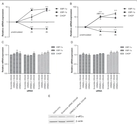
As positive controls for UPR activation, we stimulated 1HAE cells with tunicamycin and thapsigargin. Both are inducers of ER stress–tunicamycin inhibits N-linked gly-cosylation and thapsigargin inhibits the SERCA pump causing ER calcium stores to be depleted [37]. Quantita-tive measurement of transcript levels showed that both XBP-1s and CHOP increased, while XBP-1u decreased upon stimulation with either tunicamycin or thapsigargin (Figure 3A-B). This confirmed the utility of measuring these genes to monitor UPR activation.
At baseline (unstimulated cells), variation inORMDL3
expression did not induce UPR activation in A549 or 1HAE cells (Additional file 2). 1HAE cells with ORMDL3knockdown were also stimulated with tunica-mycin or thapsigargin (Figure 3C-D). In both
condi-tions, ORMDL3 knockdown did not show increased
UPR activation compared to the negative control.
Fur-thermore, levels of phosphorylated-eIF2α were
indis-tinguishable between ORMDL3 knockdown cells and
controls (Figure 3E).
A B
C D
E
Figure 3Unfolded protein response activation in cells withORMDL3knockdown.ER stress was induced in 1HAE cells by stimulation with A) tunicamycin orB) thapsigargin for 2 or 4 hours. Relative expression levels ofXBP-1u,XBP-1s, andCHOPwere quantified and compared to unstimulated cells. 1HAE cells withORMDL3knockdown were stimulated withC) tunicamycin orD) thapsigargin for 4 hours. Relative gene expression levels were quantified and compared to cells transfected with scramble (a non-specific) siRNA. Data represent the mean ± SEM of three experimental repeats. Statistical analysis was performed using one-way ANOVA with Bonferroni post-test. *** p < 0.001, ** p < 0.01, * p < 0.05.E) Western blot analysis of p-eIF2αwithβ-actin loading control. Lysates are from unstimulated 1HAE cells 24 hours afterORMDL3
Impact ofORMDL3knockdown on the expression of multiple genes involved in inflammation, asthma & allergy To expand our search for immune functions potentially
altered by ORMDL3, the expression of 168 genes was
determined at two time points (2 and 24 hours)
follow-ing stimulation with TNF-α. We performed PCR arrays
to profile expression of cytokines, chemokines, and key genes involved in asthma and allergy (a complete list of genes that were studied can be found in Additional file 1).
Gene expression was compared between 1HAE cells with high (plasmid + 500 nM scramble siRNA) and low
(plasmid + 500 nM of ORMDL3 siRNA) ORMDL3
ex-pression that were stimulated for 24 hours with TNF-α. Stimulation with TNF-αinduced a robust inflammatory re-sponse in the cells, enabling us to observe whether
vari-ation in basal ORMDL3 levels impacts the immune
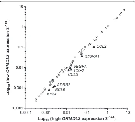
response. These arrays identified eight genes (CCL2, TSLP, CSF2, CCL5, VEGFA, ADRB2, IL1RL1, and IL13RA1), shown in Figure 4, that appeared to be differentially regu-lated by more than 1.5 fold and that were expressed at rela-tively high levels (average threshold cycle <30). However, upon replication to validate these results, we determined that the differences were not statistically significant.
The same arrays were performed on cells stimulated for 2 hours with TNF-α. The same conditions for“high” and “low” ORMDL3expression were used. From our results, we identified only one gene,IL23A, that was differentially regulated by more than 1.5 fold (fold regulation −2.37)
and amplified at cycle <30. Replication and comparison of IL23Aexpression between knockdown conditions yielded no significant difference. No other genes, as identified in the previous arrays, were found to be differentially regu-lated at this time point. These results suggest that ORMDL3variation does not have a meaningful impact on expression of a large panel of immune-related genes in air-way epithelial cells.
Discussion
Asthma is a complex disease affecting many individuals in the developed world. Genome-wide association stud-ies have recently been used to identify genetic causes for
such complex diseases. One particular gene, ORMDL3,
is of interest because of its association with asthma, IBD,
and Type I diabetes – all of which are caused by
immune-mediated pathology [6,10,22,38,39]. The gene ORMDL3 is an ER-membrane protein and is potentially
involved in Ca2+-signaling in the ER and sphingolipid
synthesis [20,21,40]. It has also been correlated to activa-tion of the UPR, though the mechanisms remain unclear [21]. Activation of the UPR may be biologically relevant, as ER stress, the UPR, and inflammation have all been
linked [23]. However, the functional role ORMDL3 in
the pathogenesis of asthma has yet to be elucidated. Airway epithelial cells play an important role in innate immunity and in the development of asthma. Current
findings in literature indicate that ORMDL3 is involved
in immunity and that asthmatics have higher expression of the gene than non-asthmatics [18,21,22]. A recent
study by Miller et al. also investigated the role of
ORMDL3 in airway epithelial cells. They reported that in vitro overexpression ofORMDL3 activated the ATF6 pathway of the UPR and induced expression of several genes with potential importance in the pathogenesis of asthma [28]. Our investigation, in contrast, focuses on the effect of variation ofORMDL3expression levels, at base-line, on the innate immune responsiveness of airway epi-thelial cells. By manipulatingORMDL3expressionin vitro to mimic differences in gene expression established be-tween asthmatics and healthy individuals, we aimed to
understand the role of ORMDL3on the innate immune
response and UPR activation status in airway epithelial cells. This method ensured control and the confidence that any effect on the innate immune response was in fact correlated with a change inORMDL3expression levels. If
the same experiments were performed on ex vivoairway
cells of patients, genetic and other differences between individuals could have affected the results.
After knockdown ofORMDL3 in vitro,cells were
stimu-lated with cytokines (TNF-α, IL-1β) or common microbial components (LPS, flagellin). We monitored production of interleukin-6 (IL-6) and interleukin-8 (IL-8) (alias CXCL8), two pro-inflammatory cytokines produced by airway cells
Figure 4PCR array.Results shown are 168 genes profiled from two PCR arrays: Allergy & Asthma and Human Cytokines & Chemokines (Qiagen). 1HAE cells with high or lowORMDL3
expression were stimulated with TNF-αfor 24 hours and differences in gene expression were compared. Comparing lowORMDL3
that are relevant in asthma pathogenesis. Specifically, IL-6 is elevated in individuals with asthma [41] and is also regu-lated by ATF6 during activation of the UPR [42]. Similarly, transfection of ORMDL3 into human airway epithelial cells triggers ATF6 activation and IL-8 secretion [28]. However,
in our experimental system, variation inORMDL3
expres-sion levels did not affect NF-κB-induced innate immune production of IL-6 and IL-8 in airway epithelial cells.
We next explored the effects ofORMDL3expression on
activation of the UPR. UPR signaling cascades are initiated in response to ER stress, and restoration of homeostasis is achieved by attenuating translation, restoring protein fold-ing, or degrading misfolded proteins [24]. Although often associated with abnormal physiological conditions, the UPR plays a central beneficial role in normal physiology; as illustrated by the role of the UPR in terminal B cell dif-ferentiation which requires a massive increase in the bio-synthetic capacity to synthesize antibodies in response to infection [43]. However, the ER stress response and UPR can also initiate inflammation through induction of cyto-kine production or activation of transcriptional regulators of inflammatory genes. Cytokines IL-6 and IL-8 are exam-ples of genes that may be induced by UPR activation [23]. ER stress and the UPR have been implicated in many immune-related diseases including IBD, diabetes, chronic obstructive pulmonary disease (COPD), arthritis, and neu-rodegenerative inflammatory diseases [44]. It is poorly understood whether ER stress is an underlying cause of disease or if its induction is a result of chronic inflamma-tion. Indeed, it is possible that environment factors such as infection or inhalation of smoke particles can activate the UPR, triggering the onset of lung disease in genetically predisposed individuals [45]. However, it is also possible that ER stress is exacerbated by inflammation and contri-butes to the perpetuation of the disease.
Cantero-Recasens et al. previously reported that
ORMDL3overexpression activated the PERK pathway, but did not affect the IRE1 pathway of the UPR [21]. In con-trast, Milleret al.reported thatORMDL3overexpression activated the ATF6 pathway, but not the PERK or IRE1 pathways [28]. In our study, we chose four markers of
UPR activation: XBP-1u, XBP-1s, CHOP, and p-eIF2α.
With activation of the UPR, we expect downregulation of XBP-1uand upregulation ofXBP-1sandCHOP.However,
our results demonstrate that knockdown of ORMDL3
does not activate the UPR, in either unstimulated or sti-mulated cells. Immunoblot analysis also showed no
change in p-eIF2αlevels withORMDL3knockdown.
Fur-thermore, downstream markers of UPR activation, IL-6 and IL-8 cytokines, were produced at similar levels in un-stimulated cells with varyingORMDL3levels. This further
supports our results that ORMDL3does not activate the
UPR. Differences in our results compared to previous work might be due to the different types of cells,
conditions, or markers used. It is possible that the effects
of variation in ORMDL3 expression are a cell
type-dependent phenomenon. While no effect on the inflam-matory response was detected in airway cells, other cells types such as dendritic cells or T cells may be affected by
alteredORMDL3expression. Observations made by Lluis
et al.suggest that the 17q21 locus may potentially play a role in T-cell development [18].
Taking a broader approach, PCR arrays looking at ex-pression of 168 common immunity genes were
per-formed. We reasoned that althoughORMDL3levels may
not affect the production of IL-6 or IL-8 cytokines, per-haps they were impacting gene expression of other
im-portant immune genes, such as IL-33, IL-25 and TSLP,
which have all been implicated in asthma pathogenesis [46]. Verification of differential expression of these genes at a transcript level, however, did not show any significant
changes between the ORMDL3 knockdown conditions.
This suggests that alteringORMDL3 expression does not
have a profound effect on the expression of innate im-mune genes upon stimulation in the airway epithelia. However, there may be other genes that are affected that were not investigated in this study. Pfeifer et al. recently showed that IL-17C cytokine is expressed by human bron-chial epithelial cells and is induced by bacterial infection [47]. It may be worthwhile in future experiments to inves-tigate a broader range of immune-related genes. Interest-ingly, we did not observe changes to expression of the
genes reported by Miller et al., MMP-9, CCL-20,
CXCL-10, CXCL-11, orIL-8.This variance may be explained by differences in experimental conditions. Our study exam-ined outcomes in gene expression after stimulation of cells
co-transfected with ORMDL3 and ORMDL3-specific
siRNA, while the other study used a different experimental approach.
Although this study focused exclusively on the
poten-tial role of ORMDL3 in asthma pathogenesis, it is
pos-sible that neighboring genes such asGSDMLcontribute
to disease susceptibility at this locus. Many groups
con-sider ORMDL3 as an ‘asthma gene’; however, it should
be acknowledged that the identified SNPs associating this gene to asthma susceptibility are not located in the gene itself. Even so, these polymorphisms have been consistently correlated with increased odds of asthma risk, highlighting the importance of this locus in disease susceptibility [6-11,13,14].
Our data show that variation inORMDL3expression is
not correlated with differential innate immune responses to stimuli or activation of the UPRin vitroin airway epi-thelial cells. Taken together, our results are biologically relevant because they suggest that normal human
vari-ation of ORMDL3 expression is not likely an important
gene remains an important candidate for asthma suscepti-bility. More research is required to elucidate its role in asthma pathogenesis and its potential role as an initial trigger of inflammation. By increasing our understanding of the mechanisms responsible for allergic and atopic dis-ease development, new treatments can then be developed. Thus, we can reduce inflammatory responses by targeting the potential triggers, rather than the symptoms, of the disease. In doing so, we will ultimately reduce the morbid-ity, mortalmorbid-ity, and socio-economic burden of asthma and related allergic diseases.
Additional files
Additional file 1:Genes analyzed by PCR array.
Additional file 2:Additional figures.
Competing interests
The authors declare that they have no competing interests.
Authors’contributions
KJH performed the research. All authors designed the research, analyzed the data, and drafted the manuscript. Both authors read and approved the final manuscript.
Acknowledgements
KJH was funded by the Child & Family Research Institute and AllerGen Networks of Centers of Excellence. SET holds the Aubrey J Tingle
Professorship in Pediatric Immunology and is a clinical scholar of the Michael Smith Foundation for Health Research. This work was supported by funding from the AllerGen Networks of Centers of Excellence.
Received: 3 December 2012 Accepted: 7 January 2013 Published: 1 February 2013
References
1. Cho SH:Pharmacogenomic approaches to asthma treatment.Allergy Asthma Immunol Res2010,2(3):177–182.
2. Davies DE, Djukanovic R, Holgate ST:Application of functional genomics to study of inflammatory airways disease.Thorax1999,54(1):79–81. 3. Gu ML, Dong XQ, Zhao J:New insight into the genes susceptible to
asthma.J Asthma2010,47(2):113–116.
4. Ober C, Hoffjan S:Asthma genetics 2006: the long and winding road to gene discovery.Genes Immun2006,7(2):p. 95–p. 100.
5. Vercelli D:Discovering susceptibility genes for asthma and allergy.
Nat Rev Immunol2008,8(3):169–182.
6. Moffatt MF,et al:Genetic variants regulating ORMDL3 expression contribute to the risk of childhood asthma.Nature2007, 448(7152):470–473.
7. Galanter J,et al:ORMDL3 gene is associated with asthma in three ethnically diverse populations.Am J Respir Crit Care Med2008, 177(11):1194–1200.
8. Halapi E,et al:A sequence variant on 17q21 is associated with age at onset and severity of asthma.Eur J Hum Genet2010,18(8):902–908. 9. Hirota T,et al:Genetic polymorphism regulating ORM1-like 3
(Saccharomyces cerevisiae) expression is associated with childhood atopic asthma in a Japanese population.J Allergy Clin Immunol2008, 121(3):769–770.
10. Moffatt MF,et al:A large-scale, consortium-based genomewide association study of asthma.N Engl J Med2010,363(13):1211–1221. 11. Ferreira MA,et al:Association between ORMDL3, IL1RL1 and a deletion on
chromosome 17q21 with asthma risk in Australia.Eur J Hum Genet2011, 19(4):458–464.
12. Sleiman PM,et al:ORMDL3 variants associated with asthma susceptibility in North Americans of European ancestry.J Allergy Clin Immunol2008, 122(6):1225–1227.
13. Tavendale R,et al:A polymorphism controlling ORMDL3 expression is associated with asthma that is poorly controlled by current medications.
J Allergy Clin Immunol2008,121(4):860–863.
14. Wu H,et al:Genetic variation in ORM1-like 3 (ORMDL3) and gasdermin-like (GSDML) and childhood asthma.Allergy2009,64(4):629–635. 15. Barrett JC,et al:Genome-wide association study and meta-analysis find
that over 40 loci affect risk of type 1 diabetes.Nat Genet2009, 41(6):703–707.
16. Barrett JC,et al:Genome-wide association defines more than 30 distinct susceptibility loci for Crohn’s disease.Nat Genet2008,40(8):955–962. 17. Neurath MF, Finotto S, Glimcher LH:The role of Th1/Th2 polarization in
mucosal immunity.Nat Med2002,8(6):567–573.
18. Lluis A,et al:Asthma-associated polymorphisms in 17q21 influence cord blood ORMDL3 and GSDMA gene expression and IL-17 secretion.
J Allergy Clin Immunol2011,127(6):p. 1587–p. 1594. e6.
19. Jin R,et al:Mechanisms elevating ORMDL3 expression in recurrent wheeze patients: role of Ets-1, p300 and CREB.Int J Biochem Cell Biol
2012,44(7):1174–1183.
20. Hjelmqvist L,et al:ORMDL proteins are a conserved new family of endoplasmic reticulum membrane proteins.Genome Biol2002, 3(6):p. RESEARCH0027.
21. Cantero-Recasens G,et al:The asthma-associated ORMDL3 gene product regulates endoplasmic reticulum-mediated calcium signaling and cellular stress.Hum Mol Genet2009,19(1):111–121.
22. McGovern DP,et al:Genome-wide association identifies multiple ulcerative colitis susceptibility loci.Nat Genet2010,42(4):332–337. 23. Hotamisligil GS:Endoplasmic reticulum stress and the inflammatory basis
of metabolic disease.Cell2010,140(6):900–917.
24. McGuckin MA,et al:ER stress and the unfolded protein response in intestinal inflammation.Am J Physiol Gastrointest Liver Physiol2010, 298(6):G820–G832.
25. Cullinan SB, Diehl JA:Coordination of ER and oxidative stress signaling: the PERK/Nrf2 signaling pathway.Int J Biochem Cell Biol2006, 38(3):317–332.
26. Deng J,et al:Translational repression mediates activation of nuclear factor kappa B by phosphorylated translation initiation factor 2.Mol Cell Biol2004,24(23):10161–10168.
27. Hu P,et al:Autocrine tumor necrosis factor alpha links endoplasmic reticulum stress to the membrane death receptor pathway through IRE1alpha-mediated NF-kappaB activation and down-regulation of TRAF2 expression.Mol Cell Biol2006,26(8):3071–3084.
28. Miller M,et al:ORMDL3 is an inducible lung epithelial gene regulating metalloproteases, chemokines, OAS, and ATF6.Proc Natl Acad Sci USA
2012,109(41):16648–16653.
29. Kim HY, DeKruyff RH, Umetsu DT:The many paths to asthma: phenotype shaped by innate and adaptive immunity.Nat Immunol2010,11(7):577–584. 30. Umetsu DT,et al:Asthma: an epidemic of dysregulated immunity.Nat
Immunol2002,3(8):715–720.
31. Palmberg L,et al:Induction of IL-8 production in human alveolar macrophages and human bronchial epithelial cells in vitro by swine dust.Thorax1998,53(4):260–264.
32. Shanks KK,et al:Interleukin-8 production by human airway epithelial cells in response to Pseudomonas aeruginosa clinical isolates expressing type a or type b flagellins.Clin Vaccine Immunol2010,17(8):1196–1202. 33. Yang Y,et al:Regulation of interleukin-1beta and interleukin-1beta
inhibitor release by human airway epithelial cells.Eur Respir J2004, 24(3):360–366.
34. Nagaraju K,et al:A variety of cytokines and immunologically relevant surface molecules are expressed by normal human skeletal muscle cells under proinflammatory stimuli.Clin Exp Immunol1998,113(3):407–414. 35. Pfaffl MW:A new mathematical model for relative quantification in
real-time RT-PCR.Nucleic Acids Res2001,29(9):e45.
36. Isler JA, Skalet AH, Alwine JC:Human cytomegalovirus infection activates and regulates the unfolded protein response.J Virol2005,79(11):6890–6899. 37. Samali A,et al:Methods for monitoring endoplasmic reticulum stress and
the unfolded protein response.Int J Cell Biol2010,2010:830307. 38. Lees CW,et al:New IBD genetics: common pathways with other diseases.
Gut2011,60(12):1739–1753.
40. Han S,et al:Orm1 and Orm2 are conserved endoplasmic reticulum membrane proteins regulating lipid homeostasis and protein quality control.Proc Natl Acad Sci USA2010,107(13):5851–5856.
41. Broide DH,et al:Cytokines in symptomatic asthma airways.J Allergy Clin Immunol1992,89(5):958–967.
42. Shi Y,et al:Role of GRP78/BiP degradation and ER stress in
deoxynivalenol-induced interleukin-6 upregulation in the macrophage.
Toxicol Sci2009,109(2):247–255.
43. Ma Y, Hendershot LM:The stressful road to antibody secretion.Nat Immunol2003,4(4):310–311.
44. Hasnain SZ,et al:The interplay between endoplasmic reticulum stress and inflammation.Immunol Cell Biol2012,90(3):260–270.
45. Adair-Kirk TL, Atkinson JJ, Senior RM:Smoke particulates stress lung cells.
Nat Med2008,14(10):1024–1025.
46. Paul WE, Zhu J:How are T(H)2-type immune responses initiated and amplified?Nat Rev Immunol2010,10(4):225–235.
47. Pfeifer P,et al:IL-17C is a Mediator of Respiratory Epithelial Innate Immune Response.Am J Respir Cell Mol Biol2012, [Epub ahead of print]. doi:10.1186/1710-1492-9-4
Cite this article as:Hsu and Turvey:Functional analysis of the impact of
ORMDL3expression on inflammation and activation of the unfolded
protein response in human airway epithelial cells.Allergy, Asthma &
Clinical Immunology20139:4.
Submit your next manuscript to BioMed Central and take full advantage of:
• Convenient online submission
• Thorough peer review
• No space constraints or color figure charges
• Immediate publication on acceptance
• Inclusion in PubMed, CAS, Scopus and Google Scholar
• Research which is freely available for redistribution