Giulia Malfatti
NEW EVIDENCE OF FUNCTIONAL INTERACTIONS WITHIN
THE HAND MOTOR SYSTEM
PhD thesis
University of Trento – Center for Mind/Brain Sciences (CIMeC)
The studies described in this thesis were carried out at the Center for Mind/Brain Sciences of the
University of Trento, Italy.
University of Trento
Center for Mind/Brain Sciences (CIMeC)
Doctoral School in Cognitive and Brain Sciences
Cycle XXX-‐XXXI | 2015-‐2018
PhD Thesis
NEW EVIDENCE OF FUNCTIONAL INTERACTIONS WITHIN
THE HAND MOTOR SYSTEM
Doctoral Student:
Giulia Malfatti
Center for Mind/Brain Sciences, University of Trento, Italy
Advisor:
Dr. Luca Turella
a Nonna Rosetta,
a chi ha creduto in me,
e a chi continuerà a farlo.
Contents
Chapter 1 ... 11
1.1 Hand motor network in non-‐human primates and human brain ... 13
1.1.1 Action-‐related areas within the NHP brain ... 16
1.1.1.1 Neurophysiological studies ... 16
Box 1: Motor, visuomotor and visual neurons in hand motor networks (following the classification of Sakata et al., 1995) ... 20
1.1.1.2 Lesion perspective ... 23
1.1.1.3 Monkey’s fMRI studies ... 23
1.1.1.4 Connectivity evidence ... 25
Box 2: Learning to use a tool: Orchestrating complex hand actions in a monkey’s brain ... 27
1.1.1.5 Summary ... 28
1.1.2 From monkey to human brain: Hand motor network within the Human brain ... 29
Box 3: Lesion studies ... 30
1.1.2.1 Neuroimaging studies of the human brain ... 31
1.1.2.2 Neurostimulation studies ... 35
1.1.2.3 Multivariate Pattern Analysis of fMRI data ... 38
1.1.2.4 The neural representation of complex actions: tool use ... 40
1.1.2.5 Connectivity analysis ... 42
1.1.3 Open questions and thesis rationale ... 44
Chapter 2 ... 47
2.1 Abstract ... 47
2.2 Introduction ... 48
2.3 Materials and Methods ... 50
2.3.1 Participants ... 50
2.3.2 Experimental protocol ... 50
2.3.3 Experimental task and setup ... 51
2.3.4 Experimental design and paradigm ... 52
2.3.5 TMS Neuronavigation ... 55
2.3.6 TMS stimulation parameters ... 56
2.3.7 Behavioural analysis ... 57
2.3.8 Magnetic Resonance (MR) Data Acquisition ... 57
2.3.9 MR Data pre-‐processing ... 58
2.3.10 Experimental Design and Statistical Analysis: Univariate approach ... 58
2.3.11 Experimental Design and Statistical Analysis: MVPA ... 59
2.3.11 Testing univariate and “multivariate” interactions ... 60
2.4 Results ... 61
2.4.1 Behavioural data (RTs & MTs) ... 61
2.4.2 Testing univariate and ‘multivariate’ interactions ... 62
2.4.3 Exploratory univariate analysis ... 63
2.5 Discussion ... 66
2.5.1 Evidence of interplay between dorsomedial and dorsolateral pathways during action planning ... 66
2.5.2 One extended hand motor network ... 68
2.5.3 TMS-‐induced modifications: Univariate and MVPA evidence ... 69
2.5.4 Conclusions ... 69
Chapter 3 ... 71
3.1 Abstract ... 71
3.3.1 Participants ... 74
3.3.2 Experimental task and design ... 74
3.3.3 Experimental session and trial ... 75
3.3.4 Tool Localizer ... 77
3.3.5 MR Data Acquisition ... 77
3.3.6 Behavioural analysis ... 78
3.3.7 MR Data pre-‐processing ... 78
3.3.8 Tool localizer: univariate analysis ... 79
3.3.9 ROIs selection for MVPA ... 79
3.3.10 Multivariate pattern analysis (MVPA) ... 81
3.3.11 Connectivity analysis: Rationale ... 84
3.3.12 Connectivity analysis: Anatomo-‐functional constrains for Dynamic Causal Modelling (DCM) ... 86
3.3.13 Connectivity analysis: DCM implementation ... 87
3.4 Results ... 90
3.4.1 Behavioural results ... 90
3.4.2 Univariate analysis: tool localizer ... 90
3.4.3 ROI based MVPA results ... 91
3.4.4 DCM results ... 95
3.4.5 Limitations for interpreting MVPA and DCM analysis ... 98
3.5 Discussion ... 99
3.5.1 Concrete and abstract representations within the tool network ... 100
3.5.2 Functional communication between ventral and dorsal streams ... 102
3.5.3 Conclusion ... 103
Chapter 4 ... 105
4.1 Abstract ... 105
4.2 Introduction ... 106
4.3 Material and Methods ... 108
4.3.1 Participants ... 108
4.3.2 Experimental design and setup ... 108
4.3.3 Experimental sessions and trials ... 109
4.3.4 Tool Localizer ... 111
4.3.5 MR Data Acquisition ... 112
4.4 Analyses ... 112
4.4.1 Behavioural analysis ... 112
4.4.2. MR data pre-‐processing ... 113
4.4.3. Univariate analysis ... 113
4.4.5. ROIs selection ... 114
3.4.4. Multivariate pattern analysis (MVPA) ... 116
4.5 Results ... 118
4.5.1. Behavioural results ... 118
4.5.2 Univariate analysis ... 118
4.5.2.1 Tool localizer ... 118
4.5.3 ROI based MVPA analysis ... 119
4.5.3.1 Representational content within the ‘classic’ tool network ... 119
4.5.3.1 Representational content ‘outside’ the tool network. The role of the right hemisphere in intention/action encoding ... 121
4.6 Discussion ... 123
4.6.1 A flexible representation of action goal information ... 123
4.6.2 The right hemisphere in tool pantomime ... 125
4.6.4 Conclusions ... 128
Chapter 5 ... 129
5.2 The dynamic organization of the hand motor network ... 131
Study 1: main findings ... 132
Implications of Study 1: Dorso-‐dorsal interactions ... 133
Study 2: main findings ... 135
Implications of Study 2: Flexible encoding within the tool network ... 136
Implications of Study 2: Ventro-‐dorsal connectivity ... 137
Study 3: main findings ... 139
Implications of Study 3: Flexible encoding within and outside the classical tool network ... 140
5.3 Future directions ... 142
5.4 Conclusion ... 143
Chapter 6 ... 145
6.1 Supplementary materials: chapter 4 ... 145
6.1.1 Main univariate contrasts ... 145
6.1.2 Interpretation ... 146
6.1.3 Execution phase results: left hemisphere ROIs ... 147
6.1.4 Execution phase results: right hemisphere ROIs ... 149
6.1.5 Searchlight analysis ... 150
Chapter 1
Introduction
The ability to manipulate objects to interact efficaciously with the surrounding world is one of the capabilities that allowed human beings to stand out from other species. The characteristics of the human hand, with its opposable thumb, as well as the creation of tools to facilitate everyday life, represented a turning point in the history of our evolution. From this perspective, there is no doubt that an impairment in controlling hand actions, e.g. following a brain lesion, has dramatic social consequences and a devastating impact on most of the everyday life activities, such as eating, driving, getting dressed, etc. Understanding the organization of the neural substrates underlying hand actions is not only a very fascinating scientific question per se, but also it can positively affect the development of rehabilitative programs for patients with motor deficits, tangibly affecting their quality of life.
with the object? Does he want to move it, to use it, to throw it? All these different types of information are processed and possibly combined already at a neural level to produce the final
motor output that allows a successful interaction with the environment.
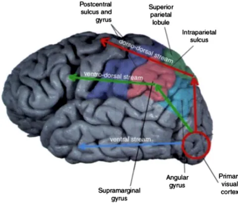
In the past decades, hand actions have been widely explored both using qualitative (e.g. clinical observation of patients’ behavior) and quantitative observations (e.g. neuroimaging investigation). The first insights about the neural correlates of hand movements came from neurophysiological studies in monkeys and patient studies in humans. These studies have been essential in describing the role of brain areas in processing information leading to hand motor outputs, and helped in defining a general model of the main functional paths subtending visual processing within the brain (Goodale and Milner, 1992).
The influential model by Milner & Goodale proposed the presence of two main visual routes: the ventral stream, where visual information is used to perceive the surrounding world (“Vision for Perception”), and the dorsal stream, where visual inputs are processed to guide actions (“Vision for Action”). These two streams were considered specialized and independent (Goodale et al., 1991).
of the hand motor network, revealing a specialized but also integrated neural system, where information processed by different areas are exchanged and integrated.
Thanks to the advent of more sophisticated methodologies (i.e. multivariate analysis), not only low-‐level motor features of hand actions (e.g. grip, direction of movement, etc.) can be explored, but also the representations at a higher hierarchical level (e.g. action’s goal) can be studied.
With this thesis, I will firstly provide a general review of the main literature on the hand motor system, considering both animal models and the human brain (Chapter 1). Secondly, I will introduce the unanswered scientific questions in the field that we have tackled in three experimental studies. They will be presented in the central chapters (Chapters 2, 3,4). Lastly, the discussion about the main results and their relevance with respect to the current knowledge of the hand motor system will be provided.
1.1
Hand motor network in non-‐human primates and human brain
2015), researchers managed to describe similar encoding within the human brain (see paragraph 1.1.2.3).
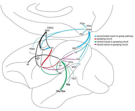
As previously mentioned, the dorsal stream comprises two distinct parieto-‐frontal pathways that subtend hand movements (Rizzolatti and Matelli, 2003). These two routes are: (i) the dorsolateral pathway that starts in the extrastriate visual areas of the occipital lobe and continues through the parietal lobe (anterior intraparietal area (AIP), inferior parietal areas (PF and PFG)) arriving at the frontal regions (ventral premotor area, area F5); (ii) and the dorsomedial pathway that starts in the same occipital visual areas and runs through the medial portion of the parietal lobe (V6A), reaching the frontal regions of the brain (dorsal premotor cortex, area F2). This subdivision has been found to be true both in human and monkey’s brain. Figure 1.1 represents the two main dorsal pathways of the hand motor system in the human brain.
Figure 1.1: A classical subdivision of the hand motor network: Prehension, our capability to grab an object, has been traditionally decomposed into a reaching component, which consists in moving the arm toward the target, and a grasping component, in which the fingers of the hand are shaped to match the object size. Red arrows show the path of the dorsomedial pathway (from V1 to V6 and to the superior parietal lobule -‐ SPL) traditionally associated with the reaching aspects of hand action. The green line shows the dorsolateral pathway (from V1 to the middle temporal -‐ MT and intra-‐parietal lobule -‐ IPL) traditionally associated with the grasping component of a prehension movement. Blue arrows show the ventral visual stream running through the temporal lobe. Adapted from Binkofski & Buxbaum (2013)
capability to approach and grasp an object. This movement has been traditionally decomposed into a reaching component, which consists in moving the arm toward the target, and a grasping
component, in which the fingers of the hand are shaped to match the object size. The dorsomedial pathway has been considered involved in the reaching component of the prehension movement, while the dorsolateral pathway in the grasping component (Rizzolatti and Matelli, 2003) (see Figure 1.2). Recent discoveries seem to challenge this view of two independent pathways, supporting a more integrated view based on the reciprocal interplay between these two streams (Nelissen and Vanduffel, 2011; Galletti and Fattori, 2017). However, this distinction is useful for an initial definition of the properties of the areas constituting the hand motor network.
Figure 1.2: The dorsal pathway. A. Dorsomedial pathway: Dorsomedial/reaching (red) pathway including area V6A and F2, associated with reaching movements and B. Dorsolateral pathway: dorsolateral/grasping (blue) pathway including the anterior intraparietal area (AIP) and the (area PF and PFG) within the frontal lobe, and the ventral premotor cortex (area F5), associated with grasping movements. Adapted from Galletti and Fattori (2017).
In the following paragraphs, I will summarize the main functional characteristics of the areas constituting the monkey’s hand motor network, and I will describe the homologies with respect to
the human brain.
1.1.1 Action-‐related areas within the NHP brain
1.1.1.1Neurophysiological studies
A series of monkey’s studies explored the neural circuits involved in hand actions (for review see Rizzolatti et al., 2014; Janssen and Scherberger, 2015; Galletti and Fattori, 2017); here, I will focus on those studies relevant to frame the topic of this thesis.
In the late nineties, an increasing number of neurophysiological studies classified the neurons constituting some of the monkey’s brain regions involved in hand actions and graspable object perception (see supplementary box 1). The main method used to investigate the properties of neurons in specific areas of the hand motor network is single-‐cell recording. With the insertion of a number of electrodes in a cytoarchitectonically and anatomically defined brain area, it is possible to register the firing rate of the neurons recorded during different experimental conditions. This method allows understanding which properties of the stimulus and/or of the action are preferentially processed.
For example, the properties of neurons within the lateral part of the dorsal stream, in particular within the intra-‐parietal sulcus (IPS) were investigated by training monkeys to fixate or to grasp different types of objects (Sakata et al., 1995; Murata et al., 2000). Within the anterior portion of IPS, the anterior intra-‐parietal area (AIP) has been described as the crucial hub involved in the
and/or grasping objects with different shapes, sizes and orientations (Murata et al., 2000) (Figure 1.3). Visuomotor neurons, responding both for the visual observation of one object and for the
grasping action performed over the same object, have been defined ‘canonical’ neurons, and have been proposed to be at the basis of the transformation of visual inputs into motor programs. Moreover, AIP activity is modulated by external cues triggering upcoming actions already during the planning phase of a delayed task (Baumann et al., 2009). Overall, AIP has been described as sensitive to the intrinsic features (i.e. size and shape) of the grasped objects, but also to the information extracted from the external context in which the action is performed.
Figure 1.3: Object and grip representation in area AIP. A. Firing rate of visuo-‐dominant neurons. Objects were medium size (A. horizontal ring, B. horizontal plate, C. cube). Neurons showed strong preference for the horizontal ring. B. Firing rate of AIP visuomotor neurons. Objects were medium size (A. vertical plate, B. vertical ring, C. cube). Neurons showed strong preference for the vertical plate. Adapted from Murata et al. (2000).
manipulation. It has been shown that PFG activity in grasping actions can be modulated also during the planning phase of the action, and not only by the visual perception of the object (Bonini
et al., 2011). Following studies demonstrated that PFG is also involved in high-‐level processing: the activity of half of PFG neurons is modulated by the final goal of the action (e.g. grasp-‐to-‐eat or grasp-‐to-‐place) and by the context in which the action is performed (e.g. external cues) (Bonini et al., 2012). Based on these findings, PFG has been described as a relevant region in action organization processes since both grip type information and high-‐level representations (i.e. goal of the action) are integrated here.
In the frontal lobe, other studies investigated the properties of the neurons within the ventral premotor area F5, finding similarities with AIP neurons in processing context-‐specific cues (Fluet et al., 2010) and grip type (Raos et al., 2006). This evidence suggested a pivotal role of F5 in selecting the most appropriate hand orientation and grip type.
In F5, two categories of visuomotor neurons with different peculiarities have been also described: (i) the ‘canonical neurons’, already found in AIP, which respond selectively to the visual presentation of 3D objects with specific size, shape and orientation, also when no motor response is required. This population seems to transform an object’s features into potential motor acts (Jeannerod et al., 1995; Bonini et al., 2014);
(ii) the extensively debated ‘mirror neurons’, present also in PFG and in AIP, that fire also when the monkey observed the execution of a particular action performed by a different agent (Rizzolatti et al., 2014). For this characteristic, mirror neurons have been considered responsible for action understanding and action recognition.
The primary motor area (M1) represents the principal area involved in motor output, as it generates neural impulses that allow the contraction of peripheral muscles and the execution of
movements. Recently, Schaffelhofer et al. (Schaffelhofer et al., 2015) showed that different hand configurations can be decoded in AIP, in F5 and in M1, both during the planning and the execution phase of a grasping action. The simultaneous recording within these three regions (Schaffelhofer and Scherberger, 2016) showed the cooperation of these nodes in creating the correct motor output, but each region maintained a certain degree of specialization. In general, AIP seems more related to processing visual information; M1 is dedicated to the execution of the prepared motor command; F5 functions as a hub where visual AIP-‐like information is stored temporarily and then transformed to motor information to be sent to M1, confirming the central role of F5 in visuomotor transformation.
Overall, the dorsolateral pathway seems to be included in a more extended lateral grasping system comprising additional regions within the fronto-‐parietal and temporal cortex with specific functional specialization (e.g. inferior frontal gyrus IFG, interior temporal IT). This complex temporo-‐parieto-‐frontal system supports the integration and exchange of different action-‐related information (e.g. perception of special relationships, storage of complex representation of actions). We focused on the main regions of the dorsolateral pathway, however, for a complete overview of all the involved areas please refer to Borra et al. (Borra et al., 2017).
The second route constituting the dorsal stream is the dorsomedial pathway, whose main nodes are: the parietal area V6A and the dorsal premotor cortex, area F2 (see Figure 2, Fattori et al., 2015; Galletti and Fattori, 2017).
et al., 2010; Fattori et al., 2012, 2015; Breveglieri et al., 2016). However, V6A activity is not only involved in reaching, but it shows modulation to hand orientation and grip type both during the
planning and the execution of a grasping movement (Fattori et al., 2009, 2010; Galletti and Fattori, 2017). This finding suggests the processing of grasp–related information also within the dorsomedial pathway. As in F5 and AIP, there are ‘canonical neurons’ also in V6A, particularly selective for shape, orientation and 3D features of the object even before the actual execution of a movement (Fattori et al., 2012).
In the dorsal premotor cortex, F2 neurons discriminate between different types of hand configurations (grip and hand orientation) during the execution of a movement (Raos et al., 2004). Together with AIP and F5, F2 contributes to process visually guided information sent from the dorsolateral areas and spatial inputs from posterior areas of the dorsomedial pathway (Raos et al., 2006).
These findings showed an involvement of dorsomedial and dorsolateral pathways in processing both reaching and grasping information, suggesting an integrated system of areas interacting with each other. It is more and more evident that the two dorsal pathways are not segregated and specialized in processing specific information. Different areas are involved in the same process and, according to the task, interact with each other to orchestrate the generation of a correct motor output (Galletti and Fattori, 2017).
Box 1: Motor, visuomotor and visual neurons in hand motor networks (following the classification of Sakata et al., 1995)
According to their neural response, neurons within the intraparietal sulcus (IPS) were classified in three different categories: i) ‘Motor-‐dominant’, if neurons showed maximum activity when
grasping in the light and in the dark, but do not show activity in the fixation condition; ii) ‘Visual-‐ dominant’, also subdivided in ‘object-‐type’, if neurons were fully activated by object fixation and object manipulation; and ‘Non-‐object-‐type’, if neurons were not activated during object fixation in the light nor during manipulation in the dark; iii) ‘Visual and motor’, subdivided in ‘object-‐type’ when showing selectivity for the object to grasp, particularly when grasping in the light compared to the dark condition, and discharging during the fixation of the preferred object; and ‘Non-‐object-‐ type’, highly active during the manipulation of a specific object, more in the light than in the dark. This last group of neurons does not show activity during the fixation of the object.
Referring to this classification, and considering following studies investigating different brain regions, it is possible to describe the functional properties of the main areas of the monkey’s prehension network based on this first classification.
SAKATA LIKE NEURONS CLASSIFICATION: F5
Somatotopically organized; represent specific hand (i.e. PG-‐WH
grip), mouth movements and
object features (i.e. orientation);
respond to 3D objects fixation
(‘canonical neurons’); involved in distal
movements and specific goal-‐ directed actions
AIP Sensorimotor transformation (i.e.
size, shape, orientation, 3D
features) for object-‐oriented
actions (i.e. grasping, hand and
fingers movements).
Mirror-‐like properties of
neurons
PFG Sensorimotor transformation.
Fine control of object manipulation. Integration hub for high (i.e. goal)
and low (i.e. physical features)
levels of action representations.
V6A Represent reach
and grasp movements. Its
activity is modulated by hand orientation
F2 Represent both reach and grasp movements planning and
execution. Neurons are modulated by wrist orientation and type of grasp.
MOTOR NEURONS
Active only during grasp execution
MOTOR DOMINANT
NEURONS Active for both grasping in the light
MOTOR DOMINANT
NEURONS Selectivity for
hand grip
MOTOR DOMINANT
NEURONS Active during reach-‐to-‐grasp
dark. Silent during
object fixation the light and in the dark VISUO-‐MOTOR
NEURONS Active during grasp execution
and during the visual presentation of specific objects
VISUO-‐MOTOR NEURONS More active for
grasping in the light, less active for
grasping in the dark. Respond when the object is
presented
VISUO-‐MOTOR NEURONS Active during reach-‐to.-‐grasp task both in the light and in the dark but with different strength
VISUO-‐MOTOR NEURONS
Active during grasp execution and during the visual
presentation of specific objects
VISUAL DOMINANT
NEURONS
Active during grasping in the light
and during object fixation. Not active
when grasping in the dark
VISUAL DOMINANT
NEURONS
Active during the execution of reach-‐to-‐grasp task only in the
light
VISUALLY MODULATED
NEURONS They lose their grip
and wrist orientation specificity when
grasping in the dark Anatomically
connected with AIP, M1, S2
thalamus, cerebellum, prefrontal cortex
F2
Anatomically connected with parietal F5, c-‐IPS,
IPL, CIP, V6A S2, temporal MTG,
TEa, TEm, ITC, frontal area 46 and
12, FEF
Anatomically connected with
AIP and F5
Anatomically connected with parietal MIP, PEc,
AIP, LIP, area46, S1, V6, frontal F2
Anatomically connected with F5, V6A, thalamus, M1
A lesion caused the incapacity to
preshape the hand using visual
feedback. Reaching preserved
A lesion caused the incapacity to correctly preshape
the hand using visual feedback
A lesion caused misreaching and
misgrasping, exaggerated fingers extension
and erroneous wrist orientation
References
AIP: (Sakata et al., 1995; Murata et al., 2000; Baumann et al., 2009); PFG: (Bonini et al., 2011, 2012); F5: (Jeannerod et al., 1995; Murata et al., 1997; Raos et al., 2006; Fluet et al., 2010; Bonini et al., 2014; Rizzolatti et al., 2014; Schaffelhofer et al., 2015; Schaffelhofer and Scherberger, 2016; Borra et al., 2017); V6A: (Marzocchi et al., 2008; Fattori et al., 2009, 2010, 2012, 2015; Bosco et al., 2010; Breveglieri et al., 2016); F2: (Raos et al., 2004).
1.1.1.2Lesion perspective
The functional properties of the areas within the dorsal pathways have been confirmed by lesion studies. Different types of motor deficits have been described depending on the lesioned cortical site. With respect to the dorsolateral pathway, both AIP and F5 are involved in coding different types of grips to match with the object features. A temporary lesion (muscimol injection) in AIP leads to the incapacity of pre-‐shaping the hand using visual feedback (Gallese et al., 1994; Janssen and Scherberger, 2015); as well, a lesion in F5 leads to similar deficits (Fogassi, 2001). With respect to the dorsomedial pathway, surgical removal of V6A determines misreaching and erroneous grasping movements (Battaglini et al., 2002; Galletti et al., 2003). This result was one of the first suggesting a possible processing of grasping information within the dorsomedial pathway, which was later demonstrated using neurophysiological recording (Fattori et al., 2004, 2009, 2010).
1.1.1.3Monkey’s fMRI studies
Functional magnetic resonance imaging (fMRI) allows measuring metabolic changes in the brain during different experimental conditions (univariate approach). Recently, novel decoding methods permitted also to compare the neural pattern of activity subtending the representation of various experimental conditions (MVPA approach) (see paragraph 1.1.2.3). These two different approaches give different, but complementary, information about the function and organization of the hand motor network.
There are only a few fMRI studies investigating the neural correlates of hand actions in
dark) with reaching-‐only (Figure 1.4). These results supported the central role of the dorsolateral pathway in the execution of grasping movements.
Figure 1.4. Monkey fMRI. Neural correlates of grasping: Activation for the univariate contrast [reach-‐and-‐grasp vs reach-‐only] activation map is overlaid on a 3D representation of a macaque brain. Adapted from Nelissen et al. (2011).
A second study (Nelissen et al., 2017) used MVPA to investigate how grasping is encoded within the hand motor network. They found that AIP, PFG and F5 significantly decode grasping related information. Moreover, significant decoding was also evident within the dorsomedial pathway in V6A and F2. These MVPA results confirmed previous neurophysiological findings showing the processing of similar information in both dorsal pathways, and supported the idea of an integrated motor network, which relies on the interplay between dorsomedial and dorsolateral areas.
1.1.1.4Connectivity evidence
Different studies investigating the connectivity profiles within the hand motor network showed that dorsomedial and dorsolateral pathways do not only share similar functional properties, but they are also anatomically connected (Davare et al., 2011) (Figure 1.5).
Areas belonging to the same pathway are anatomically connected one with the other, and anatomical connections have been identified also between the two pathways. The presence of reciprocal anatomical connections between dorsomedial and dorsolateral streams support their possible functional interaction, as suggested by recent investigations showing similar encoding within the two streams (Galletti and Fattori, 2017).
Figure 1.5. Anatomical connections between nodes of the monkey‘s hand motor network. Adapted from Davare et al. (2011).
circuit where AIP process object’s features mainly relying on visual feedback, while F5 is more engaged in translating the inputs processed by AIP into motor-‐related information (Rizzolatti and
Matelli, 2003; Raos et al., 2006; Janssen and Scherberger, 2015). F5 is also linked, among others, with the dorsal premotor cortex (F2), the somatosensory area (SII), the primary motor cortex (M1). Recently, Bonini et al. (Bonini et al., 2012) suggested to include to the AIP-‐F5 hand-‐grasping circuit also the intraparietal PFG area. This area is connected with AIP and F5, and the properties of its neurons are similar to those observed in F5.
Within the dorsomedial pathway, V6A is anatomically connected with the adjacent visual area V6, the premotor area F2, the dorsolateral AIP and with the primary somatosensory area (SI) (Gamberini et al., 2009). Area F2, in addition to the connections with V6A and F5, has direct projections to M1.
It is evident that there are strong connections among areas of the dorsolateral and dorsomedial pathways, as there are anatomical pathways linking their parietal and premotor nodes. This allows the transfer and the integration of different information between different cerebral nodes of the hand motor network through these specific anatomical pathways.
Anatomical connections have also been found between dorsal and ventral streams (Figure 1.5), linking temporal regions with areas of the dorsolateral pathway within the inferior parietal lobule (IPL). The dorsolateral pathway might represent a crucial hub where functional interactions between the dorsal and ventral streams occur (Davare et al., 2011). For example, Premereur et al. electrically stimulated different regions of monkey IPS (e.g. anterior and posterior AIP) and, using fMRI, described effective connectivity between IPS and the ventral stream (Premereur et al., 2015). Nonetheless, very few is known about the timing and the dynamic of these interactions.
dorsal and the ventral streams. The anatomical connections between these different brain pathways suggest the possible exchange of information between cortical regions, supporting a
more integrated and dynamic view of the hand motor system (Galletti and Fattori, 2017), as opposite to the traditional one asserting segregated pathways with a modular organization (Goodale and Milner, 1992). However, what is encoded within these pathways and how information is transferred is still poorly understood and investigated. Starting to answer this question is the main aim behind the present thesis.
In the supplementary Box 2, I will introduce a specific type of action (tool actions) used to investigate the functional interaction between dorsal and ventral stream. This information will be useful later on to clarify the role of temporal areas in motor control.
Box 2: Learning to use a tool: Orchestrating complex hand actions in a monkey’s brain
superior temporal sulcus, second somatosensory area and intraparietal sulcus of the right hemisphere, and to a lesser extent also in the left hemisphere. These results show that both the
dorsal and ventral streams are structurally modified when the brain ‘learns’ to perform tool actions.
When a monkey learns to use a tool, the neural representation of tool-‐related actions seems to be integrated into the existing hand motor network. In a single-‐cell recording study, after a training in which monkeys learnt to use simple and reverse pliers, Umiltà et al. showed that some neurons within F5 and to a less extent within M1 discharging for hand grasping actions, started to fire also when grasping with a tool (Umilta et al., 2008). The authors stated that the monkey motor system is based on a goal-‐centered functional organization, which allows primates to achieve a goal either with their hand or with a tool. Following studies on the human brain partially confirmed these findings, showing a common neural substrate recruited when using tools and when performing hand actions (see paragraph 1.1.2.4) (Lewis, 2006; Gallivan and Culham, 2015).
1.1.1.5Summary
pathways suggests their functional interaction. Moreover, anatomical connections between dorsal and ventral pathways might be used to transfer action information. Based on this evidence the
ventral stream might be considered as part of the hand motor network.
Based on their connectivity profiles, it is plausible to assume that these three pathways interact. However, the dynamics of these interactions are still poorly understood.
Understanding the functioning of hand motor network in monkey represents a useful starting point to explore the neural correlates subtending the same functions in the human brain. However, the approach adopted to investigate this issue in humans is radically different to the ones adopted in monkey studies. Indeed, human scientific investigation is performed mainly with non-‐invasive neurophysiological methods (i.e. transcranial magnetic stimulation -‐ TMS), neuroimaging techniques (i.e. fMRI) and machine learning approaches. As a consequence, possible homologies between the two species are not always straightforward to draw.
1.1.2 From monkey to human brain: Hand motor network within the Human brain
and those recruited in the human brain, giving a starting point for the interpretation of the results of human studies.
In the following sections, I will summarize the most relevant investigations for the present thesis about hand action processing in the human. A particular focus will be given to functional imaging (i.e. fMRI), transcranial magnetic stimulation (i.e. TMS) and connectivity studies.
Box 3: Lesion studies
In the second half of ‘800, medical doctors described the association between the location of a brain lesion and deficits in producing motor outputs, inferring the properties and the information processed by a cerebral area (Liepmann, 1905). One of the classical tests administered to clinical patients consists in imitating, pantomiming or performing the use of a familiar tool. Clinical studies of patients with focal brain lesions demonstrated that tool-‐manipulation and
predominant involvement of the left hemisphere when evaluating the site of lesions associated with apraxia and/or tool use deficits. Interestingly, patients with left posterior temporal gyrus
lesions (Buxbaum et al., 2014), showed deficits in tool related gestures performed with both right and left hand, supporting the role of the left hemisphere in controlling both hands when pantomiming. However, findings coming from often older clinical populations can be difficult to interpret due to the traumatic nature of the deficit and the comorbidity with other impairments. Only recently, thanks to imaging studies on healthy subjects, it has been possible to describe the neural basis of tool manipulation (Lewis, 2006; Gallivan et al., 2013a; Brandi et al., 2014; Valyear et al., 2017) (see paragraph 1.1.2.4) and new evidence suggested a functional interaction between dorsal and ventral stream, particularly when performing tool-‐related actions.
1.1.2.1Neuroimaging studies of the human brain
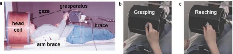
Figure 1.6: Experimental setup. A. MR setup: head of the subject tilted, grasparatus with target object placed on subject’s pelvis. B.
Grasping action. C. Reaching action. Adapted from Culham et al. (2003)
fMRI data can be analyzed in different ways. The first approach consists in univariate analysis, which is based on the comparison between neural activity evoked by a specific experimental condition with the baseline or on the comparison of different conditions. This approach showed that the execution of hand movement recruited a parieto-‐frontal motor network within the human brain similar to the one described with neurophysiological recordings in monkeys (Culham et al., 2003) (Figure 1.7a). Moreover, it demonstrated that grasping an object, compared to reaching it, induce greater activity within parietal areas, particularly within the anterior intraparietal sulcus (aIPS, the possible human homologous of monkey’s AIP) and the superior parietal lobule (SPL) (see figure 1.7b). These findings suggested the possibility of establishing homologies between the monkey and human brains based on specific motor features, e.g. a similar selectivity for grasping.
A similar selectivity for aIPS function has been confirmed in a more recent study (Cavina-‐ Pratesi et al., 2010). Moreover, the neural substrates of reaching compared to grasping were
investigated and, for the first time, the dissociation between the two components was documented. This study showed that the cortical areas more involved in the reaching task are the superior parieto-‐occipital cortex (SPOC, the human homologous of monkey’s V6A) and SPL. The study showed a possible integration of reaching and grasping information within the dorsal premotor cortex (PMd), putative homologous of the monkey area F2, the supplementary motor area (SMA), primary and secondary somatosensory cortices (SI-‐SII) and in M1. Overall, these first studies on the dorsal stream supported the specialization in processing specific information (grasping vs. reaching) within the two dorsal pathways; however, following investigations overstepped this description showing the processing of specific information both in the dorsomedial and in the dorsolateral pathway. For instance, hand orientation selectivity for grasping was demonstrated also using fMRI adaptation paradigm (fMRA), which consists in presenting the same stimulus repeatedly, leading to the reduction of BOLD response to the same type of process. This approach allows describing the selectivity to specific characteristics of a certain neural population. Monaco et al. showed that SPOC (part of the dorsomedial pathway) is engaged in processing wrist orientation for grasping an object (Monaco et al., 2011). The processing of grasp-‐related information (i.e. wrist orientation) and not only of reach-‐related information in the dorsomedial pathway, mirrors the finding of grasping response in monkey parietal area V6A and dorsal premotor area F2vr.
information processing within homologous areas (e.g. human PMv homologous to monkey’s F5 in the dorsolateral pathway; human PMd homologous to monkey’s F2 in the dorsomedial pathway).
Monkey fMRI studies (see paragraph 1.1.1.3) (Nelissen and Vanduffel, 2011; Nelissen et al., 2017) provided a clear demonstration that the neuroimaging investigations in human were looking at a similar network, reconciling decades of non-‐human primate neurophysiological evidence with human fMRI evidence.
Figure 1.8: Anatomical location of human grasping regions. A. Visual representation of dorsomedial (blue) and dorsolateral (red) pathways. B. Areas coding both reach and grasp actions. Adapted from Turella & Lingnau (2014).
different experimental conditions. The following paragraph will consider neurostimulation studies on humans used to describe the properties of specific brain areas and their involvement in specific
motor tasks.
1.1.2.2 Neurostimulation studies
Neurostimulation techniques in human, and particularly transcranial magnetic stimulation (TMS), can be adopted as a complementary tool to test the specific causal role of the dorsomedial and dorsolateral networks in hand movements. TMS is used to transitorily alter the normal activity of a brain region by inducing an electrical current, which influences the activity of a population of neurons underlying the TMS coil. This approach can be used to test the causal role of a brain region in a specific cognitive or motor task (Miniussi et al., 2013).
TMS studies tried to assess the role of the areas within the hand motor network: for example, it was used to investigate the characteristics of the posterior parietal cortex (PPC), stimulating different regions of the intraparietal sulcus, the superior and the inferior parietal lobule (Desmurget et al., 1999; Vesia et al., 2008, 2010).
In one study (Desmurget et al., 1999), participants had to reach a target located in the periphery of the visual field, the spatial position of the target could change or be kept invariant during the task, and TMS pulse was delivered soon after the movement onset. This study demonstrated that PPC is involved in detecting and correcting hand trajectory errors that occur after a change in the location of the target. On the contrary, when the target was kept to a stable location, TMS did not affect the reaching performance. Overall, PPC has an active role in guiding reaching movements particularly in the online control and monitoring of hand actions.
aIPS (65ms after the visual perturbation of the orientation of the target) affected the adjustment of fingers aperture for grasping (Tunik et al., 2005). Given these results, the visuomotor
TMS stimulation over these two regions was used to investigate the contribution of the premotor cortices in a grasp and lift task (Davare, 2006). The perturbation of left and right PMv altered the
positioning of the fingers on the object to lift, impairing the grasping action, while left PMd perturbations caused an effect in the timing of the lifting phase. These findings confirmed the role of PMv in operating visuomotor transformation necessary to perform efficient precision grasping movement; and support the idea of a functional loop between PMv, PMd and aIPS as proposed from monkeys ‘studies’ (Raos et al., 2006). Moreover, the contribution of left aIPS in force scaling (Davare et al., 2007; Dafotakis et al., 2008), suggests an interaction between PMv and aIPS during precision grasping. This hypothesis was supported by recent findings, showing that the perturbation of aIPS influences the interaction between PMv and M1 in the planning phase of a grasping movement, as evident from electromyographic (EMG) signal modifications in the specific muscles recruited for the subsequent grasping task (Davare et al., 2010).
Overall, TMS studies showed that hand action information might be present at a specific time and in specific tasks (e.g. online control) within the dorsal pathways. These studies refined our understanding about hand motor system, showing how regions within the two pathways might process specific aspects of hand actions in a time-‐dependent manner. However, the interactions between the pathways of the dorsal streams are still poorly investigated. The role of different areas in specific motor tasks, the timing and location of exchange of information that can occur between the two dorsal pathways it is still unclear. To shed light on what is represented within a specific area, new analytical approaches can be adopted (e.g. multivariate pattern analysis, MVPA, of fMRI data). I am going to describe this approach in the following paragraph.
1.1.2.3Multivariate Pattern Analysis of fMRI data
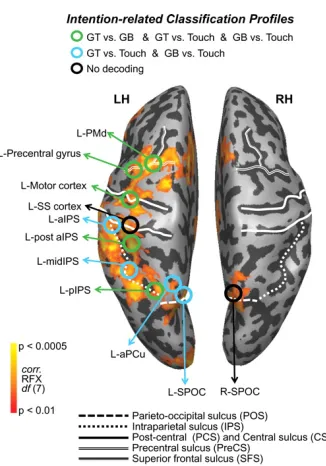
Recently, multivariate pattern analysis (MVPA) has been introduced to investigate the modifications in neural patterns produced by specific experimental conditions. In this analysis, we train a classifier to distinguish between the neural patterns produced by different experimental conditions (e.g. precision grip vs. whole hand prehension) and we then test if the same classifier can recognize to which category a new set of data belongs. MVPA allowed describing properties of the motor system that could not be defined with a univariate approach (Gallivan and Culham, 2015). The description of many of these properties were previously accessible only in monkey with single-‐cell-‐recording. Moreover, MVPA allows exploring, non-‐invasively, the representational content within brain areas during different phases of an action, comprising the planning phase of a movement and its execution. For example, recent studies investigated the basic properties within the parieto-‐frontal motor network such as grip selectivity or directional tuning (i.e. selectivity for movement towards specific directions/spatial positions). In different studies Gallivan et al. showed that it is possible to decode the direction (Gallivan et al., 2011a) and the type of action (Gallivan et al., 2011b) the subject is going to perform over an object (i.e. grasp top, grasp bottom or reach) even before the action is executed (planning phase). The regions circled in Figure 1.9, show a significant above chance decoding accuracy (i.e. how well the classifier can discriminate between experimental conditions) when comparing between grasp vs reach conditions during the planning phase of the action. Despite the reduced sample size (N=8), these results showed that areas of both dorsomedial (SPOC, SMA, PMd) and dorsolateral (aIPS, PMv) pathways stored the same type of information, i.e. grip type.
Figure 1.9: ROIs’ decoding accuracy during planning phase. Activation map contrast [plan > object fixation]. Green circles represent ROIs decoding significantly for [grasping small object vs grasping large object], [grasping small vs reach] and [grasping large vs reach]; blue circles represent ROIs decoding for [grasping small vs reach] and [grasping large vs reach]; black circles show no decoding. Adapted from Gallivan et al. (2011b).
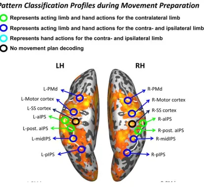
Figure 1.10: Planning phase, decoding of hand used and type of action planned. The activation map represents the contrast [execute>preview]. The considered ROIs are circled and the colors represent the general pattern of pairwise discriminations made during movement planning. Adapted from Gallivan et al. 2013b.
1.1.2.4The neural representation of complex actions: tool use
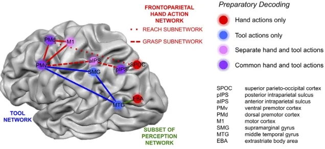
As described for monkeys (see paragraph 1.1.1.5), tool-‐related actions rely on a subset of areas within the hand motor network (Lewis, 2006). Nevertheless, tool manipulation includes a series of additional cognitive processing compared to simple hand actions, such as the identification of the tool, understanding its function and visuo-‐spatial integration that allows moving toward the to-‐be-‐used tool. All these processes are performed by a dedicated tool-‐ network, classically considered left-‐lateralized, that includes multiple areas located in the dorsomedial, dorsolateral, and in the temporal cortex (Lewis, 2006). The main areas involved in tool use are pMTG in the temporal lobe, SMG, pIPS and aIPS in the parietal lobe and PMd and PMv in the frontal lobe. The same network has been shown to be recruited both when using a real object and when pantomiming the use of the object (Hermsdörfer et al., 2007).
involved in processing body’s parts) (Figure 1.11). These results are referring to a concrete level of representation, where low-‐level features (e.g. tool features, hand used) are represented. A higher level of encoding, that we could define a more abstract representation of the goal of the action, was described within the areas located in the frontal lobe, i.e. PMv, PMd, and in the parietal lobe, i.e. pIPS, in which the type of action (grasp vs reach) is represented independently from the effector used (tool or hand).
Figure 1.11: Areas involved in action plan decoding of hand and tool movements. SMG and MTG decode for actions performed with a tool (blue). SPOC and EBA decode for actions performed with the hand (red). M1 and aIPS decode actions performed with the hand or with a tool but maintaining the representation separate (pink). PMd, PMv and pIPS show a shared representation for an action performed either with the hand or with a tool (purple).


![Figure
1.4.
Monkey
fMRI.
Neural
correlates
of
grasping:
Activation
for
the
univariate
contrast
[reach-‐and-‐grasp
vs
reach-‐only]
activation
map
is
overlaid
on
a
3D
repr](https://thumb-us.123doks.com/thumbv2/123dok_us/465510.2045012/24.595.137.449.173.436/figure-correlates-grasping-activation-univariate-contrast-activation-overlaid.webp)