R E S E A R C H
Open Access
The oncogenic Golgi phosphoprotein 3 like
overexpression is associated with cisplatin
resistance in ovarian carcinoma and
activating the NF-
κ
B signaling pathway
Shanyang He
1*†, Gang Niu
1†, Jianhong Shang
2†, Yalan Deng
1, Zhiyong Wan
1, Cai Zhang
1, Zeshan You
1and Hongwei Shen
1*Abstract
Background:Chemo-resistance is a leading cause of tumor relapse and treatment failure in patients with ovarian cancer. The identification of effective strategies to overcome drug resistance will have a significant clinical impact on the disease.
Methods:The protein and mRNA expression of GOLPH3L in ovarian cancer cell lines and patient tissues were determined using Real-time PCR and Western blot, respectively. 177 human ovarian cancer tissue samples were analyzed by IHC to investigate the association between GOLPH3L expression and the clinicopathological characteristics of ovarian cancer patients. Functional assays, such as MTT, FACS, and Tunel assay used to determine the oncogenic role of GOLPH3L in human ovarian cancer progression. Furthermore, western blotting and luciferase assay were used to determine the mechanism of GOLPH3L promotes chemoresistance in ovarian cancer cells.
Results:The expression of GOLPH3L was markedly upregulated in ovarian cancer cell lines and tissues, and high GOLPH3L expression was associated with an aggressive phenotype and poor prognosis with ovarian cancer patients. GOLPH3L overexpression confers CDDP resistance on ovarian cancer cells; however, inhibition of GOLPH3L sensitized ovarian cancer cell lines to CDDP cytotoxicity both in vitro and in vivo. Additionally, GOLPH3L upregulated the levels of nuclear p65 and phosphorylated inhibitor of nuclear factor Kappa-B kinase-βand IκBα, thereby activating canonical nuclear factor-κB (NF-κB) signaling.
Conclusions:Our findings suggest that GOLPH3L is a potential therapeutic target for the treatment of ovarian cancer: targeting GOLPH3L signaling may represent a promising strategy to enhance platinum response in patients with chemoresistant ovarian cancer.
Keywords:GOLPH3L, Chemo-resistance, Ovarian cancer, Nuclear factor-κB (NF-κB) signaling
Background
Epithelial ovarian cancer is the most lethal gynecologic cancer worldwide, and accounts for 4% of all cancers in women [1, 2]. Despite advances in surgical management and cytotoxic therapy, the overall 5-year survival rate for women with advanced ovarian cancer is just 20% because
of a lack of new diagnostic and treatment methods [3–5]. Currently, the recommended management is primary cytoreductive surgery followed by platinum–paclitaxel combination chemotherapy; however, more than 75% of treated patients experience tumor relapse and ultimately die of the disease [6, 7]. Poor understanding of the mech-anism of chemo-resistance in ovarian cancer poses a crit-ical research challenge. Elucidation of the molecular mechanisms underlying the chemo-resistance and recur-rence of ovarian cancer is necessary to improve clinical outcomes.
* Correspondence:hsy5g777@sina.com;doctorshen@163.com †Equal contributors
1Department of Obstetrics and Gynecology, The First Affiliated Hospital, Sun
Yat-sen University, Zhongshan Second Road 58, Guangzhou 510700, People’s Republic of China
Full list of author information is available at the end of the article
The transcription factor nuclear factor-κB (NF-κB) is activated in multiple cell-survival scenarios, and pro-motes survival and chemo-resistance in solid-tumor can-cers [8]. The survival cascades initiated by NF-κB are a key component of cellular apoptotic resistance. Report-edly, TGM2-NFKB/NF-κB signaling enhances lymphoma progression in both mice and humans; disruption of this network may increase the efficacy of current therapies and reduce MCL drug resistance [9]. Peng et al. [10]found that activation of NF-κB signaling confers chemo-resistance on tongue squamous cell carcinoma cells and promotes their survival, whereas inhibition of NF-κB signaling dramatically reduces the proliferation of oral squamous cell carcinoma cells. Canino and col-leagues [11] showed that the addition of a dual signal transducer and activator of transcription NF-kB inhibitor to cultured pemetrexed-treated and cisplatin-treated mesothelioma cells abolishes their chemo-resistance. Blocking NF-κB signaling using an aurora kinase A in-hibitor decreases the proliferation of epithelial ovarian cancer (EOC) stem cells by inducing cell-cycle arrest, which suggests that NF-κB inhibition may prevent recur-rence and chemo-resistance in ovarian cancer [12]. Fur-thermore, the growth of xenografts of MCF-7TN-R cells is blocked following treatment with ABC294640, a pharmacologic inhibitor of sphingosine kinase-2 that diminishes NF-κB survival signaling through decreased activation of the Ser536 phosphorylation site on the p65 subunit [13]. These results indicate that pharmacologic inhibition of NF-κB has therapeutic potential for the treatment of therapy resistant breast cancer. The NF-κB pathway may play an important role in chemo-resistance; therefore, the discovery of novel molecules capable of regulating aberrant activation of the NF-κB pathway may facilitate the treatment of chemo-resistant cancers.
GOLPH3L, is a novel gene which highly homologous to Golgi phosphoprotein 3 (GOLPH3), the protein encoded by GOLPH3L localizes to the Golgi stack and may have a regulatory role in Golgi trafficking [14]. Re-portedly, increased expression of GOLPH3L is associated with a poor prognosis in patients with EOC and may act as a novel, useful, and independent prognostic indicator [15]. Kunigou et al. [16] showed increased expression of GOLPH3L in human rhabdomyosarcoma, and that GOLPH3L knockdown by short-interfering RNA prevents the proliferation of human rhabdomyosarcoma cell lines. These findings suggest that GOLPH3L repression may be an effective treatment for rhabdomyosarcoma. Although the two isoforms are highly homologous in their amino-acid sequences and GOLPH3 is upregulated in various malignancies, the function and mechanism of action of GOLPH3L in cancer, particularly in ovarian cancer, were rarely reported.
In this study, we demonstrate that GOLPH3L expres-sion is significantly upregulated in cisplatin-resistant ovarian cancer cells and clinical tissues, and is associated with ovarian cancer recurrence. Moreover, we show that GOLPH3L overexpression enhances cisplatin resistance, and that GOLPH3L silencing restores the sensitivity of ovarian cancer cells to cisplatin, through regulation of the NF-κB signaling pathway. Our findings suggest that GOLPH3L plays a critical oncogenic role in ovarian can-cer progression, and highlight its potential as a thera-peutic target for overcoming cisplatin resistance in ovarian cancer therapy.
Materials & methods
Cell lines and cell culture.
Immortalized normal ovarian surface epithelial cell line (IOSE80) was purchased from Shanghai Ai Rui Bio-logical Technology Co., Ltd., this cells were grown in 1:1 combination of two media, Medium 199 (Invitrogen) and MCDB 105 (Cell Applications Inc., San Diego, CA) with 10% FBS in a humidified atmosphere containing 5% CO2 at 37 °C. The ovarian cancer cell lines, including SKOV3, CAOV3, OV56, A2780, A2780/cis, COV362, EFO-27, TOV21G, EFO-21 and OV90 was purchased from The European Collection of Authenticated Cell Cultures (ECACC), were grown in Dulbecco’s modified Eagle’s medium (DMEM, Invitrogen, Carlsbad, CA, USA) supplemented with 10% fetal bovine serum (FBS, Invitrogen), at 37 °C in a 5% CO2 atmosphere in a hu-midified incubator. A2780/cis was grown in RPMI 1640 + 2 mM Glutamine +1 μM cisplatin +10% Foetal Bovine Serum (FBS), at 37 °C in a 5% CO2 atmosphere in a humidified incubator. All cell lines were authenti-cated by short tandem repeat (STR) fingerprinting at Medicine Lab of Forensic Medicine Department of Sun Yat-Sen University (Guangzhou, China).
Patient information and tissue specimens
A total of 177 paraffin-embedded and archived ovarian can-cer samples, which were histopathologically and clinically diagnosed at the First Affiliated Hospital, Sun Yat-sen University from 2005 to 2010, were examined in this study. Clinical information on the samples is summarized in Additional file 1: Table S1. All tumors were staged accord-ing to the International Federation of Gynaecology and Ob-stetrics standards (FIGO). Ten freshly collected ovarian cancer tissues were frozen and stored in liquid nitrogen until further use. Prior patient consent and approval from the Institutional Research Ethics Committee were obtained for the use of these clinical materials for research purposes.
Vectors, retroviral infection and transfection
cDNA into the pMSCV retrovirus plasmid, and human GOLPH3L-targeting short hairpin RNA (shRNA) oligonu-cleotides sequences were cloned into pSuper-retro-puro to generate pSuper-retro-GOLPH3L-RNAi(s). The shRNA sequences were: RNAi#1, GOLPH3L; and RNAi#2,
TATAATGGTCAAGGTCTATGG (synthesized by
Invitrogen). pNF-κB-luc and control plasmids (Clontech) were used to examine NF-κB activity. pBabe-Puro-IκBα-mut (plasmid#15291) expressing IκBαdominant-negative
mutant (IκBα-mut) was purchased from Addgene
(Cambridge, MA). Transfection of siRNA or plasmids was performed using the Lipofectamine 3000 reagent (Invitro-gen, Carlsbad, CA) according to the manufacturer’s instruction. Stable cell lines expressing GOLPH3L or GOLPH3L RNAi were selected for 10 days with 0.5μg/ml puromycin 48 h after infection.
Western blot analysis
Western blot was performed using anti- GOLPH3L (Abcam, 1:500), anti-p-IκBα, IκBαand anti-p-IKKβ, IKKβ, anti-p65, anti-p84 antibodies (Cell Signaling, Danvers, MA, USA). The membranes were stripped and re-probed with an anti-α-tubulin antibody (Sigma, Saint Louis, MI) as a loading control.
Xenografted tumor model, IHC, and H&E staining
In the subcutaneous tumor model, the BALB/c nude mice were randomly divided into four groups (n = 6/group). Four groups of mice were inoculated subcutaneously with 2 × 10⁶ A2780-Vector, A2780- GOLPH3L, A2780-cis/ shRNA-Vector, A2780-cis/ GOLPH3L -shRNA#1 cells, re-spectively, in the left dorsal flank per mouse. Mice bearing established A2780 xenograft were established as men-tioned above. After xenografts reached 0.5 cm in diameter, CDDP (5 mg/kg) was given intraperitoneally every 4 days for 28 days. Tumor growth was monitored by measure-ments of length and width and the tumor volume was cal-culated using the eq. (L × W2)/2. Tumors were detected by an IVIS imaging system, and animals were euthanized, tumors were excised, weighed and paraffin-embedded. TUNEL assay was performed on paraffin-embedded tissue section according to the manufacturer’s instructions (Pro-mega). Apoptotic index was measured by percentage of TUNEL-positive cells.In the intraperitoneal tumor model, therapeutic effectiveness of GOLPH3L siRNA was evalu-ated in combination with cisplatin(5 mg/kg, every 4 days for 28 days). The BALB/c nude mice were divided into four groups (10 mice per group). Two groups of mice were inoculated intraperitoneally with 2 × 10⁶ A2780-Vector, A2780- GOLPH3L cells, respectively. Another two groups of mice were intraperitoneally injected with 2 × 10⁶A2780. Treatment was initiated 21 days after the cell suspension injection, when tumors could be detected by palpation. Mice injected intraperitoneally with 2 × 10⁶
A2780-Vector, A2780- GOLPH3L, A2780-cis/shRNA-Vector, A2780-cis/ GOLPH3L -shRNA#1 cells, respect-ively treated with CDDP (5 mg/kg) every 4 days for 28 days. Mice bearing established A2780 xenograft were established as mentioned above. Tumors were detected by an IVIS imaging system twice a week. Survival was evalu-ated from the first day of treatment initiation until death and tumors were excised and paraffin-embedded. Apop-totic index was measured by percentage of TUNEL-positive cells.
Cytotoxicity assay
The sensitivity to cisplatin of ovarian canccer cells was determined using the MTT assay as previously described (Landen CN, et al. Efficacy and antivascular effects of EphA2 reduction with an agonistic antibody in ovarian cancer. J Natl Cancer Inst. 2006; 98(21):1558–1570.). Briefly, 2 × 10³cells were seeded onto 96-well plates and incubated at 37 °C overnight. Cells were then transfected with different concentrations of cisplatin (0–200 μM). After incubation for 72 h, 50 μl of the MTT solution (0.15%) was added to each well, and the plates were fur-ther incubated for 2 h. One hundred microliters of DMSO was added to solubilize the MTT formazan prod-uct. Absorbance at 540 nm was measured with a Falcon microplate reader (BD-Labware). Dose-response curves were plotted on a semilog scale as the percentage of the control cell number, which was obtained from the sam-ple with no drug exposure. IC50 was determined by the intersection of the cisplatin concentration and the mid-point of the 570-nm reading.
Apoptosis assay
For evaluation of apoptosis, PE Annexin V Apoptosis De-tection Kit I (BD Pharmingen) was used. Briefly, 1 × 10⁶ ovarian cancer cells were plated in 10-cm plates and incu-bated for 24 h. Treatment was started with cisplatin (10μM) for 24 h. Cell morphology was assessed by phase-contrast microscopy. Then, cells were removed from plate by trypsin-EDTA, washed twice with PBS, and resuspended with binding buffer at 106cells/ml. FITC Annexin V and propidium iodide were added (each at 5μl/105cells). Cells were incubated for 15 min at room temperature in the dark. Percentage of apoptosis was analyzed with an EPICS XL flow cytometer (Beckman-Coulter). Each sample was analyzed in triplicate.
Transient luciferase assay
according to the manufacturer’s instruction. 48 h after transfection, cells were harvested and Luciferase and renilla signals were measured using the Dual Luciferase Reporter Assay Kit (Promega) according to a protocol provided by the manufacturer. The luciferase activity was normalized by the Renilla luciferase activity of each trans-fection to normalize the transtrans-fection efficiency.
Nuclear and cytoplasmic extraction assay
Nuclear fractions were prepared by using the nuclear ex-traction kit (Active Motif, Carlsbad, CA). Briefly, after drug treatment, cells were pelleted and lysed by vigorous vortex in hypotonic buffer for 15 min. The samples were then centrifuged at 14,000×g for 1 min; the supernatant was considered cytoplasmic. Insoluble pellets were further lysed in complete lysis buffer for 30 min, and nuclear extracts (supernatant) were collected after a 10-min centrifugation at 14,000×g. Both cytoplasmic and nuclear fractions were quantified and subjected to Western blot analysis.
Statistical analysis
Statistical tests for data analysis included Fisher’s exact test, log-rank test, Chi-square test, and Student’s 2-tailed t test. Multivariate statistical analysis was performed using a Cox regression model. Statistical analyses were performed using the SPSS 11.0 statistical software pack-age. Data represent mean ± SD.P< 0.05 was considered statistically significant.
Microarray data process and visualization
Microarray data were downloaded from the GEO database: (http://www.ncbi.nlm.nih.gov/geo/).
GSEA was performed using GSEA 2.0.9:(http://
www.broadinstitute.org/gsea/).
Results
GOLPH3L overexpression correlates with progression and poor prognosis in human ovarian cancer
By analyzing the two published mRNA expression profiles (GSE 66957, GSE 27651) obtained from NCBI, (https:// www.ncbi.nlm.nih.gov/geo/), we found that GOLPH3L mRNA was significantly upregulated in ovarian cancer tis-sues compared with normal tistis-sues (Fig. 1a-b). Analysis of TCGA datasets showed that ovarian cancer patients with higher GOLPH3L expression had a shorter survival time and demonstrated an earlier relapse disease-free survival time (P< 0.05; Fig. 1c).
Consistently, real-time PCR and western blotting ana-lyses revealed that GOLPH3L was markedly overex-pressed in all nine ovarian cancer cell lines at both the protein and mRNA levels, compared with Immortalized normal ovarian surface epithelial cell line (IOSE80)
(Fig. 1d and Additional file 2: Figure S1A). Further-more, comparative analyses showed that GOLPH3L ex-pression were elevated in the twenty ovarian cancer samples compared with two non-tumor ovarian speci-mens (Fig. 1e and Additional file 2: Figure S1B), sug-gesting that GOLPH3L is upregulated in human ovarian cancer.
To determine the clinical relevance of GOLPH3L in ovarian cancer, GOLPH3L expression was examined in 177 paraffin-embedded, archived ovarian cancer tissues by IHC assay. As showed in Fig. 1f and Additional file 1: Ta-bles S1-S2, GOLPH3L levels were correlated with the FIGO stage (P< 0.001), and Histology (P< 0.001) in pa-tients with ovarian cancer. The increased expression of GOLPH3L was also detected in approximately three-fifths of the clinical ovarian cancer tissue samples, but not de-tectable in normal ovarian surface epithelial cell speci-mens (Fig. 1f). Importantly, statistical analysis showed that ovarian cancer patients with high GOLPH3L expres-sion had significantly worse overall and disease-free sur-vival than those with low GOLPH3L expression (Fig. 1g). These results suggest that GOLPH3L has potential clinical value as a predictive biomarker for disease outcome in ovarian cancer.
Upregulation of GOLPH3L confers CDDP resistance in ovarian cancer in vitro
Upregulation of GOLPH3L confers CDDP resistance in ovarian cancer in vivo
Cancer recurrence is mainly attributed to resistance to chemotherapy. Interestingly, we found that GOLPH3L overexpression was correlated with progression-free sur-vival of ovarian cancer patients with CDDP-resistance in multiple GEO datasets (Fig. 3a). GSEA analysis also re-vealed that GOLPH3L overexpression was strongly corre-lated with gene signatures associated with CDDP-based chemotherapy signatures, suggesting that GOLPH3L over-expression may contribute to CDDP-resistance in ovarian cancer (Fig. 3b). In order to explore the function of GOLPH3L in ovarian cancer chemo-resistance, we used two mouse models to assess the anti-tumor effect of GOLPH3L in ovarian cancer. In this two model, A2780/ cis cell line(cisplatin-resistant) and A2780 was used be-cause A2780/cis was resistance to CDDP and expressed a relatively higher level of GOLPH3L, but A2780 expressed a relatively lower level of GOLPH3L (Fig. 1d). Nude mice were subcutaneously inoculated with A2780/cis, A2780/
cis-GOLPH3L -shRNA, A2780 and A2780 –GOLPH3L
respectively, mouse were treated with CDDP when the
treatment with drugs started as soon as the tumor became palpable. As shown in Fig. 3c, treatment with GOLPH3L-shRNA plus cisplatin resulted in a significantly reduction, but overexpression GOLPH3L resulted in a significantly increase in tumor growth compared with that in the control group (Tumor weight: Vector vs GOLPH3 L = 373.4 ± 6.1 g vs 763 ± 11.2 g; control vs GOLPH3L-shRNA = 683.4 ± 9.2 g VS 58.7 ± 2.1 g,P< 0.01). Next, we determined if the suppression of GOLPH3L expression was effective in suppressing intraperitoneal tumor growth in an in vivo. Consistently, analyzing in an in vivo intraperi-toneal mice model also showed that genetically engineered GOLPH3L conferred great resistance to chemotherapy-induced apoptosis on intraperitoneal growth of A2780 cell, as determined by proportion of TUNEL+-cells, active caspase 3 cells compared with that in the control group (Fig. 3d-f) (proportion of TUNEL + −cells: Vector vs GOLPH3L = 13.5% vs 3.3%; control vs GOLPH3L-shRNA = 5.1% VS 27.7%. P < 0.01; proportion of active cas-pase 3 cells: Vector vs GOLPH3L = 14.7% vs 2.1%; control vs GOLPH3L-shRNA = 7.1% VS 29.9%. P < 0.01). However, silencing GOLPH3L via GOLPH3L -shRNA enhance the
cytotoxic effect of CDDP on ovarian cancer cells, which re-sulted in remission tumor progression and increased TUNEL+-cells, active caspase 3 cells compared with that in the control group (Fig. 3d-f). Therefore, these results dem-onstrated that overexpression of GOLPH3L contributes to ovarian cancer chemo-resistance.
Upregulation of GOLPH3L activates the NF-κB signaling pathway in ovarian cancer
In order to better understand the mechanism underlying GOLPH3L overexpressed induced chemo-resistance, TCGA dataset and mRNA microarray (GSE66957) were performed. Gene ontology (GO) enrichment analysis showed that NF-κB signaling pathway was enriched in GOLPH3L up-regulated genes in TCGA and GSE 66957 dataset (Fig. 4a). By analyzing GOLPH3L mRNA ex-pression levels and NF-κB-regulated gene signatures
from published ovarian cancer patient profiles, we found that GOLPH3L expression was positively corre-lated with NF-κB signaling gene signatures (Fig. 4b). These results suggest that GOLPH3L may play crucial roles in NF-κB signaling pathway regulation. As ex-pected, overexpression of GOLPH3L significantly en-hanced, whereas silencing of GOLPH3L reduced, the activity of NF-κB luciferase reporter activity in A2780 and SKOV3 cancer cells (Fig. 4c). Moreover, western blotting revealed that the levels of nuclear p65 and phosphorylated-IKK-βand -IκBαwere dramatically up-regulated in GOLPH3L -overexpressing cells but were downregulated in GOLPH3L -silenced cells (Fig. 4d-e). Furthermore, the expression levels of numerous well-characterized NF-κB downstream genes were showed to be increased in GOLPH3L overexpressing cells, but were lower in GOLPH3L-silenced cells (Fig. 4f). These results
Fig. 2Upregulation of GOLPH3L conferred ovarian cancer to CDDP resistance in vitro.aIC50 of CDDP in the indicated cells.bAnnexin V-FITC and PI staining of the indicated cells treated with cisplatin (10μM) for 24 h. Each bar represents the mean ± SD of three independent experiments.c
Fig. 3Upregulation of GOLPH3L conferred ovarian cancer to CDDP resistance in vivo.aThe Kaplan-Meier survival curves compare ovarian cancer patients with low and high GOLPH3L expression levels.bGSEA plot, indicating a significant correlation between the mRNA levels of GOLPH3L expression in ovarian cancer and the CDDP resistance gene signatures in published datasets.cRepresentative tumor-bearing mice and tumor weights of the subcutaneous tumor model treated as indicated.d-eThe luminescence of the intraperitoneal tumor xenografts from different treatment groups at the indicated weeks.f
suggesting that GOLPH3L plays an important role in acti-vating the NF-κB signaling pathway in ovarian cancer.
NF-κB signaling pathway is required for GOLPH3L-induced chemoresistance
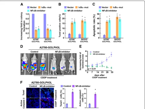
Next, we investigated whether GOLPH3L-mediated ovarian cancer progression occurs through NF-κB activation. As shown in Additional file 4: Figure S3, the stimulatory effect of GOLPH3L on NF-κB activation was significantly in-hibited by transfection of an IκBαdominant-negative mu-tant (IκBα-mut) or treatment with a NF-κB inhibitor.
Meanwhile, we found that blockade of the NF-κB pathway significantly abrogates the effect of GOLPH3L on ovarian cancer aggressiveness in both in vitro (Fig. 5a-c). Strikingly, IκBα dominant-negative mutant (IκBα-mut) or treatment with a NF-κB inhibitor significantly enhanced the effects of CDDP in vivo, as determined by quantification of biolu-minescence signal and proportion of TUNEL+-cells, active caspase 3-cells compared with that in the control group (Fig. 5d-f). Taken together, these results indicate that activa-tion of the NF-κB signaling pathway exerted funcactiva-tional ef-fects of GOLPH3L on ovarian cancer progression.
Clinical relevance of GOLPH3L -induced NF-κB activation in human ovarian cancer
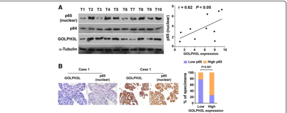
The clinical relevance of GOLPH3L expression and NF-κB activation was further characterized in human ovarian cancer. As showed in Fig. 6a, GOLPH3L levels in ten freshly collected clinical ovarian cancer samples were positively correlated with nuclear p65 signals (r = 0.62, P < 0.05). The correlation of GOLPH3L expression and nuclear p65, as indicator of NF-κB signaling activation, was further confirmed in a cohort of clinical ovarian cancer tissues by IHC analysis (P < 0.01; Fig. 6b). These data further support the no-tion that GOLPH3L up-regulano-tion promotes ovarian cancer aggressiveness and activation of the NF-κB sig-naling pathway, which may lead to a poor clinical out-come for patients with ovarian cancer.
Discussion
Our results provide evidence that GOLPH3L plays an im-portant role in cisplatin resistance in ovarian cancer and the regulation of the NF-κB signaling pathway. GOLPH3L gene expression was substantially increased in cisplatin-resistant cells and GOLPH3L overexpression enhanced cisplatin resistance, but GOLPH3L silencing restored the sensitivity of ovarian cancer cells to cisplatin. Moreover, we found that GOLPH3L enhanced cisplatin resistance by upregulating downstream target genes that regulate the anti-apoptosis effect of the NF-κB signaling pathway both in vitro and in vivo. These findings identify GOLPH3L as a potential target for overcoming cisplatin resistance in patients with ovarian cancer.
Chemo-resistance has a considerable influence on the ef-ficacy of cancer therapy, and involves anti-apoptotic
transduction pathways that prevent cell death [17, 18]. Intrinsic or acquired resistance of cancer to current treat-ment protocols is associated with apoptosis resistance in cancer cells and treatment failure [19, 20]. Despite this, the currently recommended management is primary cytoreduc-tive surgery followed by platinum–paclitaxel combination chemotherapy, but more than 75% of treated patients ex-perience tumor relapse. Current and future efforts toward designing new therapies must include strategies that specif-ically target cancer cell resistance to current chemother-apies [21, 22]. Therefore, we will discuss the potential roles of small-molecule candidates that target apoptosis signaling to enhance the sensitivity of tumors to conventional cancer therapies and improve the survival and quality of life of cancer patients.
Activation of the transcription factor NF-κB is fre-quently encountered in tumor cells and contributes to chemoresistance during cancer treatment [8, 23, 24]. Suppression of NF-κB by genetic or chemical inhibitors induces apoptosis and restores the apoptotic response after treatment with chemotherapeutic agents or radi-ation in various tumor cells, thus overcoming NF-κB-mediated chemoresistance. It is established that the inhibition of NF-κB activation abolishes tumor che-moresistance [25–29]. Suppression of NF-κB through adenoviral delivery of a modified form of IκBα markedly sensitizes chemoresistant tumors to the apoptotic poten-tial of tumor necrosis factor-α and the chemotherapeutic compound CPT-11, resulting in tumor regression [30]. Treatment with the proteasome inhibitor MG132 in-creases the apoptotic effects of etoposide or doxorubicin on Capan-1 and A818–4 cells through the inhibition of NF-κB [31]. Furthermore, using reporter assays and
reverse-transcription PCR analysis, Li et al. [32] demon-strated that abrogation of NF-κB activation by a dominant-negative IκBα adenoviral construct triggered paclitaxel-induced cell death, suggesting that suppression of the activation of NF-κB blocks paclitaxel-induced apop-totic signaling pathways. In chemoresistant cancer cells, both inhibitors of apoptosis and NF-κB play a pivotal role in preventing apoptosis triggered by a variety of stresses, highlighting them as potential targets for cancer treatment [33–37]. Collectively, these findings establish a strong ra-tionale for therapeutic targeting of the NF-κB pathway in cancer therapy. Although current therapeutic approaches, such as the use of NF-κB or IKK-βinhibitors, may abro-gate the cancer-promoting activities of NF-κB, they fail to preserve its pleiotropic physiologic functions in normal cells, such as in immunity and inflammation. Therefore, there is an urgent need to identify more effective th-erapeutic targets that regulate NF-κB in an appropriate manner as alternatives to global NF-κB blockade. Here, we reported that GOLPH3L expression was significantly upregulated in cisplatin-resistant ovarian cancer and associated with ovarian cancer recurrence. Moreover, GOLPH3L overexpression enhanced cisplatin resistance, but GOLPH3L silencing restored the sensitivity of ovarian cancer cells to cisplatin by regulation of the NF-κB signal-ing pathway, suggestsignal-ing that GOLPH3L contributes to ovarian cancer progression and thereby represents a novel target for overcoming cisplatin resistance in ovarian can-cer therapy.
GOLPH3L is a GOLPH3 paralog found in all verte-brate genomes. Like GOLPH3, GOLPH3L binds to PI4P, localizes to the Golgi as a consequence of PI4P binding, and is required for efficient anterograde trafficking [14].
Although the two isoforms are highly homologous in their amino-acid sequences, the function of GOLPH3L has yet to be determined. We showed that GOLPH3L overexpression enhanced the resistance of ovarian can-cer cells to cisplatin treatment through regulation of the NF-κB signaling pathway. However, the underlying mechanism by which GOLPH3L activates NF-κB signal-ing remains unclear. Interestsignal-ingly, Tsignal-ing Dai et al. [38] showed that GOLPH3 promotes K63-linked polyubiqui-tination of Tumor necrosis factor receptor-associated factor 2, receptor interacting proteins, and NF-κB essen-tial modulator and substanessen-tially sustained the activation of NF-κB in hepatocellular carcinoma (HCC) cells. It is likely that GOLPH3L activates NF-κB signaling via the same mechanism as GOLPH3 activation of NF-κB sig-naling in HCC cells. Therefore, the underlying mechan-ism by which GOLPH3L activates NF-κB signaling requires further investigation.
Although GOLPH3L is reportedly overexpressed in sev-eral cancers, including EOC and human rhabdomyosar-coma, the mechanism of GOLPH3L upregulation in ovarian cancer remains unknown. Interestingly, we found that large amounts of NF-κB were recruited to the pro-moter region of GOLPH3L, according to chromatin im-munoprecipitation sequencing tracks in the University of California Santa Cruz Genome Browser (http://geno-me.ucsc.edu/cgi-bin/hgGateway). Furthermore, according to TCGA data, we found that GOLPH3L exhibited a high amplification rate of 61.8% in ovarian cancer, suggesting that the overexpression of GOLPH3L in ovarian cancer is associated with genomic amplification. Further studies are necessary to determine whether GOLPH3L upregulation in ovarian cancer is attributable to genomic amplification or NF-κB-mediated transcriptional upregulation.
In summary, GOLPH3L was markedly upregulated in ovarian cancer cells and clinical ovarian cancer samples, and a positive correlation was evident between GOLPH3L expression and the recurrence-free survival of ovarian cancer patients. Overexpression of GOLPH3L augmented the cisplatin resistance of ovarian cancer both in vitro and in vivo, and activated the NF-κB signaling pathway. Elucidation of the biologic function of GOLPH3L during ovarian cancer progression will advance our knowledge of the mechanisms underlying ovarian cancer chemo-resistance and establish GOLPH3L as a potential thera-peutic target for overcoming drug resistance in patients with ovarian cancer.
Conclusions
GOLPH3L is a potential therapeutic target for the treat-ment of ovarian cancer: targeting GOLPH3L signaling may represent a promising strategy to enhance platinum response in patients with chemoresistant ovarian cancer.
Additional files
Additional file 1:Table S1.Clinicopathological characteristics and expression of GOLPH3L in studied ovarian cancer patients.Table S2.
Correlation between the clinicopathological features and expression of GOLPH3L (DOC 54 kb)
Additional file 2:Figure S1.mRNA expression analysis shows that GOLPH3L was up-regulated in ovarian cancer cell lines and tissues. (A). Real-time PCR analysis of GOLPH3L expression in Immortalized normal ovarian surface epithelial cell line (IOSE80) and in ovarian cancer cell lines (SKOV3, CAOV3, OV56, A2780, A2780/cis, COV362, 27, TOV21G, EFO-21 and OV90). Transcript levels were normalized toGAPDHexpression. (B). Real-time PCR analysis of GOLPH3L expression in 8 ovarian cancer tissues (T) with 2 non-tumor ovarian tissues (N). Transcript levels were normalized to GAPDHexpression. Each bar represents the mean ± SD of three independent experiments. *P< 0.05 (TIFF 160 kb)
Additional file 3:Figure S2.(A-B) Western blot analysis of GOLPH3L in the indicated ovarian cancer cells. (C). Annexin V-FITC and PI staining of the indicated cells treated with cisplatin (10μM) for 24 h. Each bar represents the mean ± SD of three independent experiments (TIFF 545 kb) Additional file 4:Figure S3.Relative of luciferase reporter activity in the indicated ovarian cancer cells transfected with vector or IκBα-mut or treated with an NF-κB inhibitor (JSH-23) (TIFF 47 kb)
Acknowledgments
This project was supported by the National Natural Scientific Foundation of China (No.81772764);The Guangdong Natural Science Foundation, China (No.S2016A030313820) and the Science and Technology Planning Project of Guangzhou City, China (No.201704020163).
Authors’contributions
SYH, HWS, GN conceived and designed the experiments.; HWS, GN, JHS and YLD conducted the experiments; JHS and YLD performed the statistical analysis; ZYW and CZ supported the experiments and helped to draft the manuscript.ZSY and GN and SYH rote the manuscript. All authors read and approved the final manuscript.
Ethics approval and consent to participate
This study was approved by the Institutional Ethics Committee of the First Affiliated Hospital, Sun Yat-sen University for the use of clinical materials for research purpose. And animal use and experiment protocol were approved by the Institutional Animal Care and Use Committee of the First Affiliated Hospital, Sun Yat-sen University.
Consent for publication
Not applicable.
Competing interests
The authors declare that they have no competing interests.
Publisher’s Note
Springer Nature remains neutral with regard to jurisdictional claims in published maps and institutional affiliations.
Author details
1
Department of Obstetrics and Gynecology, The First Affiliated Hospital, Sun Yat-sen University, Zhongshan Second Road 58, Guangzhou 510700, People’s Republic of China.2Department of Ultrasonic Medicine, Fetal Medical Center, the First Affiliated Hospital, Sun Yat-sen University, Guangzhou, People’s Republic of China.
Received: 20 June 2017 Accepted: 25 September 2017
References
2. Chang SJ, Bristow RE. Surgical technique of en bloc pelvic resection for advanced ovarian cancer. J Gynecol Oncol. 2015;26(2):155.
3. Wright JD, Chen L, Tergas AI, Patankar S, Burke WM, Hou JY, Neugut AI, Ananth CV, Hershman DL. Trends in relative survival for ovarian cancer from 1975 to 2011. Obstet Gynecol. 2015;125(6):1345–52.
4. Menon U, Jacobs IJ. Ovarian cancer screening in the general population: current status. International journal of gynecological cancer : official journal of the International Gynecological Cancer Society. 2001;11(Suppl 1):3–6. 5. Kumar A, Bakkum-Gamez JN, Weaver AL, McGree ME, Cliby WA. Impact of
obesity on surgical and oncologic outcomes in ovarian cancer. Gynecol Oncol. 2014;135(1):19–24.
6. Bender E. Trials show delayed recurrence in ovarian cancer. Cancer discovery. 2013;3(6):OF8.
7. Amate P, Huchon C, Dessapt AL, Bensaid C, Medioni J, Le Frere Belda MA, Bats AS, Lecuru FR. Ovarian cancer: sites of recurrence. International journal of gynecological cancer : official journal of the International Gynecological Cancer Society. 2013;23(9):1590–6.
8. Dai Y, Lawrence TS, Xu L. Overcoming cancer therapy resistance by targeting inhibitors of apoptosis proteins and nuclear factor-kappa B. Am J Transl Res. 2009;1(1):1–15.
9. Zhang H, McCarty N. Tampering with cancer chemoresistance by targeting the TGM2-IL6-autophagy regulatory network. Autophagy. 2017:1–2. 10. Peng B, Gu Y, Xiong Y, Zheng G, He Z. Microarray-assisted pathway analysis
identifies MT1X & NFkappaB as mediators of TCRP1-associated resistance to cisplatin in oral squamous cell carcinoma. PLoS One. 2012;7(12):e51413. 11. Canino C, Luo Y, Marcato P, Blandino G, Pass HI, Cioce M. A STAT3-NFkB/
DDIT3/CEBPbeta axis modulates ALDH1A3 expression in chemoresistant cell subpopulations. Oncotarget. 2015;6(14):12637–53.
12. Chefetz I, Holmberg JC, Alvero AB, Visintin I, Mor G. Inhibition of Aurora-A kinase induces cell cycle arrest in epithelial ovarian cancer stem cells by affecting NFkB pathway. Cell Cycle. 2011;10(13):2206–14.
13. Antoon JW, White MD, Slaughter EM, Driver JL, Khalili HS, Elliott S, Smith CD, Burow ME, Beckman BS. Targeting NFkB mediated breast cancer chemoresistance through selective inhibition of sphingosine kinase-2. Cancer biology & therapy. 2011;11(7):678–89.
14. Suter B, Fontaine JF, Yildirimman R, Rasko T, Schaefer MH, Rasche A, Porras P, Vazquez-Alvarez BM, Russ J, Rau K, et al. Development and application of a DNA microarray-based yeast two-hybrid system. Nucleic Acids Res. 2013; 41(3):1496–507.
15. Feng Y, He F, Wu H, Huang H, Zhang L, Han X, Liu J. GOLPH3L is a Novel Prognostic Biomarker for Epithelial Ovarian Cancer. J Cancer. 2015;6(9):893–900. 16. Kunigou O, Nagao H, Kawabata N, Ishidou Y, Nagano S, Maeda S, Komiya S,
Setoguchi T. Role of GOLPH3 and GOLPH3L in the proliferation of human rhabdomyosarcoma. Oncol Rep. 2011;26(5):1337–42.
17. Ponder BA. Cancer genetics. Nature. 2001;411(6835):336–41. 18. Kumar MV, Shirley R, Ma Y, Lewis RW. Role of genomics-based strategies in
overcoming chemotherapeutic resistance. Curr Pharm Biotechnol. 2004;5(5):471–80. 19. Xu L, Frederik P, Pirollo KF, Tang WH, Rait A, Xiang LM, Huang W, Cruz I, Yin Y, Chang EH. Self-assembly of a virus-mimicking nanostructure system for efficient tumor-targeted gene delivery. Hum Gene Ther. 2002;13(3):469–81. 20. DiPaola RS, Patel J, Rafi MM. Targeting apoptosis in prostate cancer.
Hematol Oncol Clin North Am. 2001;15(3):509–24.
21. Devi GR. XIAP as target for therapeutic apoptosis in prostate cancer. Drug news & perspectives. 2004;17(2):127–34.
22. Watson RW, Fitzpatrick JM. Targeting apoptosis in prostate cancer: focus on caspases and inhibitors of apoptosis proteins. BJU Int. 2005;96(Suppl 2):30–4. 23. Hoesel B, Schmid JA. The complexity of NF-kappaB signaling in
inflammation and cancer. Mol Cancer. 2013;12:86.
24. Weldon CB, Burow ME, Rolfe KW, Clayton JL, Jaffe BM, Beckman BS. NF-kappa B-mediated chemoresistance in breast cancer cells. Surgery. 2001; 130(2):143–50.
25. Uetsuka H, Haisa M, Kimura M, Gunduz M, Kaneda Y, Ohkawa T, Takaoka M, Murata T, Nobuhisa T, Yamatsuji T, et al. Inhibition of inducible NF-kappaB activity reduces chemoresistance to 5-fluorouracil in human stomach cancer cell line. Exp Cell Res. 2003;289(1):27–35.
26. Wang CY, Cusack JC, Jr., Liu R, Baldwin AS, Jr.: Control of inducible chemoresistance: enhanced anti-tumor therapy through increased apoptosis by inhibition of NF-kappaB. Nat Med 1999, 5(4):412–417. 27. Patel NM, Nozaki S, Shortle NH, Bhat-Nakshatri P, Newton TR, Rice S,
Gelfanov V, Boswell SH, Goulet RJ, Jr., Sledge GW, Jr.et al: Paclitaxel sensitivity of breast cancer cells with constitutively active NF-kappaB is
enhanced by IkappaBalpha super-repressor and parthenolide. Oncogene 2000, 19(36):4159–4169.
28. Scholz-Pedretti K, Eberhardt W, Rupprecht G, Beck KF, Spitzer S, Pfeilschifter J, Kaszkin M. Inhibition of NFkappaB-mediated pro-inflammatory gene expression in rat mesangial cells by the enolized 1,3-dioxane-4, 6-dione-5-carboxamide, CGP-43182. Br J Pharmacol. 2000;130(5):1183–90. 29. Arlt A, Vorndamm J, Breitenbroich M, Folsch UR, Kalthoff H, Schmidt WE,
Schafer H. Inhibition of NF-kappaB sensitizes human pancreatic carcinoma cells to apoptosis induced by etoposide (VP16) or doxorubicin. Oncogene. 2001;20(7):859–68.
30. Cusack JC Jr, Liu R, Houston M, Abendroth K, Elliott PJ, Adams J, Baldwin AS Jr. Enhanced chemosensitivity to CPT-11 with proteasome inhibitor PS-341: implications for systemic nuclear factor-kappaB inhibition. Cancer Res. 2001; 61(9):3535–40.
31. Arlt A, Gehrz A, Muerkoster S, Vorndamm J, Kruse ML, Folsch UR, Schafer H. Role of NF-kappaB and Akt/PI3K in the resistance of pancreatic carcinoma cell lines against gemcitabine-induced cell death. Oncogene. 2003;22(21): 3243–51.
32. Flynn V, Jr., Ramanitharan A, Moparty K, Davis R, Sikka S, Agrawal KC, Abdel-Mageed AB: Adenovirus-mediated inhibition of NF-kappaB confers chemo-sensitization and apoptosis in prostate cancer cells. Int J Oncol 2003, 23(2): 317–323.
33. Salvesen GS, Duckett CS. IAP proteins: blocking the road to death's door. Nat Rev Mol Cell Biol. 2002;3(6):401–10.
34. Hunter AM, LaCasse EC, Korneluk RG. The inhibitors of apoptosis (IAPs) as cancer targets. Apoptosis : an international journal on programmed cell death. 2007;12(9):1543–68.
35. Holcik M, Gibson H, Korneluk RG. XIAP: apoptotic brake and promising therapeutic target. Apoptosis : an international journal on programmed cell death. 2001;6(4):253–61.
36. Wang CY, Mayo MW, Korneluk RG, Goeddel DV, Baldwin AS, Jr.: NF-kappaB antiapoptosis: induction of TRAF1 and TRAF2 and c-IAP1 and c-IAP2 to suppress caspase-8 activation. Science 1998, 281(5383):1680–1683. 37. Campbell KJ, Rocha S, Perkins ND. Active repression of antiapoptotic gene
expression by RelA(p65) NF-kappa B. Mol Cell. 2004;13(6):853–65. 38. Dai T, Zhang D, Cai M, Wang C, Wu Z, Ying Z, Wu J, Li M, Xie D, Li J,
et al. Golgi phosphoprotein 3 (GOLPH3) promotes hepatocellular carcinoma cell aggressiveness by activating the NF-kappaB pathway. J Pathol. 2015;235(3):490–501.
• We accept pre-submission inquiries
• Our selector tool helps you to find the most relevant journal
• We provide round the clock customer support
• Convenient online submission
• Thorough peer review
• Inclusion in PubMed and all major indexing services
• Maximum visibility for your research
Submit your manuscript at www.biomedcentral.com/submit