J Res Dentomaxillofac Sci
http://www.jrdms.dentaiau.ac.ir e(ISSN):2383 -2754 p(ISSN):2588-4166
Journal of Research in Dental and Maxillofacial Sciences
Comparison between self-etching and conventional primers in
re-peated bracket bonding
khosravanifard B1, Fetrati A2, Asadi E3
1Associate Professor, Department of Orthodontics, Dental Branch of Tehran, Islamic Azad University, Tehran, Iran 2Orthodontist, Private Practice, Tehran, Iran
3 Assistant Professor, Department of Orthodontics, Dental Branch of Tehran, Islamic Azad University, Tehran, Iran
ABSTRACT ARTICLE INFO
Article Type
Original Article
Article History
Received: July 2017 Accepted: Sep 2017 ePublished: Oct 2017
Keywords:
Dental Bonding, Orthodontic Brackets, Self-etch Adhesive, Aging
Background and Aim: This study aimed to evaluate the effect of repeated bonding by self-etching primers (SEPs) and a conventional phosphoric acid-etchant on shear bond strength (SBS), adhesive remnant index (ARI), and enamel morphology at dif-ferent debonding time points.
Materials and Methods:In this experimental study, 120 premolars were randomly divided into six groups of 20. In the first three groups,the brackets were bonded by Transbond XT, Transbond Plus, and Beauty OrthoBond, and were debonded after 30minutes. Adhesive remnants were removed from the enamel surface by a tungsten carbide bur. Rebonding was done with new brackets as described. The remaining three groups were debonded after aging. The SBS, ARI, and enamel surface morphol-ogy were evaluated. The SBS data were analyzed by two-way analysis of variance (ANOVA). The ARI scores were compared by using Mann-U-Whitney and Kruskal-Wallis tests.
Results:The SBS of Transbond XT in the first debonding was significantly higher than that of Transbond Plus. Transbond Plus showed a higher SBS than Beauty Ortho Bond. In the second debonding, the SBS values of Transbond XT and Transbond Plus were not significantly different, but their SBS values were significantly higher than that of Beauty Ortho Bond. SEPs showed a higher bond strength in the second bond-ing compared to the first bondbond-ing. Scannbond-ing electron microscopy (SEM) showed more porosity in the enamel surface before the second bonding compared to the first bond-ing. The SBS of Beauty Ortho Bond significantly decreased after aging, and SEM images showed a gap at the resin-enamel interface.
Conclusion: SEPs are recommended for secondary bonding in the clinical setting due to a decreased chair time, less damage to enamel, and an adequate bond strength.
*Corresponding author:
Introduction:
Bracket debonding from the tooth surface is a problem encountered by clinicians in orthodontic dentistry. This usually occurs as the result of accuracy in the process of bonding and due to in-adequate moisture control by dentists. However, in some cases, this may be due to excessive oc-clusal forces. Bracket debonding can prolong the course of treatment, increase the chair time, and damage the dental enamel.(1-4) On the other hand,
in many cases, orthodontists have to rebond the bracket in order to correct its position, especially in the preadjusted system where the correct posi-tion of brackets is the most important factor for treatment success. Thus, achieving an acceptably strong rebond between the bracket and tooth that has lost part of its fluoride-rich enamel due to pri-mary bonding and removal of resin remnants is an important goal for clinicians.(5,6)
Since 1950, when etching was first introduced by Buonocore, great advancements have been made in the field of bonding.(7) Researchers seek
to achieve an effective bond in the shortest time possible and with the least damage to the dental enamel. By the recent introduction of self-etching primers (SEPs) into the market, as an alternative to etch-and-rinse techniques, the post-etch rins-ing and dryrins-ing steps have been eliminated, and the etchant and the primer are used simultaneous-ly in one combined step. This decreases the chair time for bracket bonding and reduces the risk of perioperative errors by clinicians. Studies have shown an adequately high bond strength achieved by the use of the mentioned technique.(8-10)In this
method, iatrogenic damages to the enamel and its demineralization are prevented as well;(8-11)this
is especially important in rebonding. Thus, self-etching adhesives are recommended for this pur-pose to save time and to prevent demineralization and loss of enamel.(12-14)
The bond strength and efficacy of SEPs have been the subject of numerous investigations; however, the success rate of rebonding by the use of self-etching adhesives is still questionable. Studies on this subject are scarce, and the avail-able ones by Nicolas et al,(13) Montasser et al,(14)
and Bishara et al (15) are controversial, indicating
the need for further investigations. Bishara et al reported the rebond strength to be lower than that in primary bonding.(15) Montasser et al reported
this rate to be equal to that in primary bonding,(14)
while Nicolas et al found no significant difference between the two values.(13) Similar to the primary
bond, a rebond has the risk of immediate failure after the bonding and placement of the wire in the bracket or late failure due to occlusal forces. In this study, we tried to simulate the clinical setting and to assess the rebond strength of brackets after 30 minutes and after aging (storage of specimens in artificial saliva at 37°C for 3 months and sub-sequent thermocycling).(16,17)
This study sought to assess the effect of re-bonding with self-etching adhesives on the shear bond strength (SBS) of metal orthodontic brack-ets in 2016-2017.
Materials and Methods:
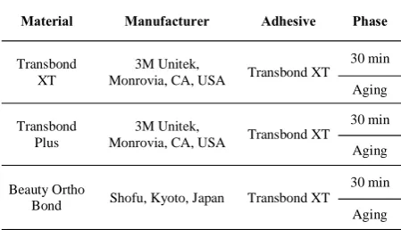
Material Manufacturer Adhesive Phase
Transbond XT
3M Unitek,
Monrovia, CA, USA Transbond XT 30 min
Aging
Transbond Plus
3M Unitek,
Monrovia, CA, USA Transbond XT 30 min
Aging
Beauty Ortho
Bond Shofu, Kyoto, Japan Transbond XT
30 min
Aging
A thin coat of Transbond XT was applied to the enamel, which was gently air-dried from a 30-cm distance and was cured for 10 seconds. Transbond XT adhesive was ap-plied to the bracket base, and the bracket was placed on the buccal surface of the tooth in the middle of the clinical crown, exactly on the height of contour. Care was taken to ensure the placement of the bracket slot perpendicu-lar to the long axis of the tooth. The bracket was pressed on the tooth surface by using a gauge with a 300-g load and was light-cured for 40 seconds (10 seconds from each side) by using OptiLux 501 light-curing unit (Kerr, Or-ange, CA, USA) with 600 mW/cm2 intensity.
In group 2, Transbond Plus adhesive (3M Unitek, Monrovia, CA, USA) was used. The SEP was applied to the enamel surface and was gently air-dried from a 30-cm distance. The adhesive was applied to the bracket sur-face, the same as in group 1, and was light-cured.
In group 3, Beauty Ortho Bond adhesive (Shofu, Kyoto, Japan) was used. One drop of primer A was mixed with one drop of primer B and was applied to enamel surface for 3 sec-onds. The next steps were similar to those de-scribed for group 1. For correct mounting of teeth into cylindrical molds, a 0.18×0.25-mm stainless steel wire, parallel to the horizon and fixed at a specific height, was passed through the brackets, and then, the roots of the teeth (with brackets and wires on their surfaces) were mounted and fixed into a self-curing acrylic resin.
The bracket slot was parallel to the horizon, and during the SBS testing, the load was applied perpendicular to the bracket base.
The specimens in each group were divided into two subgroups of 22 each. The bond strength in one subgroup was measured after 30 minutes and in the other after aging. The specimens in the aging subgroup were stored in artificial saliva at 37°C for 3 months, and then, they were subjected to thermocycling for 6000 cycles between 5-55°C according to the method described by Faltermeier et al.(18) The SBS was measured in a Zwick
uni-versal testing machine (Instron M500, Ulm, Ger-many) with a 1-kN load cell attached to a metal rod at a crosshead speed of 0.5 mm/minute. The teeth were fixed on the base of the device, and the shear load was applied by the 30° beveled tip of the metal rod at the bracket-tooth interface along the incisogingival axis and parallel to the tooth surface until fracture (Figure 1).
Figure 1. Specimen under shear bond strength test-ing
The amount of the applied load in Newton (N) was divided by the bracket’s cross-sectional area in mm2 (12.33) to calculate the bond strength in
Megapascal (MPa).
The Adhesive Remnant Index (ARI) was evaluated according to the method described by Artun and Bergland (19) and by using a
digital stereomicroscope (Motic
DMW-143-Triple Output, Hong Kong) at ×10
magnification and was scored from 0 to 3
based on the amount of adhesive remnant
on the enamel surface:
0: No adhesive remnant on the enamel
surface.
1: Less than 50% resin on the enamel
surface.
2: More than 50% resin on the enamel
surface.
3: The entire adhesive left on the enamel
surface.
(19)After debonding, adhesive
remnants were removed from the enamel
surface by using a carbide bur (Komet
FGH 22 GK016, Germany) until no
ad-hesive remnant was seen on the surface
with the naked eye.
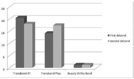
figure 2-Primary and Secondary shear bond strengths in the three group 30 minutes after bonding the orthodontic brackets.
Bonding in each group was
repeat-ed as before. In the subgroups that had undergone the primary bond strength measurement after 30 minutes, the second bond strength measurement was done after 30 minutes. The same was applied to the subgroups that un-derwent aging. The ARI was evaluated again as well.
Preparation of the specimens for scanning electron microscopy (SEM):
Figure 3. Primary and secondary shear bond strengths in the three groups after aging
The crowns were cut from the roots. The enamel surface was prepared with a phosphoric acid-etch-ant in the first specimen and with the two SEPs in the second and third specimens. Acetone was used for eliminating the primer in the SEPs. After debonding and removal of resin remnants with the carbide bur, the next three specimens were prepared the same as the first three specimens, were stored in an incubator for 10 days, and were observed under the SEM at ×2000 magnification (Figure 2).
Statistical analysis:
Two-way analysis of variance (ANOVA) was used to assess the variables related to the adhesive system, the phases of debonding, and the time of debonding, which indicated significant interac-tions between the variables. Thus, one-way ANO-VA was applied whenever required followed by Scheffe post-hoc
test for multiple comparisons.
Kruskal-Wallis test or Mann-U-Whitney and
Dunn's tests, as
a post-hoc test, were also applied for the comparison of the ARI and the mode of fail-ure among the three adhesive groups, between the first and second debonding processes, and after 30 minutes and thermocycling.Results:
Univariate ANOVA revealed significant differences in the first debonding among the three adhesives and also between different debonding time points (P<0.05). However, the interaction of the adhesives and differ-ent time points was not significant (P=0.06), which means that the differences among the adhesives 30 minutes after debonding were similar to those after aging. As observed in Table 2, the post-hoc test demonstrated that in the primary bond strength testing, the SBS of Transbond XT was significantly higher than that of Transbond Plus.
Table 2. Comparison of the primary shear bond strength of the adhesives
Primer Primer Std.
Error Sig.
XT Plus 1.25347 .000
Beauty Ortho Bond 1.00499 .000
Plus XT 1.25347 .000
Beauty Ortho Bond 1.06953 .000
Beauty Ortho Bond XT 1.00499 .000
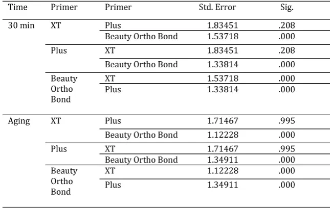
The SBS of Transbond Plus was also significant ly higher than that of Beauty Ortho Bond. Univariate ANOVA showed significant differenc-es among the adhdifferenc-esivdifferenc-es at different time points and in the interaction of the adhesives and the time points in the second debonding (P<0.05). The differences among the adhesives are shown in Table 3 (the post-hoc test).
At 30 minutes after debonding, the bond strengths of Transbond XT and Transbond Plus were significantly higher than that of Beauty Or-tho Bond, whereas the bond strengths of Trans-bond XT and TransTrans-bond Plus were not signifi-cantly different (P=0.208).
The situation was the same after thermocy-cling. However, it should be noted that the chang-es in the bond strengths of Transbond XT and Transbond Plus were not significant (P=0.995). How
ever, the obtained values were more similar to one another compared to the values after 30 min-utes; this can explain the significant interaction of the two variables.
Moreover, each adhesive was separately evalu ated:
Transbond XT:
The secondary bond strength of Transbond XT was not significantly different from its prima-ry bond strength (P=0.43). The interaction of the first and second bondings and the time points was not significant either (P=0.23). In other words, the secondary bond of Transbond XT was not sig-nificantly different from its primary bond after 30 minutes or after aging. The interaction with the time points was not significant either (P=0.26). The bond strength of Transbond XT was not sig-nificantly different after 30 minutes and after ag-ing.
Transbond Plus:
The secondary bond strength of Transbond Plus was significantly greater than its primary bond strength (P<0.05). The interaction of the bonding phase and the time points was not signif-icant (P=0.25). After 30 minutes and after aging, the secondary bond strength of Transbond Plus was higher than its primary bond strength. The interaction with the time points was not signifi-cant (P=0.59). The bond strength of Transbond Plus after aging was not significantly different from the value after 30 minutes. Thus, Trans-bond XT and TransTrans-bond Plus maintain their Trans-bond
strength after aging. Beauty Ortho Bond:
The secondary bond strength of Beauty Ortho Bond after 30 minutes was significantly higher than its primary bond strength; however, after aging, no significant difference was found be-tween the primary and secondary bond strengths (both were too low).
The bond strength of Beauty Ortho Bond sig-nificantly decreased after thermocycling. Thus, for this adhesive, the effects of time (P<0.05) and debonding phase (P<0.05) and the interaction be tween them were significant (P<0.05).
Comparison of the variables of time and primer in the first and second bondings is shown in Figures 2 and 3.
ARI:
Dunn’s test demonstrated that in the primary and secondary bondings, the ARI in the Trans-bond XT group was significantly higher than that in
the Beauty Ortho Bond group after 30 minutes and after aging (P<0.017). After aging, Trans-bond Plus showed a higher ARI than Beauty Or-tho Bond (P<0.017). However, these two SEPs showed no significant difference 30 minutes after the first (P=0.06) and second (P=0.77) bondings. The ARI was not significantly different in the first and second bondings and at different time points in the Transbond Plus and Transbond XT groups (P>0.017).
Comparison of the 30-minute and post-aging time points in the three groups of adhesives with Mann-U-Whitney test showed no significant dif-ferences in this respect after the first and sec-ond bsec-ondings (P>0.017), which means that the amounts of adhesive remnants on the enamel were not significantly different after 30 minutes or post-aging.
finding is in complete agreement with the bond strength test results and SEM images. However, after thermocycling, the bond strength of this adhesive was so weak. There was little adhesive remnant on the enamel surface after the first and second debondings, and the values were not sig-nificantly different (P=0.705).
Discussion:
Cleaning and preparation of enamel surface for new bracket bonding not only increase the clinician’s working time but also traumatize the enamel. Cleaning the composite remnants from enamel surface by the use of burs and rotary in-struments results in the loss of 11.3-19.2 μm of the enamel surface. Re-etching leads to further loss of 10-50 μm of the enamel and changes the surface structure to the depth of 200 μm.(20,21)
The shear rebond strength tests aim to im-prove the bonding system, decrease the damage to enamel, and reduce the clinical working time. For rebonding, in addition to a conventional ad hesive (Transbond XT), two SEPs, namely Trans-bond Plus and Beauty Ortho Bond were used in this study. The latter has the ability to release and uptake fluoride.(22) For a comprehensive
evalua-tion of these adhesives, aging was done before and after debonding of brackets.
Table 3. Comparison of the secondary shear bond strength of the adhesives at two time points
The results showed that in primary bond-ing, Transbond XT with a separate etchant had a higher bond strength than Transbond Plus which is a SEP. Transbond Plus also showed a higher bond strength than Beauty Ortho Bond. These results are in accord with the findings of Aljubouri et al (23)and Grubisa et al (24)who
be-lieved that adhesives with separate etchants have a higher bond strength than SEPs. The authors believe that the higher bond strength of total-etch adhesives is attributed to the greater penetration of resin into the enamel demineralized through separate-etching. SEPs form a hybrid layer and decrease the penetration of resin tags due to the higher pH of the acid and simultaneous etching and priming.(23,24)These results are in contrast to
those of Scougall Vilchis et al (25)and Iijima et
al (9) who reported the bond strength of SEPs to
In comparison of the two SEPs, Transbond Plus showed a higher bond strength than Beauty Ortho Bond, which confirms the findings of Endo et al
(22)and Iijima et al. (9)Although these two
adhe-sives are both SEPs, as observed in SEM images, Beauty Ortho Bond adhesive, due to a higher pH than Transbond Plus, creates less demineraliza-tion and a smoother enamel surface, which justify its weak bond to enamel. However, it should be noted that it has a bond strength of 6 MPa, which is within the clinically acceptable range.(26,27)
Our study showed that the bond strength of Transbond XT adhesive was not significantly different in primary and secondary bondings. This result is in accord with the findings of Endo et al,(22)Nicolas et al,(13) and Grunheid and
Larson,(28) and in contrast with the findings of
Bishara et al who reported that there was a sig-nificant decrease in the SBS of Transbond XT be-tween the debonding sequences 1 and 2.(29)
An interesting point is that Transbond Plus had a higher strength in the secondary bond than in the primary bond, equal to that of Transbond XT and the conventional bonding system. Beauty Ortho Bond had a lower bond strength compared to the mentioned two adhesives. However, its secondary bond strength was greater than its pri-mary bond strength.
The intact enamel surface is rich in minerals and has a greater fluoride content than ground enamel. After tooth eruption, some changes oc-cur in the enamel’s outer surface, and the saliva saturated with calcium phosphate hypermin-eralizes the enamel. Fluoride ions convert hy-droxyapatite to fluoroapatite.(30) Thus, the
prism-less enamel may prevent the penetration of SEPs, which may leave some enamel areas unetched, decreasing the penetration of resin into the micr-oporosities of the intact enamel. (31)Resin tags are
short and barely detectable. They are structurally incomplete. Since bond strength is due to micro-mechanical retention, a decreased resin penetra-tion decreases the bond strength.(32) In a
compre-hensive study by Kanemura et al in 1999, SEPs showed greater penetration into prepared enamel compared to intact enamel.(33) By removing the
superficial fluoride-rich enamel, the penetration of SEPs was enhanced, leading to their greater bond strength to a more porous surface.(33)
In 2010, Karan et al quantitatively evalu-ated the enamel roughness after debonding by
an atomic force microscope (AFM) and showed that the use of a tungsten carbide bur for remov-al of adhesive remnants caused enamel surface roughness.(34)In our study, a low-speed tungsten
carbide bur was used for removal of adhesive remnants. This bur also removes a small amount of fluoride-rich enamel in addition to adhesive remnants, enhancing the penetration of SEPs and increasing the secondary bond strength. This explains the higher secondary bond strength of Beauty Ortho Bond and Transbond Plus, which are both SEPs, compared to their primary bond strengths. This finding is in accord with that of Montasser et al.(14)
However, this finding is in contrast to that of Nicolas et al (13) and Endo et al (22)who reported
that the secondary bond strength was not signifi-cantly different from the primary bond strength. Such differences in bond strength may be attrib-uted to the technique of the operator and to the methodology of the study. Sample size may also play a role in this regard. Nicolas et al(13) used
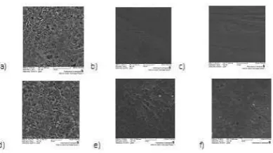
bo-vine teeth, which are different from human teeth. Figure 4 shows the SEM images of the enamel surface; a, b, and c show enamel surfaces before the first bond, while d, e, and f show them before the second bond. As expected and observed, the porosity of enamel was greater before the second bond because the enamel surface was ground by the carbide bur for removal of adhesive remnants. Figures 4a and 4d show the enamel surface after etching with 37% phosphoric acid (conventional technique). The clear etched pattern and honey-combing are evident. Figures 4b and 4e show the enamel surface structure after treatment with Transbond Plus SEP. The etched pattern is irregu-lar and less prominent in the second bond com-pared to the first bond. Figure 4c and 4f show the enamel surface treated with Beauty Ortho Bond. The surface is smooth without adequate reten-tion, explaining a weak enamel bond.
Another factor influencing the bond strength is the adhesive remnants on the enamel sur-face. Resin remnants decrease the porosity and roughness of enamel surfaces (35,36) and can
form a chemical bond with the new resin.(14) In
af-ter debonding was evaluated under the SEM by using silicon (Si) and carbon (C) mapping.
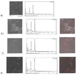
Figure 5 shows that although resin remnants were removed by the carbide bur, some resin remnants were still present on the enamel sur-face. In Figure 5a, the etched enamel before bonding was searched for elements. The dia-gram only shows the presence of calcium (Ca) and phosphate (P). Figures 5b, 5c, and 5d il-lustrate the enamel surface after debonding of Transbond XT, Transbond Plus, and Beauty Or-tho Bond, respectively, and after resin removal by the carbide bur. In all three groups, in addi-tion to Ca and P, C and Si were found in dif-ferent percentages; this indicates the presence of resin remnants which are invisible to the naked eye.
In this study, in order to simulate the clini-cal setting, the bond strength was measured 30 minutes after bonding (the time of load appli-cation to brackets in the clinic) and after aging. Many adhesives undergo structural changes due to thermal alterations after exposure to the oral cavity. A comprehensive study should compare the bond strength while taking these conditions into account.(16,37,38)
Figure 4. Evaluation of enamel surface with scanning electron microscopy (SEM) at ×2000 magnification. (a) Etched with phosphoric acid, (b) Treated with Trans-bond Plus, (c) Treated with Beauty Ortho Bond before the first bond. (d) Etched with phosphoric acid, (e) Treated with Trans-bond Plus, (f) Treated with Beauty Ortho Bond before the second bond.
The results of our study demonstrated that the bond strengths of Transbond Plus and Transbond XT, whether in the first or second bond, were not significantly different at 30 minutes and after ag-ing. However, the bond strength of Beauty Ortho Bond significantly decreased after aging follow-ing both the first and second bonds. These results are in agreement with those of Yuasa et al in 2009 regarding Transbond XT and Transbond Plus.(16)
Yuasa et al mentioned leakage at the enamel-adhesive interface following aging in the Beauty Ortho Bond group.(16)
The decreased bond strength in the Beauty Ortho Bond group after aging may be attributed to the hydrolysis and degradation of interface components.(39)Also, water sorption may weaken
the polymer matrix.(40)
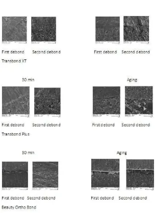
Figure 6 shows SEM images of the enamel interface. The gap formed at the resin-enamel interface after aging in the Beauty Ortho Bond group can be seen following the first and second bonds, explaining the significant reduc-tion in the bond strength.
Evaluation of the ARI:
In restorative dentistry, the highest bond strength to tooth structure is desirable; however, in orthodontics, the bond strength should be high enough to maintain the bracket on the tooth sur-face and at the same time has to be low enough to allow for easy cleaning of enamel following removal of brackets.(4,9,10)
The results of our study demonstrated that the adhesive remnants on the enamel after 30 min-utes and after aging were significantly greater in the Transbond XT group than in the Beauty Ortho Bond group.
The weak bond of Beauty Ortho Bond adhesive leads to bond failure at the adhesive-enamel in-terface, whereas Transbond XT with separate etching forms a strong bond to enamel, and bond failure mostly occurs at the bracket-adhesive interface.(9,22) Moreover, based on the results,
the Transbond Plus group, indicating the weak bond and insufficient retention of Beauty Ortho Bond after aging.
Transbond XT and Transbond Plus were not significantly different in terms of adhesive rem-nants, and this finding is in agreement with the results of previous studies.(9,13,14,22,27) Transbond
Plus is a SEP that forms a bond equal in strength to that of Transbond XT.
Transbond XT and Transbond Plus were not significantly different in terms of the ARI in com-paring the first and second bonds; however, in the Beauty Ortho Bond group, at 30 minutes after debonding, the ARI following the second bond was greater than that after the first bond. This finding can be attributed to the increased enamel roughness due to resin removal by a carbide bur leading to a significant increase in the second bond strength. However, the Beauty Ortho Bond
Figure 5. Searching for elements on the enamel surface by scanning electron microscopy (SEM) at ×2000 magnification. (a) Etched surface. (b) Enamel surface after the removal of Transbond XT resin by a carbide bur. (c) Enamel surface after the removal of Transbond Plus resin by a carbide bur. (d) Enamel surface after the removal of Beauty Ortho Bond resin by a carbide bur. Red points are Carbon and green points are Silicon.
bond strength However, the Beauty Ortho Bond group showed no significant difference in the bond strength in comparing the two bonds after aging, and practically no adhesive was left on the enamel in the two groups after debonding.
Conclusion:
SEPs are recommended for secondary bonding in the clinical setting due to a de-creased chair time, less damage to enamel, and an adequate bond strength.
Acknowledgement:
References:
1. Bishara SE, VonWald L, Laffoon JF, Warren JJ. Ef-fect of a self-etch primer/adhesive on the shear bond strength of orthodontic brackets. Am J Orthod Dentof-acial Orthop. 2001 Jun;119(6):621-4.
2. Cacciafesta V, Sfondrini MF, De Angelis M, Scrib-ante A, Klersy C. Effect of water and saliva contami-nation on shear bond strength of brackets bonded with conventional, hydrophilic, and self-etching primers. Am J Orthod Dentofacial Orthop. 2003 Jun;123(6):633-40.
3. Wickwire NA, Rentz D. Enamel pretreatment: a critical variable in direct bonding systems. Am J Or-thod. 1973;64:499-512.
4.Fleming PS, Johal A, Pandis N. Self-etch primers and conventional acid-etch technique for orthodontic bonding: a systematic review and meta-analysis. Am J Orthod Dentofacial Orthop. 2012 Jul;142(1):83-94. 5. Fitzpatrick DA, Way DC. The effects of wear, acid etching and bond removal on human enamel. Am J Orthod. 1977 Dec;72(6):671-81.
6. Chu CH, Ou KL, Dong DR, Huang HM, Tsai HH, Wang WN. Orthodontic bonding with self-etching primer and self-adhesive systems. Eur J Orthod. 2011 Jun;33(3): 276-81.
7. Buonocore MG. A simple method of increasing the adhesion of acrylic filling materials to enamel surfac-es. J Dent Rsurfac-es. 1955 Dec;34(6):849-53.
8. Buyukyilmaz T, Usumez S, Karaman AI. Effect of self-etching primers on bond strength--are they reli-able? Angle Orthod. 2003 Feb;73(1):64-70.
9. lijima M, Ito S, Yuasa T, Muguruma T, Saito T, Mizoguchi I. Bond strength comparison and scanning electron microscopic evaluation of three orthodontic bonding systems. Dent Mater J. 2008 May;27(3):392-9.
10. Hosein I, Sherriff M, Ireland AJ. Enamel loss dur-ing bonddur-ing, debonddur-ing, and cleanup with use of a self-etching primer. Am J Orthod Dentofacial Orthop. 2004 Dec;126(6):717-24.
11. Cehreli ZC, Kecik D, Kocadereli I. Effect of self-etching primer and adhesive formulations on the shear bond strength of orthodontic brackets. Am J Orthod Dentofacial Orthop. 2005 May;127(5):573-9.
12. dos Santos JE, Quiocab J, Loguercioc AD, Reisc A. Six-month bracket survival with a self-etch adhe-sive. Angle Orthod. 2006;76(5):863-8.
13.Nicolas AI, Vicente A, Luis LA. The in vitro effect of repeated bonding on the shear bond strength with different enamel conditioning procedures. Eur J Or-thod. 2010 Jun;32(3):291-6.
14.Montasser MA, Drummond JL, Evans CA. Rebond-ing of orthodontic brackets. Part I, a laboratory and clinical study. Angle Orthod. 2008 May;78(3):531-6.
15.Bishara SE, Vonwald L, Laffoon JF, Warren JJ. The effect of repeated bonding on the shear bond strength of a composite resin orthodontic adhesive. Angle Or-thod. 2000 Dec;70(6):435-41.
16.Yuasa T, Iijima M, Ito S, Muguruma T, Saito T, Mizoguchi I. Effects of long-term storage and thermo-cycling on bond strength of two self-etching primer adhesive systems. Eur J Orthod. 2010 Jun;32(3):285-90.
17.Turk T, Elekdag-Turk S, Isci D. Effects of self-etching primer on shear bond strength of orthodon-tic brackets at different debond times. Angle Orthod. 2007 Jan;77(1):108-12.
18.Faltermeier A, Behr M, Müssig D. A comparative evaluation of bracket bonding with 1-, 2-, and 3-com-ponent adhesive systems. Am J Orthod Dentofacial Orthop. 2007 Aug;132(2):144.e1-5.
19. Artun J, Bergland S. Clinical trials with crys-tal growth conditioning as an alternative to ac-id-etch enamel pretreatment. Am J Orthod. 1984 Apr;85(4):333-40.
20. Legler LR, Retief DH, Bradley EL. Effects of phosphoric acid concentration and etch duration on enamel depth of etch: an in vitro study. Am J Orthod Dentofacial Orthop. 1990 Aug;98(2):154-60.
21. Zentner A, Duschner H. Structural changes of acid etched enamel examined under confocal laser scanning microscope. J Orofac Orthop. 1996 Aug;57(4):202-9. 22. Endo T, Ozae R, Shinkai K, Aoyagi M, Kurok-awa H, Katoh Y, et al. Shear bond strength of brackets rebonded with a fluoride-releasing and -recharging adhesive system. Angle Orthod. 2009 May;79(3):564-70.
23. Aljubouri YD, Millett DT, Gilmour WH. Labora-tory evaluation of a self-etching primer for orthodon-tic bonding. Eur J Orthod. 2003Aug;25(4):411-5 24. Grubisa HS, Heo G, Raboud D, Glover KE, Ma-jor PW. An evaluation and comparison of orthodon-tic bracket bond strengths achieved with self-etch-ing primer. Am J Orthod Dentofacial Orthop. 2004 Aug;126(2):213-9.
25. Scougall Vilchis RJ, Yamamoto S, Kitai N, Hotta M, Yamamoto K. Shear bond strength of a new flu-oride-releasing orthodontic adhesive. Dent Mater J. 2007 Jan;26(1):45-51.
26. Pandis N, Eliades T. A comparative in vivo assess-ment of the long-term failure rate
of 2 self-etching primers. Am J Orthod Dentofacial Orthop. 2005 Jul;128(1):96-8.
27. Rüger D, Harzer W, Krisjane Z, Tausche E. Shear bond strength after multiple bracket bonding with or without repeated etching. Eur J Orthod. 2011 Oct;33(5):521-7.
Orthod. 2014 Sep;3(3):102-5.
29. Bishara SE1, Laffoon JF, Vonwald L, Warren JJ. The effect of repeated bonding on the shear bond strength of different orthodontic adhesives. Am J Or-thod Dentofacial Orthop. 2002 May;121(5):521-5. 30. Brudevold F. A study of the phosphate solubil-ity of the human enamel surface. J Dent Res. 1948 Jun;27(3):320-9.
31.Gwinnett AJ. The ultrastructure of the “prism-less” enamel of permanent teeth. Arch Oral Biol. 1967 Mar;12(3):381-6.
32.Di Hipólito V, de Goes MF, Carrilho MR, Chan DC, Daronch M, Sinhoreti MA. SEM evaluation of contemporary self-etching primers applied to ground and unground enamel. J Adhes Dent. 2005 Au-tumn;7(3):203-11
33.Kanemura N, Sano H, Tagami J. Tensile bond strength to and SEM evaluation of ground and intact enamel surfaces. J Dent. 1999 Sep;27(7):523-30. 34.Karan S, Kircelli BH, Tasdelen B. Enamel sur-face roughness after debonding. Angle Orthod. 2010 Nov;80(6):1081-8.
35.Leas TJ, Hondrum S. The effect of rebonding on the shear bond strength of orthodontic brackets-a comparison of two clinical techniques. Am J Orthod Dentofacial Orthop. 1993 Feb;103(2):200-1.
36.Khosravanifard B, Nemati-Anaraki S, Nili S, Ra-khshan V. Assessing the effects of three resin removal methods and bracket sandblasting on shear bond strength of metallic orthodontic brackets and enamel surface. Orthod Waves. 2011 Mar;70(1):27-38. 37. Bishara SE1, Ostby AW, Laffoon JF, Warren J. Shear bond strength comparison of two adhesive sys-tems following thermocycling. A new self-etch primer and a resin-modified glass ionomer. Angle Orthod. 2007 Mar;77(2):337-41.
38.Trites B, Foley TF, Banting D. Bond strength com-parison of 2 self-etching primers over a 3-month stor-age period. Am J Orthod Dentofacial Orthop. 2004 Dec;126(6):709-16.
39.De Munck J, Van Landuyt K, Peumans M, Poitevin A, Lambrechts P, Braem M, et al. A critical review of the durability of adhesion to tooth tissue: methods and results. J Dent Res. 2005 Feb;84(2):118-32.