A Thermodynamic Study of the Interaction
between Urease and Copper Ions
A.A. Saboury,
1,*E. Poorakbar-Esfahani,
2and G. Rezaei-Behbehani
31
Institute of Biochemistry and Biophysics, University of Tehran, Tehran, Islamic Republic of Iran
2
Biology Department, Payam Noor University, Tehran, Islamic Republic of Iran
3
Chemistry Department, Imam Khomeini International University, Qazvin, Islamic Republic of Iran
Received:1November2009/Revised:14December2009/Accepted:24December2009
Abstract
A thermodynamic study of copper ions by jack bean urease (JBU) was carried
out at two temperatures of 27 and 37
°
C in Tris buffer (30 mM; pH=7.0) using an
isothermal titration calorimetry. There is a set of twelve identical and
non-interacting binding sites for copper ions. The intrinsic dissociation equilibrium
constant and the molar enthalpy of binding are 285 µM and −15.2 kJ/mol at 27
°
C
and 346 µM and −14.6 kJ/mol at 37
°
C, respectively. The molar entropy of
binding is 17.2 J K
-1mol
-1at 27
°
C and +19.1 J K
-1mol
-1at 37
°
C. Hence, the
binding process of copper ion to JBU is not only enthalpy driven but also it is
entropy driven, which the role of entropy driven should be more effective by
increasing the temperature.
Keywords: Urease; Copper ion; Isothermal titration calorimetry; Enthalpy of binding; Entropy of Binding
* Corresponding author, Tel.: +98(21)66409517, Fax: +98(21)66409517, E-mail: [email protected] Introduction
Jack bean urease (urea amidohydrolase; E.C. 3.5.1.5) is the first crystallized enzyme[1] and also is the first enzyme shown to contain nickel [2-3]. The crystal structure of urease has not been determined yet. The best resolution obtained at 3.5 Å only allowed to assign the octahedral crystals of this urease to a cubic spacegroup [4]. Jack bean urease has six identical subunits. Each subunit consist of a single kind of polypeptide chain containing 840 amino acid residue with relative molecular mass of 90770, excluding the two nickel ions per subunit[5]. Hence, a mass of the hexamer urease, including 12 nickel ions, being 545.340 kDa5 (590 kDa by a sedimentation method [6]). The subunit of urease from microorganisms appear to be
catalytic nucleophile [17].
Ureases are inhibited by a number of compounds. The study of urease inhibitors may have medical or agronomic significance, as well as providing insight into the urease catalytic mechanism [21-22].Substrate urea, product ammonium ions, and substrate analogues are weak inhibitors of urease [21, 23-24]. Thiols inhibit urease competitively in their thiolate anion form R-S− [22]. Amides and esters of phosphoric acid are also slowbinding inhibitors of urease, classified as the strongest inhibitors. Boric and boronic acids are rapidly binding urease inhibitors, comparatively weak. Fluoride ion was found to be a competitive inhibitor for jack bean urease.25 There are some reports on the inhibition of urease by bismuth compounds, which are of medical importance since are widely used as bactericidal agents [26-29]. The inhibition of ureases by quinones has been mainly tested for their potential application with urea fertilizers [29-30].
Inhibition of urease by heavy metal ions is important not only in view of heavy metal ion pollution, appropriate levels of urease activity in agricultural soils may be endangered, but also this inhibition may be exploited in constructing urease inhibition-based sensing systems [31-33] for in situ and real time determination of trace levels of the ions, e.g. in environmental monitoring, food control and biomedical analysis. Heavy metal ions inhibit both plant [31,34-38] and bacterial ureases[39-40] at the following order of effectiveness [36,38]:
Hg2+≈ Ag+ > Cu2+ >> Ni2+ > Cd2+ > Zn2+ > Co2+ > Fe3+ > Pb2+ > Mn2+
Hg2+, Ag+ and Cu2+ ions nearly always listed as the strongest inhibitors [31-36,38,41]. The inhibition has been habitually ascribed to the reaction of the metal ions with the thiol groups of the enzyme [33,41-42]. However, both copper and silver ions coordinate to nitrogen- (histidine) and possibly oxygen- (aspartic and glutamic acids) containing functional groups in urease [43-44]. Notwithstanding heavy metal ion binding to urease is important, there is not a comprehensive binding study in this case. Here, we applied isothermal titration microcalorimetry as a powerful tool for studying of copper ion binding to jack bean urease, which all thermodynamic parameters for the binding process can be found.
Materials and Methods
Materials
Jack bean urease (JBU) was obtained from Sigma
Chemical Co. Copper nitrate was obtained from Merck. The buffer solution used in the experiments was 30 mM Tris using double-distilled water, pH=7.0, which was obtained from Merck. Experiments were carried out at two temperatures of27 and 37 °C.
Methods
The experiments were performed with the 4-channel commercial microcalorimetric system, Thermal Activity Monitor 2277 (Thermometric, Sweden). Each channel is twin heat conduction calorimeter (multijuction thermocouple plates) positioned between the vessel holders and the surrounding heat sink. Both, sample and reference vessels were made from stainless steel. The limited sensitivity for the calorimeter is 0.1 µJ Copper ion solution (10 mM) was injected into the calorimetric titration vessel, which contained 1.8 ml JBU, 4 µM (2.2 mg/ml), in Tris buffer (30 mM), pH=7.0, using a Hamilton syringe. Thin (0.15 mm inner diameter) stainless steel hypodermic needles, permanently fixed to the syringe, reached directly into the calorimetric vessel. Injection of copper ion solution into the perfusion vessel was repeated 30 times and each injection included 20 µl copper ion solution. The calorimetric signal was measured by a digital voltmeter that was part of a computerized recording system. The heat of injection was calculated by the “Thermometric Digitam 3” software program. The heat of dilution of the copper ion solution was measured as described above except JBU was excluded. Also, the heat of dilution of the protein solution was measured as described above except that the buffer solution was injected to the protein solution in the sample cell [45]. The enthalpies of copper ion and protein solutions dilution were subtracted from the enthalpies of copper ion solutions in JBU solutions. The microcalorimeter was frequently calibrated electrically during the course of the study.
Results and Discussion
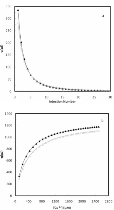
The raw data obtained from ITC at two temperatures of 27°C and 37 °C are shown in Figure 1. Figure 1a shows the heat of each injection and Figure 1b shows the cumulative heat at each total concentration of copper ion, [Cu2+]t. For a set of identical and independent binding sites, we have before shown different methods of ITC data analysis[45]. For a set of identical and independent binding sites, we have previously introduced the following equation[45-46]:
d
0 0
max
K
Δq Δq 1
M ( ) L
Figure 1. (a) The heat of copper ion binding to JBU for 30 automatic cumulative injections, each of 20 μl, 10 mM of the copper nitrate solution, into sample cell containing 1.8 ml 4
μM protein solution at 300 K (▲) and 310 K (∆). (b) The total cumulative heat of binding vs. total concentration of copper ion calculated from Fig. 1a.
where g is the number of binding sites, Kd is the dissociation equilibrium constant, M0 and L0 are total concentrations of biomacromolecule and metal ion, respectively, Δq=qmax–q, q represents the heat value at a certain L0 and qmax represents the heat value upon saturation of all biomacromolecule. If q and qmax are calculated per mole of biomacromolecule then the standard molar enthalpyof binding for each binding site (ΔH°) will be ΔH qmax
g
° = . According to the equation (1), a plot of (Δq/qmax)M0 vs. (Δq/q)L0 should be a linear plot by a slope of 1/g and the vertical-intercept of Kd/g, which g and Kd can be obtained. The related plot for the binding of Cu2+ ions by JBU is shown in Figure 2. The linearity of the plot has been examined by different estimated values for qmax to find the best value for the
Figure 2. (a) The best linear plot of 0 max
Δq
( )M
q vs. 0
Δq
( )L
q ,
according to Eq. (1), using values of −1303 µJ (■), −1308 (♦), −1313 µJ (○), −1318 µJ (∆), −1323 µJ (●) at 300 K for qmax to
obtain the best correlation coefficient value (R2=0.999) for a
linear plot. (b) The best linear plot of 0 max
Δq
( )M
q vs. 0
Δq
( )L
q ,
according to Eq. (1), using values of −1251 µJ (■), −1256 (♦), −1261 µJ (○), −1266 µJ (∆), −1271 µJ (●), at 310 K for qmax to
obtain the best correlation coefficient value (R2=0.999) for a
linear plot.
correlation coefficient (near to one). The best linear plot with the correlation coefficient (R2) value (near to one) was obtained using −1313 μJ and −1261 μJ (equal to −182.4 kJ/mol and −175.1 kJ/mol) at 27 °C and 37
°C, respectively, for qmax (Fig. 2). The lack of a suitable value for qmax to obtain a linear plot of 0
max Δq
( ) M
q vs.
0 Δq ( ) L
g=12, therefore, gives ∆H° = −15.2 kJ/mol and
−14.6 kJ/mol at 27 °C and 37 °C, respectively.
The molar enthalpy of each binding site (ΔH°) and its dissociation equilibrium constant (Kd) in a set of
biomacromolecule binding sites can also be obtained via a simple graphical nonlinear fitting method using the following equation [45, 47-48].
{
2 1/ 2}
d
ΔH° =1/ A (B K)+ −⎡⎣(B K )+ −C⎤⎦ (2)
A, B and C are constants in each injection, which have been defined as follows:
A=V/ 2 q B gM= 0+L0 C 4gM L= 0 0 (3) where V is the volume of the reaction solution in the calorimetric sample cell in each injection step. Equation (2) contains two unknown parameters, Kd and ∆H°. A series of reasonable values for Kd is inserted into equation (2) and corresponding amounts for ∆H° are calculated and the graph ∆H° versus Kd is constructed. Curves of all titration steps will intersect in one point, which represents true amounts for ∆H° and Kd. The plots of ∆H° versus Kd, according to Eq. (2) for all injections are shown in Figure 3. The intrinsic dissociation equilibrium constant and the molar enthalpy of binding were obtained 285 μM and −15.2
kJ/mol at 27 °C and 346 μM and −14.6 kJ/mol at 37
°C, respectively (see Figure 3). These results are identical with results obtained by previous method described above.
To compare all thermodynamic parameters in metal binding process for JBU, the change in standard Gibbs free energy (ΔGo) should be calculated according to the equation (4), which its value can use in equation (5) for calculating the change in standard entropy (ΔSo) of binding process.
ΔGo = −RT ln Ka (4)
ΔGo = ΔHo− TΔSo (5)
where Ka is the association binding constant (the inverse of the dissociation binding constant, Kd). The Ka values are obtained 3509 and 2891 M-1 at 27 °C and 37 °C, respectively. Hence:
ΔGo = −20.4 kJ/mol ΔSo= +17.2 J/ K mol (at 27 °C)
ΔGo = −20.6 kJ/mol ΔSo= +19.1 J/ K mol (at 37 °C) It means that the binding process is spontaneous resulted by not only enthalpic but also entropic driven.
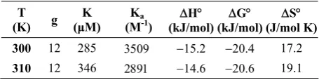
All thermodynamic parameters for the interaction between JBU and copper ion at two temperatures of 27 and 37 °C have been summarized in Table 1. There is a
set of twelve identical and non-interacting binding sites for copper ions to JBU. The binding process is exothermicat both temperatures.The binding process of is not only enthalpy driven but also it is entropy driven. The role of entropy driven in the binding process should be more effective by increasing the temperature. The molar entropy of binding means that the difference between the entropy of Cu2+−JBU complex (SCu
−JBU)
Figure 3.∆H versus Kdfor all 30 injections at 300 K (a) and
310 K (b) using data in Fig.1b. The coordinates of the intersection point of the curves give the true values for ∆H and Kd.
Table1. Thermodynamic parameters of binding for copper ions to JBU obtained by ITC
T
(K) g (μK M) (MKa-1) (kJ/mol) ΔH° (kJ/mol)ΔG° (J/mol K)ΔS°
300 12 285 3509 −15.2 −20.4 17.2
and the entropy of native JBU (SJBU): ΔS= SCu−JBU −
SJBU. Hence, the disorder of the protein structure has been increased due to the binding of copper ions.
Acknowledgement
Financial support from the University of Tehran is gratefully acknowledged.
References
1. Sumner, J.B., The isolation and crystallization of the enzyme urease ,J. Biol. Chem., 69: 435–441, (1926). 2. Dixon, N.E.; Gazzole, C.; Blakeley, R. P.; Zerner, B.,
Jack bean urease (EC 3.5.1.5), A metalloenzyme.A simple biological role for nickel. J. Am. Chem. Soc., 97: 14, 4131–4133 (1975).
3. Mamiya, G.; Takishima, K.; Masakuni, M.; Kayumi, T.; Ogawa, K., Complete amino acid sequence of jack bean urease J. Protein Chem., 6:1, 55–59(1987).
4. Jabri, E.; Lee, M. H.; Hausinger, R. P.; Karplus, P.A. , Preliminary Crystallographic Studies of Urease from Jack Bean and from Klebsiella_Aerogenes. J. Mol. Biol., 227:3, 934–937(1992).
5. Takishima, K.; Suga, T.; Mamia, G., The structure of jack bean urease. The complete amino acid sequence, limited proteolysis and reactive cysteine residues. Eur. J. Biochem., 175:1, 151–165(1988).
6. Dixon, N. E.; Hinds, J. A.; Fihelly, A. K. ; Gazzola, C.; Winzor, D. J.; Blakeley, R. L.; Zerner, B., Jack bean urease (EC 3.5.1.5). IV. The molecular size and the mechanism of inhibition by hydroxamic acids. Spectrophotometric titration of enzymes with reversible nhibitors. Can. J. Biochem., 58:1-2, 1323–1334(1980). 7. Hausinger, R. P., Purification of a nickel-containing
urease from the rumen anaerobe Selenomonas ruminantium. J. Biol. Chem., 261: 17, 7866–7870(1986). 8. Christians, S.; Hausinger, R. P., Nickel-content of urease
from Bacillus pasteurii. Arch. Microbiol., 145: 1, 51–55 (1986).
9. Mobley, H. L. T.; Hausinger, R. P., Microbial ureases: significance, regulation, and molecular characterization. Microbiol. Rev., 53:1, 85–108 (1989).
10. Varner, J. E. The Enzymes, Vol 4, 2nd Edition, Academic Press, New York, 247-256(1960).
11. Fishbein, W. N.; Winter, T. S.; Davidson, J. D., Ureae catalysis. I. Stoichiometry, specificity and kinetics of a second substrate: Hydroxyurea. J. Biol. Chem., 240, 2402–2406 (1965).
12. Fishbein, W. N.; Carbone, P. P., Urease Catalysis. J. Biol. Chem., 240, 2407–2414 (1966).
13. Blakeley, R. L.; Hinds, J. A.; Kunze, H. E.; Webb, E.C.; Zerner, B. , Jack bean urease (EC 3.5.1.5). Demonstration of a carbamoyl-transfer reaction and inhibition by hydroxamic acids. Biochemistry, 8:5, 1991– 2000 (1969).
14. Fishbein, W. N., A simple, sensitive, and specific colorimetric assay for dihydroxyurea. Anal. Chim. Acta, 40:2, 269 –275 (1968).
15. Fishbein, W. N., Urease catalysis. 3. Stoichiometry, kinetics, and inhibitory properties of a third substrate: dihydroxyurea. J. Biol. Chem., 244: 5, 1188–1193(1969). 16. Gazzole, C.; Blakeley, R. L.; Zerner, B., On the substrate
specificity of jack bean urease (urea amidohydrolase, EC 3.5.1.5). Can. J. Biochem., 51:9, 1325–1330 (1973). 17. Dixon, N. E.; Riddles, P. W. C. Gazzole, R.L. Blakeley
and B. Zerner, Jack bean urease (EC 3.5.1.5). V. On the mechanism of action of urease on urea, formamide, acetamide, N-methylurea, and related compounds.,Can. J. Biochem., 58 :12 ,1335–1344(1980).
18. Fishbein, W. N., Formamide- minimum-structure
substrate for urease. Biochem. Biophys. Acta., 484:2, 433–442 (1977).
19. Benini, S.; Rypniewski, W. R.; Wilson, K. S.; Miletti, S.; Ciurli, S.; Mangani, S., A new proposal for urease mechanism based on the crystal structures of the native and inhibited enzyme from Bacillus pasteurii: why urea hydrolysis casts two nickels. Structure, 7:2, 205–216 (1999).
20. Karplus, P. A.; Pearson, M. A.; Hausinger, R. P., 70 years of crystalline urease: What have we learned?. Acc. Chem. Res., 30:8, 330–337(1997).
21. Krajewska, B., Ureases I. Functional, catalytic and kinetic properties: A review .J. Mol. Catalysis B: Enzymatic, 59:1-3, 9–21 (2009).
22. Todd, M. J.; Hausinger, R. P., Competitive inhibitors of Klebsiella aerogenes urease. Mechanisms of interaction with the nickel active site. J. Biol. Chem., 264:27, 15835– 15842(1989).
23. Saboury, A. A.; Moosavi-Movahedi, A. A. , A Simple Novel Method For Studying The Combined Inhibitory Effects of Ethylurea and N, N-Dimethylurea on Jack Bean Urease . J. Enzyme Inhib. , 11:3, 217–22(1997). 24. Saboury, A. A., Isothermal titration microcalorimetric
method for studying the combined ligand binding with application to the binding of ethylurea and (N,N)dimethylurea on urease Thermochim. Acta, , 320:1-2, 97-100(1998).
25. Saboury, A. A.; Moosavi-Movahedi, A. A., A simple novel method for determination of an inhibition constant by isothermal titration microcalorimetry. The effect of fluoride ion on urease J. Enzyme Inhib., 12:4, 273– 279(1997).
26. Zhang, L.; Mulrooney, S. B.; Leung, A. F. K.; Zeng, Y.; Ko, B. B. C.; Hausinger, R. P.; Sun, H., Inhibition of urease by bismuth(III): Implications for the mechanism of action of bismuth drugs. Biometals, 19 :5,503 – 511(2006).
27. Asato, E.; Kamamuta, K.; Akamine, Y.; Fukami, Y.; Nukada, R.; Mikuriya, M.; Deguchi, S.; Yokota, Y. , Bismuth(III) complexes of 2-mercaptoethanol: Preparation, structural and spectroscopic characterization, antibactericidal activity toward Helicobacter pylori, and inhibitory effect toward H-pylory-produced urease Bull. Chem. Soc. Jpn., 70:3, 639–648(1997).
29. Bundy, L. G.; Bremner, J. M., Effects of substituted p-benzoquinones on urease activity in soils. Soil. Biol. Biochem., 5, 847–853 (1973).
30. Mulvaney, R. L.; Bremner, J. M., Control of urea
transformations in soils. Soil Biochem., 5: 153–196 (1981).
31. Preininger, C.; Wolfbeis, O. S., Disposable cuvette test with integrated sensor layer for enzymatic determination of heavy metals Biosens. Bioelectron., 11:10, 981–990 (1996).
32. Krawczyk, T. K. V.; Moszczynska, M.; Trojanowicz, M., Inhibitive determination of mercury and other metal ions by potentiometric urea biosensor Biosens. Bioelectron., 15: 11-12, 681–691 (2000).
33. Kuswandi, B., Simple optical fibre biosensor based on immobilised enzyme for monitoring of trace heavy metal ions . Anal. Bioanal. Chem., 376: 7, 1104–1110 (2003). 34. Shaw, W. H. R., The Inhibition of Urease by Various
Metal Ions. J. Am. Chem. Soc., 76, 2160–2163(1954). 35. Shaw, W. H. R.; Raval, D. N., The Inhibition of Urease
by Metal Ions at pH 8.9. J. Am. Chem. Soc., 83, 3184– 3187 (1961).
36. Krajewska, B., Urease immobilized on chitosan
membrane- inactivation by Heavy_metal Ions. J. Chem. Technol. Biotechnol., 52: 2, 157–162 (1991).
37. Krajewska, B.; Zaborska, W.; Chudy, M., Multi-step analysis of Hg2+ ion inhibition of jack bean urease J. Inorg. Biochem., 98: 6, 1160–1168 (2004).
38. Zaborska, W.; Krajewska, B.; Olech, Z., Heavy metal ions inhibition of jack bean urease: Potential for rapid contaminant probing J. Enzyme Inhib. Med. Chem., 19, 65–69 (2004).
39. Nakano, H.; Takenishi, S.; Watanabe, Y., Purification and
Properties of Urease from Brevibacterium ammo-niagenes. Agric. Biol. Chem., 48, 1495–1502 (1984). 40. Kenny, G. E. Inhibition of the Growth of
ureaplasma-urealyticum by a new ureases inhibitor, Flurofamide Yale. J. Biol. Med., 56: 5-6, 717–722 (1983).
41. Toren Jr., E. C.; Burger, F., Trace determination of metal ion inhibitors of the glucose-glucose oxidase system. J. Mikrochim. Acta, 56, 1049–1058 (1968).
42. Hellerman, L.; Chinard, F. P.; Deitz, V. R., Ascorbic Acid and Urease. J. Biol.Chem., 147,443–462 (1943). 43. Krajewska, B., Mono- (Ag, Hg) and di- (Cu, Hg) valent
metal ions affects on the activity of jack bean urease.Probing the modes of metal binding to the enzyme. J.Enzyme Inhib. Med. Chem., 23, 535–542 (2008).
44. Follmer, C.; Carlini, C. R., Effect of chemical
modification of histidines on the copper-induced oligomerization of jack bean urease (EC 3.5.1.5) Arch. Biochem. Biophys., 435, 15–20 (2005).
45. Saboury, A. A., A review on the ligand binding studies by isothermal titration calorimetry J. Iran. Chem. Soc., 3:1, 1–21 (2006).
46. Saboury, A. A.; Atri, M. S.; Sanati, M. H.; Moosavi-Movahedi, A. A.; Hakimelahi, G. H.; Sadeghi, M. , A thermodynamic study on the interaction between magnesium ion and human growth hormone Biopolymers, 81:2, 120–126 (2006).
47. Ghadermarzi, M. Saboury, A. A.; Moosavi-Movahedi, A. A., A microcalorimetry and spectroscopy study on the interaction of catalase with cyanide ion. Polish J. Chem., 72, 2024–2029 (1998).
48. Saboury, A. A., New methods for data analysis of