www.balimedicaljournal.com
or
www.ojs.unud.ac.id
or
www.balimedicaljournal.org
73
ANTI-MÜLLERIAN HORMONE AND INHIBIN-B LEVEL
PROFILE IN CERVICAL CANCER PATIENTS TREATED
WITH PACLITAXEL AND CISPLATIN COMBINATION
Nusratuddin Abdullah, Anggrainy Dwifitriana Kouwagam and Andina
Department of Obstetrics and Gynecology
Medical Faculty of Hasanuddin University/Dr. Wahidin Sudirohusodo Hospital
Makassar-Indonesia
Background:
The age of nulliparous women has increased in developed countries and the 5-year
survival rate for the late stage does not exceed 58%. Moreover, as more women are delaying
child-bearing, preservation of fertility and reproductive function is a major concern when young women are
counseled with regard to the effects of treatment for cervical cancer.
Objective
: To determine the
effect of the combination chemotherapy on serum levels of Anti-Mullerian Hormone (AMH) and
inhibin B.
Methods
: This study is a prospective cohort study in 16 cervical cancer patients aged 28-48
years who received Paclitaxel-Cisplatin chemotherapy. AMH and inhibin B levels were examined
before and after third series chemotherapy. Statistical analysis used the Wilcoxon test with a level of
significance selected at 0.05.
Results
: There is a significant decrease in median serum levels of AMH
and inhibin B patients between before and after Paclitaxel–Cisplatin chemotherapy with p=0.000 (P
<0.05). Similarly, a decrease in the median value of serum levels of AMH and inihibin B after
Paclitaxel-Cisplatin chemotherapy in each series of chemotherapy was significant with p=0.000 (P
<0.05).
Conclusions
: Serum levels of AMH and inhibin B in cervical cancer patients who received
the combination chemotherapy decreased dramatically after 3 months of chemotherapy and the factors
that contribute to the diminution is age.
Keywords
: Chemotherapy, Ovarian reserve, AMH, Inhibin B
INTRODUCTION
Among cancers of female reproductive system, cervical cancer became the third most common with an estimated of 529,000 new cases worldwide in 2008 and is the fourth ranked cause of cancer-related mortalities in women worldwide with an estimated of 275,100 deaths. More than 25% of women with cervical cancer are under 40 years old. The age of nulliparous women has increased in developed countries and the 5-year survival rate for the late stage does not exceed 58%.1,2 Moreover, as more women are delaying child-bearing, preservation of fertility and reproductive function is a major concern when young women are counseled with regard to the effects of treatment for cervical cancer.3
The depletion of oocytes as a result of chemotherapy is irreversible and considered to be the most critical side effect of chemotherapy related to fertility preservation. Recently, the effects of chemotherapy on reproductive capacity,
Address for correspondence: Nusratuddin Abdullah
Department of Obstetrics and Gynecology
Medical Faculty of Hasanuddin University/Dr. Wahidin Sudirohusodo Hospital Makassar-Indonesia Email: nusratuddin@gmail.com
especially on ovarian function, have become more apparent because the number of patients who had survived from hematologic malignancies and gynecologic cancer has increased.4
The ability to predict a woman's reproductive lifespan would be a considerable value to the chemotherapeutically damaged ovary and the potential risks of fertility dysfunction attributed by chemotherapy agents are important to be informed to the patients before starting the chemotherapy. Therefore, an accurate ovarian reserve marker is needed in order to know their reproductive capacity after treatment properly and may also assist in considering a particular effort of strategies of fertility preservation. These tests have the potential to estimate the reproductive lifespan of the ovaries, which would allow an accurate estimation of fertility status and the risk of premature ovarian failure. Direct products of the ovary, including inhibin B and anti-Mullerian hormone (AMH), have been investigated as markers of ovarian reserve.5
www.balimedicaljournal.com
or
www.ojs.unud.ac.id
or
www.balimedicaljournal.org
74
bone morphogenetic proteins family.6 AMH is expressed in the Sertoli cells of fetal testis from the seventh week of pregnancy, and its secretion is essential in the regression of the Mullerian ducts.7 AMH is produced by the granulosa cells of preantral and antral follicles of the ovary. AMH serum levels are undetectable in newborns, increase during childhood and adolescence, reach its peak in the early 20s, and remain stable throughout the reproductive period then decrease during the menopausal transition. During a menstrual cycle, AMH serum maintains its level and lowers slightly in early secretory phase.8,9
Inhibin B is secreted primarily during the follicular phase by the granulosa cells of smaller antral follicles, and might therefore be expected to have some value as an ovarian reserve test. However, concentration of inhibin B serum increases in response to exogenous GnRH or FSH stimulation and varies widely across and between menstrual cycles.10
METHODS
This study is a prospective cohort study to determine whether there are differences between AMH and Inhibin B levels in cervical cancer before and after the administration of paclitaxel-cisplatin chemotherapy. The difference between AMH and Inhibin B serum levels before and after Paclitaxel-Cisplatin chemotherapy will be tested using the Wilcoxon test (p ≤ 0.05 with 95% confidence intervals). This research was conducted in Wahidin Sudirohusodo Hospital, Makassar. The population in this study were women with cervical cancer which will be given Paclitaxel-cisplatin chemotherapy as much as 3 series by gynecologic oncologists at the Wahidin Sudirohusodo hospital, Makassar. The samples were all patients with cervical cancer given Paclitaxel-Cisplatin chemotherapy, aged 28-48 years old and are willing to complete the first cycle of chemotherapy series until the third series with signed informed consent in writing. The samples were not suffering from premature ovarian failure, granulosa cell tumors, had not undergone pelvic radiation therapy or oophorectomy and never received chemotherapy previously. This recruitment of the samples used consecutive sampling method conducted random sampling. Based on the sample formula of paired t-test, the numbers of samples were 16 people.
RESULTS
Sixteen cervical cancer patients met the samples inclusion criteria. The patients’ characteristics are listed in Table 1, which show that the largest age group is the age group of >35 years (75.0%). According to the category of education, elementary has 9 cases (56.3%), most jobs is housewife 13 cases (81.3%), all cases were multiparous (100%) and the most histopathology
type is squamous cell carcinoma found in 11 cases (68.8%).
Table 1
Characteristic of Patients
No Characteristic Subject,
n (%) 1 Age (years)
< 35 4 (25)
>35 12 (75)
2 Education
Elementary 9 (56.3)
Junior high school 4 (25.0) Senior high school 2 (12.4)
University 1 (6.3)
3 Occupation
Housewife 13 (81.3)
Entrepreneur 2 (12.4)
Civil servants 1 (6.3)
4 Parity
Primiparous 0 (0)
Multiparous 16 (100)
5 Histopathology Type
Adenocarcinoma 5 (31.2)
Squamous cell carcinoma 11 (68.8)
Table 2 shows patients with AMH serum and Inhibin B levels before Paclitaxel-cisplatin chemotherapy.
Table 2
AMH and Inhibin B serum levels in cervical cancer patients before and after chemotherapy
Paclitaxel-Cisplatin (first, second and third series)
Paclitaxel-Cisplatin Chemotherapy
(n=16)
AMH Serum levels (ng/ml)
p median range
Before therapy 2.54 0.18 - 9.82 After first
chemotherapy 1.99 0.22 - 9.19 0.001 After second
chemotherapy 1.49 0.14 - 7.42 0.0001 After third
chemotherapy 1.37 0.06 - 6.15 0.0001
Paclitaxel-Cisplatin Chemotherapy
(n=16)
Inhibin B Serum levels (ng/ml)
p median range
Before therapy 140.29 124.04 – 319.07 After first
chemotherapy 124.86 118.75 – 324.50 0.002
After second
chemotherapy 123.24 111.57 – 240.00 0.0001
After third
chemotherapy 112.98 92.36 – 219.98 0.0001
Wilcoxon test, significant at p < 0.05
www.balimedicaljournal.com
or
www.ojs.unud.ac.id
or
www.balimedicaljournal.org
75
Wilcoxon test showed a significance values for both AMH and inhibin B (P<0.05), which means that there are differences in median values of AMH and inhibin B serum levels before and after the first chemotherapy of paclitaxel-cisplatin. Furthermore, AMH and inhibin B serum levels after the second chemotherapy were 1.49 ng/ml and 123.24 ng/ml (p=0,000). After the third chemotherapy, the serum levels were 1.37 ng/ml and 112.98 ng/ml (p=0.000).
Table 3 shows significant values (p<0.05) of both of AMH and inhibin B serum levels after chemotherapy first and second series, after the first and third chemotherapy, and after second and third, which means there is a difference in the median AMH serum levels after each series of chemotherapy.
Table 3
Serum levels of AMH and Inhibin B in cervical cancer patients after chemotherapy
Paclitaxel-Cisplatin (first, second and third series)
Paclitaxel-Cisplatin Chemotherapy
(n=16)
AMH Serum levels (ng/ml)
p median range
After first
chemotherapy 1.99 0.22 - 9.19 After second
chemotherapy 1.49 0.14 - 7.42 0.0001 After first
chemotherapy 1.99 0.22 - 9.19 After third
chemotherapy 1.37 0.06 - 6.15 0.0001 After second
chemotherapy 1.49 0.14 - 7.42 After third
chemotherapy 1.37 0.06 - 6.15 0.0001
Paclitaxel-Cisplatin Chemotherapy
(n=16)
Inhibin B Serum levels (ng/ml)
p median range
After first
chemotherapy 124.86 118.75 – 324.50 After second
chemotherapy 123.24 111.57 – 240.00 0.007
After first
chemotherapy 124.86 118.75 – 324.50 After third
chemotherapy 112.98 92.36 – 219.98 0.0001
After second
chemotherapy 123.24 111.57 – 240.00 After third
chemotherapy 112.98 92.36 – 219.98 0.0001
Wilcoxon test, significant at p < 0.05
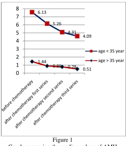
Figure 1 and 2 show the median values of AMH and inhibin B serum levels were higher in patients aged ≤ 35 years compared with patients aged > 35 years.
Figure 1
Graph comparing the median values of AMH serum levels in cervical cancer patients before and after chemotherapy based on the age of the patient
Figure 2
Graph comparing the median values of inhibin B serum levels in cervical cancer patients before and after chemotherapy based on the age of the patient
DISCUSSION
This study shows that there is a significant decrease in the levels of anti Muellerian Hormone and inhibin B after Paclitaxel-cisplatin combination chemotherapy in patients with cervical cancer. AMH may be a good predictor of ovarian reserve because the AMH production was not influenced by the feedback mechanism of the hypothalamus-pituitary-axis so that the ovary serum values will not fluctuate significantly.
In this study, 12 (75.0%) of 16 patients were in the age group of > 35 years and 4 (25.0%) were in the range of ≤ 35 years of age. The numbers of patients who were diagnosed with cervical cancer in the Wahidin Sudirohusodo hospital from 2008-2011 were 429 people (stage IIb - IV) and from
1.44
0.89 0.78
0.51 6.13
5.26
4.31 4.09
0 1 2 3 4 5 6 7 8
age < 35 year
www.balimedicaljournal.com
or
www.ojs.unud.ac.id
or
www.balimedicaljournal.org
76
2008 - 2011 aged <35 years were 122 people, while those who were > 35 years old were 327 patients. In this study, we found that the decreased levels in both serum levels of AMH and inhibin B before and after paclitaxel-cisplatin chemotherapy were significant p=0.000 (p<0.05). The median AMH level was 2.54ng/ml before chemotherapy and decreased to 1.37ng/ml after the third series. The median inhibin B level was 140.29ng/ml before chemotherapy and decreased to 112.98ng/ml after the third series. Similarly, the decreased serum levels both of AMH and inhibin B after chemotherapy in each series were also significant with P <0.05. This study also shows that the median value of AMH and inhibin B serum levels were higher in patients aged ≤ 35 years compared with patients aged > 35 year.11
Research conducted by Mehmet Sait et al concluded that the administration of the Paclitaxel-Cisplatin chemotherapy given in experimental animals demonstrated a significant reduction in the number of primordial follicles.12 Similarly, a study conducted by Xiaohuan Li et al concluded that administration of cisplatin had significantly lowered the serum levels of AMH.13
The decrease of ovarian reserve in patients with chemotherapy drugs can be caused by the damage of the primordial granulose cells. The continuous damage to the growing follicles will lead to the more rapid recruitment that will further lead to premature exhaustion of primordial follicles. Toxicity of chemotherapeutic agents in ovarian varies significantly. In vitro studies showed that paclitaxel induces apoptosis in granulose cell causes damage to the primordial follicles and then will cause a delay in the maturation of oocytes.
The most toxic chemotherapy agent to ovarian reserve is alkylating agent group. The presence of one or more alkyl groups to DNA causes apoptosis. Cisplatin is a member of alkylating agent group of chemotherapy. This group of antineoplastic agents primarily interacts with DNA, composed of unstable alkyl binding which reacts with nucleophilic parts in several important organic components such as nucleic acids, proteins and amino acids. This interaction is the main mechanism of cytotoxic effect. In vitro research showed that the administration of cisplatin chemotherapy on human ovarian cortex caused changes in apoptosis and fibrosis in granulose cells of primordial follicles, with consequent increase of damaged follicles number. In vivo research showed an increase damage primordial follicle, which brought the effects of reduced ovarian reserve, infertility and early menopause. In younger patients who have a larger initial follicle reserves, the impact of chemotherapy on fertility and estrogen production is mild. But, these young women are still at risk of early menopause due to the reduced number of follicles associated with the cumulative
dose of chemotherapy. Meirow et al reported that the use of alkylating agent increases the risk of early menopause was 4.5 fold.
The risk for ovarian failure after chemotherapy also increases with the increase of the patients’ age. Chemotherapy in cancer patients appears to show the presence of reproductive dysfunction, therefore, efforts to preserve fertility are required, especially in women who are diagnosed with cancer at young age. Efforts that can be done by doing the cryopreservation of ovarian tissue before all primordial follicles are damaged by chemotherapy. Possibility to minimize the damage to the gonad by administering the gonadotropin-releasing hormone agonists (GnRHas) during chemotherapy may also be considered. At present, scientific evidence to explain the mechanism of GnRHas protection is still limited.14
Because AMH is derived from preantral and small antral follicles, its levels are gonadotropin-independent and exhibit little variation within and between menstrual cycles. AMH is a very promising screening test for a diminished ovarian reserve. Inhibin B is generally not regarded as a reliable measure of ovarian reserve, but inhibin B serum levels still decrease in patients with chemotherapy drugs.10
CONCLUSIONS
Our research confirmed the gonadotoxic effect of combined chemotherapy by showing the significant decreased serum levels of AMH and inhibin B for 3 months since the initial treatment. Further larger and longer research with other types of histopathology and different chemotherapy regiments are required to evaluate the reversibility of ovarian function in those who still needs the fertility functions, so it can provide the basis to maintain the fertility therapy in patients receiving chemotherapy
REFERENCES
1. American Cancer Society. Global Cancer Facts & Figures 2nd Edition. Atlanta: American Cancer Society. 2011.
2. American Cancer Society. Cervical Cancer Overview. Atlanta: American Cancer Society. 2013.
3. Muraji M, Sudo T, Iwasaki S, Ueno S, Wakahashi S, Yamaguchi S, et al. The Effect of Abdominal Radical Trachelectomy on Ovarian Reserve: Serial Changes in Serum Anti-Müllerian Hormone Levels. Journal of Cancer. 2012;3:191-5.
4. Wallace WH. Oncofertility and Preservation of Reproductive Capacity in Children and Young Adults. Cancer. 2011;117(10):2301-10.
www.balimedicaljournal.com
or
www.ojs.unud.ac.id
or
www.balimedicaljournal.org
77
reserve in young women with breast cancer. British Journal of Cancer. 2007;96:1808-16. 6. LaMarca A, G.Stabile, Artenisio AC, A.Volpe.
Serum anti-Mullerian hormone throughout the human menstrual cycle. Human Reproduction. 2006;21(12):3103-7.
7. Lee M, Donahoe P, Hasegawa T, Silverman B, Crist G, Best S, et al. Mullerian inhibiting substance in humans: normal levels from infancy to adulthood. J Clin Endocrinol Metab. 1996;81(2):571-6.
8. Brougham MF, Crofton PM, Johnson EJ, Evans N, Anderson RA, Wallace WH. Anti-Müllerian hormone is a marker of gonadotoxicity in pre- and postpubertal girls treated for cancer: a prospective study. J Clin Endocrinol Metab. 2012;97(6):2059-67.
9. PawelWalentowicz, Sadlecki P, Krintus M, Sypniewska G, AnetaMankowska-Cyl, Grabiec M, et al. Serum Anti-Müllerian Hormone Levels in Patients with Epithelial Ovarian Cancer. International Journal of Endocrinology. 2013;2013.
10.Fritz MA, Speroff L. Female Infertility. Clinical gynecologic endocrinology and infertility. 8 ed: Lippincott Williams & Wilkins 2011. p. 1137-248.
11.Badawy A, Elnashar A, El-Ashry M, Shahat M. Gonadotropin-releasing hormone agonists for prevention of chemotherapy-induced ovarian damage: prospective randomized study. Fertility and Sterility. 2009;91(3).
12.YUCEBILGIN MS, TEREK MC, OZSARAN A, AKERCAN F, ZEKIOGLU O, ISIK E, et al. Effect of chemotherapy on primordial follicular reserve of rat: An animal model of premature ovarian failure and infertility. Australian and New Zealand Journal of Obstetrics and Gynaecology. 2004;44:6-9.
13.Li X, Yang S, Lv X, Sun H, Weng J, Liang Y, et al. The mechanism of mesna in protection from cisplatin-induced ovarian damage in female rats. J Gynecol Oncol. 2013;24(2):177-85.
14.Visser JA, Jong FHd, Laven JSE, Themmen APN. Anti-Mullerian hormone: a new marker for ovarian function. Society for Reproduction and Fertility. 2006;131:1-9.