ISSN 2449-8955
5(2) 2015
Volume 5
Number 2
May-August 2015
European Journal
of Biological Research
formerly
Journal of Biology and Earth Sciences
European Journal of Biological Research, Volume 5, Issue 2, May-August 2015 European Journal of Biological Research
ISSN 2449-8955
Editor-in-Chief
Tomasz M. Karpiński, Poznań, Poland
Co-Editors (Thematic Editors)
Artur Adamczak, Poznań, Poland - biology
Anna K. Szkaradkiewicz, Poznań, Poland - medicine
Statistical Editor
Paweł Zaprawa, Lublin, Poland
Language Editor
Dominik Piechocki, London, UK
Scientific Editorial Board
Tamara Bayanova, Apatity, Russia Fadi Hage Chehade, Beirut, Lebanon Alexander Ereskovsky, Marseille, France Agnieszka Gałuszka, Kielce, Poland Vittorio Gentile, Naples, Italy Stanisław Hałas, Lublin, Poland Afaf M. Hamada, Stockholm, Sweden Sven Herzog, Tharandt, Germany Liviu Holonec, Cluj-Napoca, Romania Miłosz A. Huber, Lublin, Poland Shri Mohan Jain, Helsinki, Finland Wouter Kalle, Wagga Wagga, Australia Tomasz Klepka, Lublin, Poland
Nikolaos Labrou, Athens, Greece Igor Loskutov, Sankt Petersburg, Russia Ákos Máthé, Sopron, Hungary
Ahmed El-Mekabaty, Mansoura, Egypt Artem V. Mokrushin, Apatity, Russia Shahid M. Mukhtar, Birmingham, USA Robert Pal, Pécs, Hungary
Amal K. Paul, Kolkata, India Rajiv Ranjan, Narkatia Ganj, India Antonio Tiezzi, Viterbo, Italy
Timotej Verbovšek, Ljubljana, Slovenia Vladimir K. Zhirov, Apatity, Russia
List of Peer-Reviewers
http://www.journals.tmkarpinski.com/index.php/ejbr/ pages/view/reviewers
Author Guidelines
http://www.journals.tmkarpinski.com/index.php/ejbr/ about/submissions
More information
www.journals.tmkarpinski.com/index.php/ejbr
DISCLAIMER
The Publisher and Editors cannot be held responsible for errors and any consequences arising from the use of information contained in this journal; the views and opinions expressed do not necessarily reflect those of the Publisher and Editors, neither does the publication of advertisements constitute any endorsement by the Publisher and Editors of the products advertised.
Cover: http://openwalls.com/image?id=20115, Licence Creative Commons Attribution 3.0 Unported (CC BY 3.0)
Copyright: © The Author(s) 2015. European Journal of Biological Research © 2015 T.M.Karpiński. All articles and abstracts are open-access, distributed under the terms of the Creative Commons Attribution Non-Commercial 4.0 International License, which permits unrestricted, non-commercial use, distribution and reproduction in any medium, provided the work is properly cited.
European Journal of Biological Research, Volume 5, Issue 2, May-August 2015
Contents
1-8
9-16
17-24
25-35
36-41
L-cysteine ameliorated testicular toxicity induced by acrylamide in rats
Hossam El-Din Mohamed Omar, Sary Kh. Abd-elghafar, Imhemed O. Fiedan, Emad A. Ahmed
The efficiency of Thymus laevigatus extract on the penconazole toxicity in some rabbit tissues
Hassan S. Nada, Ebtisam Y. Shikoo, Abdul R. Thabet, Elham Al-Shaibani
Participation of MEM+ bacteria in the bacterioplankton community in Ustka harbor, the River Słupia estuary, Southern Baltic Sea
Piotr Perliński, Piotr Skórczewski, Marta Zdanowicz, Zbigniew Mudryk
Physiological effects of allelopathic activity of Citrullus colocynthis on Vicia faba and Hordeum vulgare
Hediat Mohamed Salama, Hala Khaled Al Rabiah
Phytochemical profile and antibacterial activity of crude extracts of the pod of
Aframomum angustifolium (Sonn.) K. Schum.
ISSN 2449-8955
European Journal
of Biological Research
Research ArticleEuropean Journal of Biological Research 2015; 5 (2): 1-8
L-cysteine ameliorated testicular toxicity induced
by acrylamide in rats
Hossam El-Din M. Omar
1*, Sary Kh. Abd-elghafar
2, Imhemed O. Fiedan
1and Emad A. Ahmed
11
Department of Zoology, Faculty of Science, Assiut University, Egypt; 2Department of Pathology and Clinical Pathology, Faculty of Veterinary Medicine, Assiut University, Egypt,
*Corresponding author: Prof. Hossam El-Din M. Omar; Physiology Laboratory, Department of Zoology, Faculty of Science, Assiut University, Assiut, 71516, Egypt; e-mail: [email protected]
ABSTRACT
The general public is exposed to acrylamide from industrial manufacturing, laboratory work, foods rich in carbohydrates that have been cooked at high-temperature, and through cigarette smoke. The present experiment was conducted to investigate the reproductive toxicity of acrylamide exposure in male rats and the role of L-cysteine supplementation in amelioration of this toxicity. Forty eight adult male albino rats (weighing 120-140 g) were divided into four groups (16 rats/group). Group I - negative control group, drank tap water, group II - positive control, drank tap water that contains acrylamide (25 mg/kg body weight) for 28 days and group III drank tap water that contains acrylamide (25 mg/kg body weight) and L-cysteine (100 mg/kg body weight). Four rats from each group were killed at 7, 14, 21 and 28 days time intervals from the beginning of the experiment. In general, exposure to acrylamide induced a significant elevation in testes lipid peroxides and nitric oxide levels and a signi-ficant reduction in the level of glutathione and the activity of superoxide dismutase and catalase in all
periods of experiment. However, plasma testoste-rone was significantly decreased in acrylamide treated rats with congestion and interstitial edema, necrosis, calcification and degeneration of sperma-togenic cells in the seminiferous tubules and for-mation of spermatid gaint cells. Co-treatment of rats with L-cysteine reduced the changes in oxidative stress parameters and improved the pathological changes in testis. Therefore, supplementation of L-cysteine can be useful when there is a risk of acrlamide toxicity.
Keywords: Acrylamide, Testis, Oxidative stress, L-cysteine, Testosterone.
1. INTRODUCTION
Acrylamide (ACR) is formed through the Maillard reaction during the heating process by interactions of amino acids, especially asparagine, with reducing sugars like glucose [1, 2]. There is variance in literature about the levels of ACR in different foods and the potential risk from dietary exposure. The daily intakes of dietary ACR for the Received: 26 February 2015; Revised submission: 16 April 2015; Accepted: 17 April 2015
Copyright: © The Author(s) 2015. European Journal of Biological Research © T.M.Karpiński 2015. This is an open-access article distributed under the terms of the Creative Commons Attribution License, which permits
2 | Omar et al. Testicular toxicity induced by acrylamide
European Journal of Biological Research 2015; 5 (2): 1-8
general population are estimated to be in the range of 0.3-2.0 mg/kg body weight [3]. ACR can be reactive in three different ways, radical-mediated polymerization, and addition to thiol, hydroxyl, or amino groups result in alkylation of proteins or metabolized to an epoxide derivative, glycidamide being readily reactive toward DNA and other macromolecules [4-6]. The mechanism by which ACR exposure causes cellular dysfunction in experimental animals and humans is not completely clear. However, it is thought that oxidative stress was associated with ACR cytotoxicity. Reproduc-tive toxicity in rodents exposed to ACR includes alterations in gonadal and pituitary hormones associated with histopathological changes that includes formation of multinucleated giant cells, vacuolation and production of high numbers of apoptotic cells in the seminiferous tubules [7-11]. Acrylamide induced oxidative stress is capable of disrupting the steroidogenic capacity of Leydig cells [12] as well as the capacity of the germinal epithelium to differentiate into normal spermatozoa [13]. Fortunately, testes contain a complicated group of antioxidants and free radical scaven-gers to protect the spermatogenic and steroidogenic functions of testis from oxidative stress [14].
Kurebayashi and Ohno [15] reported that glutathione (GSH) precursors such as N-acetyl- L-cysteine and L-methionine increased the protec-tion against the cytotoxicity of ACR in isolated rat hepatocytes. Moreover, N-acetyl-L-cysteine as potent antioxidant protects tissues from ACR toxicity by inhibiting neutrophil infiltration, balan-cing the oxidant-antioxidant status, and regulating the generation of inflammatory mediators [8]. Also,
α-lipoic acid protect cells from oxidative stress induced by ACR exposure via enhances cellular antioxidant defense capacity [9]. In this regard, the objectives of the present study were to measures the markers of oxidative stress in testis of rats exposed to ACR and to evaluate the protective role of L-cysteine as precursor of GSH against ACR toxicity.
2. MATERIALS AND METHODS
Forty eight adult male albino rats (weighing 120-140 g) were purchased from the Animal House, Faculty of Medicine, Assiut University, Assiut,
Egypt. The animals were housed in cages at a controlled temperature (25±3°C) and ambient humi-dity (50-60%). Lights were maintained on a 12-h light-dark cycle. All animals received basal diet and water ad-libitum for one week as an adaptation period. Following one week of acclimatization, the rats were randomly divided into three groups (16 rats/group):
• Group I: Negative control, fed on basal diet and normal drinking water for 4 weeks.
• Group II: Positive control, fed on basal diet and drinking water that contains ACR (25 mg/kg body weight) according to Alturfan et al. [8].
• Group III: Fed on basal diet and drinking water that contains ACR (25 mg/kg body weight and L-cysteine (100 mg/kg body weight) according to Omar et al. [16].
Then, each week from the beginning of the experiment 4 rats from each group were killed under anesthesia with ether. The blood samples were collected directly from portal vein into centrifuge tubes for separation of serum by centrifugation at 3000 rpm for 15 minutes and were frozen at -20°C for subsequent biochemical analysis. Immediately after killing rats, small piece of testes were excised and fixed in formaline for histological studies, and the remnant was washed in cold saline, immersed in liquid nitrogen and stored at -20°C for biochemical assay. All animal experiments were carried out in accordance with Ethical Committee Acts.
2.1. Determination of oxidative stress biomarker
Lipid peroxidation (LPO) products as TBARS content were determined according to the method of Ohkawa et al. [17]. Nitric oxide (NO) content was measured as nitrate concentration colori- metrically using the method of Ding et al. [18]. Glutathione (GSH) content was determined using the method of Beutler et al. [19]. The activity of superoxide dismutase (SOD) was determined basing on its ability to inhibit the autoxidation of epinephrine at alkaline medium according to the method of Misra and Fridovich [20]. The activity of catalase (CAT) was determined basing on its ability to decompose H2O2 to H2O and O2 according to
3 | Omar et al. Testicular toxicity induced by acrylamide
European Journal of Biological Research 2015; 5 (2): 1-8
2.2. Estimation of testosterone
Testosterone hormone in plasma was deter-mine by Enzyme Immunoassay Method (ELISA), Biocheck, Inc, 323 vintage Park Dr. Forster City, CA, USA, according to the kit manufacture instruc-tions.
2.3. Statistical analysis
The data was expressed as mean ± SE. The results were analyzed statistically using column statistics and one-way analysis of variance with the Newman-Keuls multiple comparison test as a post-test. These analyses were carried out using the computer prism program for windows, version 6.0 (Graph pad software Inc., San Diego, California, USA). Differences between the groups were considered significant if P < 0.05, 0.01, or 0.001.
3. RESULTS
Compared to control rats, ACR treated rats exhibited a significant decrease in plasma testo-sterone in 2nd, 3rd, and 4th weeks and LC treatment resulted in an increase in testosterone level especially in 2nd and 4th week (Fig. 1).
In relation to control rats, ACR treated rats had greater level of LPO in testis especially in the 3rd week (P<0.001) and LC co-treatment failed to restore the elevation of LPO especially in 1st and 4th week (Fig. 2). Also, Fig. 2 showed that NO was significantly increased (P<0.001) in all periods of the experiment in comparison with control and LC treatment resulted in significant reduction in NO level (P<0.001).
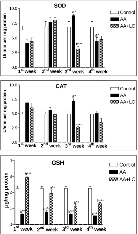
In the ACR group, GSH level in testis was decreased significantly (P<0.001) in comparison to the control group. In the LC + ACR group, GSH level was resorted to the normal level in control group (Fig. 3). Moreover, SOD and CAT activities were significantly increased (P<0.05) in the 3rd week in ACR treated rats, while they significantly decreased (P<0.001) in the same week in LC and ACR treated groups (Fig. 3).
Fig. 1. Testosterone level in plasma of control and treated
rats.
Fig. 2. Levels of LPO and NO in testis tissue of control
and treated rats.
3.1. Histopathology of testes
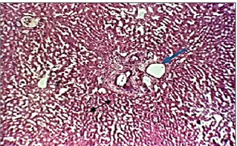
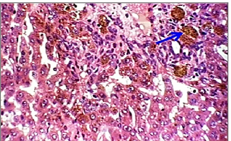
In the present study, it was observed that testis of control rats stained with hematoxylin and eosin (H&E) was formed of seminiferous tubules and interstitial cells of Leydig. Most tubules contain normal spermatogenic cell layers and spermatozoa (Fig. 4A). After one week of treatment with ACR, testes showed mild degeneration of spermatogenic cells in the seminiferous tubules, interstitial edema and degeneration in spermatogenic cells with formation of spermatid giant cells (arrows) (Fig. 4B). After two weeks, the testes showed severe degeneration in the spermatogenic cells with the presence of spermatid giant cells, the absence of
0.0 2.5 5.0 7.5 Control AA AA+LC
1st week 2nd week 3rd week 4th week
n m o l/ m g p ro te in a** b*** a* b*** b*** a*** b*** a** NO 0.0 0.5 1.0 1.5 Control AA AA+LC
1stweek 2ndweek 3rdweek 4th week
n m o l T M P /m g p ro te in a** LPO 0.00 0.25 0.50 0.75 Control AA AA+LC p g /m l
1st week 2nd week 3rd week 4th week
b**
a* a**
4 | Omar et al. Testicular toxicity induced by acrylamide
European Journal of Biological Research 2015; 5 (2): 1-8
some spermatogenic cells layers and the presence of spermatid giant cells in some cases was also noticed (Fig. 4D). After three weeks, testes showed severe coagulative necrosis of seminiferous tubules and the absence of spermatogenic cells (Fig. 4F). And after four weeks, testes showed severe necrosis with dystrophic calcification in the seminiferous tubules (Fig. 4H). In group of rats co-treated with LC for one week, testes showed congestion, interstitial edema and hyperplasia Leydig cells in the first week and mild degeneration in the spermatogenic cells (Fig. 4C). After 2 weeks of treatment with LC, testes showed mild degeneration of the spermatogenic cells with the presence of multiple spermatid giant cells (Fig. 4E). In rats co-treated with LC for 3 and 4 weeks, testes showed mild degeneration in some seminiferous tubules (Figs. 4. G and I).
Fig. 3. Level of GSH and the activities of SOD and CAT
in testis tissue of control and treated rats.
4. DISCUSSION
ACR is one of the major environmental public health problems; it induced oxidative organ damages in the brain, lung, liver, kidney, spleen, and testes tissues [8, 23-25]. The present result in (Fig. 2) revealed that administration of ACR in drinking water increased LPO and NO in testes and co-treatment of rats with LC decreased the level of LPO and NO in comparison with ACR treated rats. It is known that, NO may show either cytoprotective or cytotoxic effects and the elevated NO level has the ability to induce γ-glutamyl cycle [26]. In consistence with our results, Jiazhong et al. [27] and Abd El-Halim and Mohamed [23] mentioned that ACR is able to increase LPO by inducing oxidative stress with generation of free radicals. Garlic, resveratrol and NAC may be protected against ACR-induced oxidative injury by scaven-ging free radicals, preventing depletion of GSH and inhibiting neutrophil infiltration, and subsequent activation of inflammatory mediators that induce LPO [8, 28].
ACR is capable of interacting with vital cellular nucleophiles possessing -SH group. There-fore, it reacts with GSH in a similar manner and forms GSH S-conjugates, which is the initial step in the biotransformation of ACR [29]. Depletion of cellular GSH seems to play a pivotal role in the genotoxicity in human caused by ACR [30, 31]. In the present study, decreased GSH content in testes as presented in (Fig. 3) can be explained by the reaction of ACR with GSH, which in turn causes the depletion of GSH and the enhancement of LPO. Similarly, Abd El-Halim and Mohamed [23] found that administration of ACR caused a significant reduction in testes GSH level. However, Özturan-Özeri et al. [32] concluded that GSH is not directly capable of protecting tissues against ACR-induced oxidative stress. The present results showed that co-treatment of rats with LC attenuate GSH deple-tion by ACR. In consistence, treatment with garilic prior to ACR attenuated the depletion of GSH level [18]. Also, treatment of rats with lipoic acid caused an increase in GSH level with a decrease in LPO which might be attributed to the oxidative damage repairing ability of lipoic acid [33].
0.0 2.5 5.0 7.5 10.0 Control AA AA+LC U / m in p e r m g p ro te in a* b*** a*
1stweek 2nd week 3rd week 4th week SOD 0.0 2.5 5.0 7.5 10.0 Control AA AA+LC U /m in p e r m g p ro te in
1st week 2nd week 3rd week 4th week
CAT a* b*** 0 1 2 3 4 Control AA AA+LC µ g /m g p ro te in
1st week 2nd
5 | Omar et al. Testicular toxicity induced by acrylamide
European Journal of Biological Research 2015; 5 (2): 1-8
Fig. 4. Photomicrographs of control testis formed of seminiferous tubules and interstitial cells of leydig. A. Most tubules
contain normal spermatogenic cells layers and spermatozoa in control rats. B-I testis of ACR and ACR plus L-cysteine treated groups at different weeks of treatment. B. mild degeneration of spermatogenic cells with formation of spermatid giant cells in the seminiferous tubules (arrows). C. Congestion interstitial edema and hyperplasia of Leydig cells (arrows).
D. Severe degeneration in the spermatogenic cells with presence of spermatid giant cells (arrows). E. Mild degeneration in
the spermatogenic cells (arrows). F. Severe coagulative necrosis of seminiferous tubules and absence of spermatogenic cells (arrows). G. Moderate degeneration of the spermatogenic cells and presence of multiple spermatid gaint cells (arrows). H. Severe necrosis (star) with dystrophic calcification in the seminiferous tubules (arrows). I. mild degeneration in some seminiferous tubules (arrows). H&E X400.
Superoxide radical may oxidize SH groups and undergo dismutation to form H2O2 and singlet
oxygen [34]. This change in the redox status of the cell may modulate gene expression directly or via the transcription factors that are redox-regulated, and may lead to apoptosis, cell proliferation, or transformation (29). In the current study, (Fig. 3) showed alteration in the testicular SOD and CAT activities depending on the period of treatments. Also, it showed that co-treatments of rats with LC ameliorate these changes by different levels. These results are in agreement with Abd El-Halim and Mohamed [23] who found that administration of ACR caused a significant reduction in the activity of SOD in testes tissues. The reduction in antioxi-dant enzyme activities was increased with
increas-ing doses of ACR [35]. Treatment with Curcuma
longa L. powder and garilic prior to ACR attenuated
the reduction of SOD activity [23, 36], and admi-nistration of catechin and neem leaves extracts significantly enhanced the hepatic CAT activity [37].
Testosterone level in plasma of rats treated with ACR was significantly decreased, and co-treatment with LC elevates this decrease especially in the 4th week of treatment (Fig. 1). In this aspect, administration of ACR caused a significant reduc-tion of serum testosterone level as reported by many authors [9, 38, 39]. This significant reduction of testosterone may be a result of direct damage of ACR on the Leydig cells [40]. The previous opinion was confirmed histopathologicaly in the present
A
C
D
E
F
G
H
I
6 | Omar et al. Testicular toxicity induced by acrylamide
European Journal of Biological Research 2015; 5 (2): 1-8
study by congestion and interstitial edema, necrosis, calcification and degeneration of spermatogenic cells in the seminiferous tubules with formation of spermatid gaint cells (Fig. 4 A, C, E & G). More-over, ACR may affect the endocrine function of the testes by altering the androgen biosynthesis of interstitial cells in the testes [41] or inducing the enzymes activity of hepatic biotransformation, which is capable of metabolically transforming androgens into products with low androgen receptor binding activity [42]. Song et al. [43] found that ACR can directly damage Leydig cells and affect the endocrine function of the testis. Moreover, Yang et al. [44] found that ACR induces histopathological lesions such as formation of multinucleated giant cells, vacuolation and production of high numbers of apoptotic cells in the seminiferous tubules of the rat. In the present study, treatment with LC along with ACR resulted in moderate attenuation of the histopathological changes in testes that were induced by ACR.
From these observations, it can be concluded that LC ameliorates the toxicity of ACR in rat testes by alleviating LPO and NO through scavenging of free radicals, and enhancing the activity of SOD and CAT and GSH level.
AUTHORS CONTRIBUTION
All authors contributed equally in planning, conduct, data analysis, and editing the work. The final manuscript has been read and approved by all authors.
TRANSPARENCY DECLARATION
The author declares no conflicts of interest.
NOTES
This paper was presented in the 48th Annual Conference of Physiology and Pathology of Reproduction and simultaneously 40th Joint Congress of Veterinary and Human Medicine, Zurich. Switzerland, 11th-13th February 2015, Abstract No 74.
REFERENCES
1. Boettcher MI, Schettgen T, Kutting B, Pischetsrie-der M, Angerer J. Mercapturic acids of acrylamide and glycidamide as biomarkers of the internal exposure to acrylamide in the general population. Mutat Res. 2005; 580: 167-176.
2. Mottram DS, Wedzicha BL, Dodson AT. Acryl-amide is formed in the Maillard reaction. Nature. 2002; 419: 448-449.
3. Dybing E, Farmer PB, Andersen M, Fennell TR, Lalljie SPD, Müller DJG, et al. Human exposure and internal dose assessments of acrylamide in food. Food Chem Toxicol. 2005; 43: 365-410.
4. Park J, Kamendulis LM, Friedman MA, Klaunig JE. Acrylamide-induced cellular transformation. Toxicol Sci. 2002; 65: 177-183.
5. Besaratinia A, Pfeifer GP. DNA adduction and mutagenic properties of acrylamide. Mutat Res. 2005; 580: 31-40.
6. Paulsson B, Rannug A, Henderson AP, Golding BT, Törnqvist M, Warholm M. In vitro studies of the influence of glutathione transferases and epoxide hydrolase on the detoxification of acrylamide and glycidamide in blood. Mutat Res. 2005; 580: 53-59. 7. Hamdy SM, Bakeer HM, Eskande EF, Sayed ON.
Efect of acrylamide on some hormones and endo-crine tissues in male rats. Hum Exp Toxicol. 2011; 5: 1-9.
8. Alturfan EI, Beceren A, Şehirli AQ, Demiralp ZE,
Şener G, Omurtag GZ. Protective effect of N-acetyl-L-cysteine against acrylamide-induced oxidative stress in rats. Turk J Vet Anim Sci. 2008; 36(4): 438-445.
9. Lebda M, Gad S, Gaafar H. Effects of lipoic acid on acrylamide induced testicular damage. Mater Sociomed. 2014, 26(3): 208-212.
10. Khalil WKB, Ahmed HH, Hanan F, Aly HF, Eshak MG. Toxicological effects of acrylamide on testi-cular function and immune genes expression profile in rats. J Pharm Sci Rev Res. 2014; 24(1): 143-151. 11. Yang HJ, Lee SH, Jin Y, Choi JH, Han DU, Chae C,
et al. Toxicological effects of acrylamide on rat testicular gene expression profile. Reprod Toxicol. 2005; 19(4): 527-534.
12. Hales DB, Allen JA, Shankara T, Janus P, Bucks S, Diemer T, Hales RH. Mitochondrial function in Leydig cell steroidogenesis. Ann NY Acad Sci. 2005; 1061: 120-134.
7 | Omar et al. Testicular toxicity induced by acrylamide
European Journal of Biological Research 2015; 5 (2): 1-8
14. Aitken RJ and Roman SD. Antioxidant systems and oxidative stress in the testes. Oxid Med Cell Longev. 2008; 1(1): 15-24.
15. Kurebayashi H, Ohno Y. Metabolism of acrylamide to glycidamide and their cytotoxicity in isolated rat hepatocytes: protective effects of GSH precursors. Arch Toxicol 2006; 80: 820-828.
16. Omar HM, Ahmed EA, Abdel-Ghafar S Kh, Ragab MMS, Nasser AY. Hepatoprotective effects of vitamin C, DPPD, and L-cysteine against cisplatin-induced oxidative stress in male rats. J Biol Earth Sci. 2012; 2(1): B28-B36.
17. Ohkawa H, Ohishi N, Yagi K. Assay for lipid peroxides in animal tissue by thiobarbaturic acid reaction. Anal. Biochem. 1979; 95: 351-358. 18. Ding AH, Nathan CF, Stuchr DJ. Release of
reactive nitrogen intermediate and reactive oxygen intermediate from mouse peritoneal macrophages Comparison of activating cytokines and evidence for independent production. J Immunol. 1988; 141: 2407-2412.
19. Beutler E, Duron O, Kelly BM. Improved method for the determination of blood glutathione. J Lab Clin Meth. 1963; 61: 882-888.
20. Misra HP, Fridovich I. The role of superoxide anion in the autoxidation of epinephrine and a simple assay for superoxide dismutase. J Biol Chem. 1972; 247: 3170-3175.
21. Gregory EM, Fridovich I. Visualization of catalase on acrylamide gels. Anal Biochem. 1974; 58: 57-62. 22. Lowry OH, Rosebrough NJ, Farr AL, Randall RJ. Protein measurement with the Folin phenol reagent. J Biol Chem. 1951; 41: 1863-1870.
23. Abd El-Halim SS, Mohamed MM. Garlic powder attenuates acrylamide-induced oxidative damage in multiple organs in rat. J Appl Sci Res. 2012; 8: 168-173.
24. Taha N, Korshom M, Mandour AW, Sadek K. Effects of garlic and acrylamide on some antioxidant enzymes. Global J Med Plant Res. 2013; 1: 190-194. 25. Venkatasubbaiah K, Venkataswamy DM, Suresh KS, Rao KJ. Acrylamide induced oxidative stress in rat and chick embtyonic liver. Indo Am J Pharmac Res. 2014; 6: 2791-2798.
26. Kuo PC, Abe KY, Schroeder RA. Interleukin-1-induced nitric oxide production modulates gluta-thione synthesis in cultured rat hepatocytes. Am J Physiol. 1996; 271: 851-862.
27. Jiazhong J, Yong ZU, James EK. Induction of oxidative stressing rat brain by acrylonitrile. Toxicol Sci. 1998; 46: 333-341.
28. Nursal GK, Levent S, Ozer E, Feriha S, Serap M, Keyer-Usal S, Goksel. Long term administration of aqueous garlic extract (AGE) alleviates liver fibrosis and oxidative damage induced by biliary obstruction in rats. Life Sci. 2005; 76: 2593-2606.
29. Awad ME, Abdel-Rahman MS, Hassan SA. Acryl-amide toxicity in isolated rat hepatocytes. Toxicol In Vitro. 1998; 12: 699-704.
30. Lamy E, Völkel Y, Roos PH, Kassie F, Mersch-Sundermann V. Ethanol enhanced the genotoxicity of acrylamide in human, metabolically competent HepG2 cells by CYP2E1 induction and glutathione depletion. Int J Hyg Environ Health. 2007; 1-2: 74-81.
31. Zhang X, Cao J, Jiang L, Geng C, Zhong L. Protec-tive effect of hydroxytyrosol against acrylamide-induced cytotoxicity and DNA damage in HepG2 cells. Mutat Res. 2009; 664: 64-68.
32. Özturan-Özeri E, Ucar G, Helvacoglu F, Ery AC, Rkaydin-Aldemir D, Turkoglu S. Effect of acryl-amide treatment on arginase activities and nitric oxide levels in rat liver and kidney. Acta Med Mediter. 2014; 30: 375-382.
33. Prahalathan C, Selvakumar E, Varalakshmi P. Lipoic acid modulates adriamycin-induced testicular toxi-city. Reprod Toxicol. 2006; 211: 54-59.
34. Andreev YA, Kushnareva YV, Starkov AA. Meta-bolism of reactive oxygen species in mitochondria. Biokhimiya. 2005; 70: 246-264.
35. Swamy MV, Subbaiah KV, Aumau B, Kamala K, Rao KJ, Raju KT. Toxic effect of acrylamide on body weight, the study of antioxidants and histoarchitecture of heart in the developing chick embryo. Indian J Appl Res. 2013; 3: 27-30.
36. Abd EL-Halim SS, EL-Adawi AS. Modulation of acrylamide-induced oxidative damage in rat tissues by Curcuma longa L. Med J Cairo Univ. 2008; 76: 639-547.
37. Mansour MK, Ibrahim EM, Kholy MM, El-Madawy SA. Antioxidant and histopathological effect of catechin and neem leaves extract in acrylamide toxicity of rats. Egypt. J Comp Path Clinic Path. 2008; 21: 290-313.
38. Abd El-Mottaleb EM, Rashed AYM. Some studies on acrylamide intoxication in male albino rats. Egypt. J Comp Path Clinic Path. 2008; 21: 222-245.
8 | Omar et al. Testicular toxicity induced by acrylamide
European Journal of Biological Research 2015; 5 (2): 1-8
40. Tag El-Din HA, Abbas HE, El-Kashoury AI. Experi-mental studies of dicofol reproductive toxicity on male albino rats. Bull Fac Pharm Cairo Univ. 2003; 40: 179-188.
41. Fowler A, Mistry P, Goering L. Mechanisms of meta-induced cell injury. Res Comm Chem Pathol Pharmacol. 1987; 28: 689.
42. Sonderfen AJ, Arlotto MP, Dutton DR, McMillen SK, Parkinson A. Regulation of testosterone hydro-xylation by rat liver microsomal cytochrome P-450. Arch Biochem Biophys. 1987; 255: 27-41.
43. Song HX, Wang R, Geng ZM, Cao SX, Liu TZ. Subchronic exposure top acrylamide effects repro-duction and testis endocrine functions if rats. Zhonghua Nan Ke Xue. 2008; 14: 406-910.
ISSN 2449-8955
European Journal
of Biological Research
Research ArticleEuropean Journal of Biological Research 2015; 5 (2): 9-16
The efficiency of
Thymus laevigatus
extract
on the penconazole toxicity in some rabbit tissues
Nada S. Hassan
1*, Ebtisam Y. Shikoo
1, Abdul R. Thabet
2and Elham Al-Shaibani
31
Aden University, Faculty of Education, Zoology Department, Khormakser, Airport Street, Aden, Yemen
2
Sanaa University, Faculty of Agriculture, Plant Protection Department, Yemen 3Sanaa University, Faculty of Science, Biology Department, Yemen.
*Corresponding author: Nada S. Hassan; Aden University, Faculty of Education, Zoology Department, Khormakser, Airport Street, Aden, Yemen; e-mail: [email protected]
ABSTRACT
The Thymus laevigatus (Vahl), Lamiaceae (Labiatae), an endemic species of Yemen, is traditionally used in the treatment of various disorders including sto-mach and respiratory system diseases. Penconazole is a systemic triazole fungicide with preventive and curative properties for the control of powdery mildew. This study was designed to investigate the toxicity of penconazole fungicide and effect of the extract Thymus laevigatus on male and female rabbit tissues. The orally administered penconazole to male and female rabbits at the dose (1/10 LD50
of 380 mg/kg b.w.) for 60 days showed signs of toxicity including piloerection, subsequent fall and swollen condition some organs such as the stomach and the intestine, reduced body weight and caused death of rabbits. Histopathological changes showed in liver tissue including mild lymphocytes infiltration around bile duct in the portal area and fibrosis, and in lungs showed less inflammation and lymphoid aggregates around the bronchus, more consolidation of the alveolar space and edema. Pare with Thymus
laevigatus extract 50 and 100 mg/kg administered
orally on two different groups of animals found that
the Thymus laevigatus possess strong antioxidant properties and therefore it inhibits penconazole toxi-city effect on rabbit tissues.
Keywords: Penconazole, Thymus laevigatus, Rabbit, Mortality, Behavior, Histopathology.
1. INTRODUCTION
The genus Thymus which belongs to the family Lamiaceae (Labiatae) includes 350 species widespread all around the world. Several studies investigated earlier the chemical compositions of the essential oils of numerous species of the Thymus genus and focused on their antimicrobial activities [1-2]. Recently, the essential oils of various species of the Thymus genus have been screened for their traditional indigenous uses and investigated inten-sive as promise sources of antibacterial, antifungal, antioxidant and other natural products.
The most species of thymus contain altimol (20-55%) (thymol), a compound that most benefit in medicine, alcarvacrol (carvacrol), allinalol (lina-lool), cymene, and gamma-terpinene. The propor-tion of these components varies depending on the Received: 14 March 2015; Revised submission: 01 May 2015; Accepted: 04 May 2015
Copyright: © The Author(s) 2015. European Journal of Biological Research © T.M.Karpiński 2015.
This is an open-access article distributed under the terms of the Creative Commons Attribution License, which permits unrestricted use, distribution, and reproduction in any medium, provided the original work is properly cited.
10 | Hassan et al. The efficiency of Thymus laevigatus extract on the penconazole toxicity in some rabbit tissues
European Journal of Biological Research 2015; 5 (2): 9-16
soil and the time of harvest the plant and the surrounding geographical conditions [3]. In the flora of Yemen, this genus is represented by only the species Thymus laevigatus which is endemic to Yemen. It occurs in the higher mountains in North Yemen in Haggah (2500 m) and in Dhamar (2200 m) [4]. The Yemeni local name of the plant is Za’tar. In Yemeni folk medicine, the fresh and dried leaves of T. laevigatus are used as powder in warm milk, sesame oil or olive oil to treat different stomach diseases, cough, tonsillitis, pharangitis, and renal colic [5]. It also treats respiratory infections, such as chronic bronchitis, whooping cough and asthma [6] and is also working on prevention teeth from decay and treats inflammation of the tonsils, gums and teeth [7] and is thyme useful in the treatment of emphysema and inflammation of the stomach, diarrhea, headache, migraine, nocturnal enuresis in children [8- 9].
Penconazole is a systemic triazole fungicide with the preventive and curative properties for the control of powdery mildew disease of different crops. The mode of action is by stopping the development of fungi by interfering with the biosynthesis of sterols in cell membranes. It is used on fruit, especially apples and grapes and vegetables [10]. FAO and WHO [11] report that penconazole oral acute LD50 for rats is 2125 mg/kg, for mice
2444 mg/kg. Skin and eye acute percutaneous LD50
for rats is >3000 mg/kg. It is not a skin irritant; is irritating to eyes (rabbits). Not a skin sensitize inhalation LC50 (4 h) is >4000 mg/m
3
[12]. The penconazole is particularly effective against powde-ry mildew and feedbacks caused by fungi
Deutero-mycetes, Basidiomycotiona and Ascomycetes as Ery-siphe cichoracearum, Microspharea alin and Phyll-actinia sp. [13].
The objective of this study was to evaluate protective role of Thymus laevigatus leaves extract against the effect of penconazole fungicide. Studies were conducted of the behavior changes and histo-pathological changes such as liver and lungs of male and female rabbits.
2. MATERIALS AND METHODS
2.1. Fungicide, plant materials and test animals
Penconazole fungicide EC 10% was from
Ministry of Agriculture, Department of Plant Pro-tection (Producer Shenzhen Pisticides Ltd., China). The leaves of Thymus laevigatus were collected in October 2012 from Ibb Yemen. Forty local domestic rabbits have been bought from Lahej, Alseilla, Sheikh Othman markets.
2.2. Experimental animals
Forty local rabbits weighting 800-1200 grams have been used in this study. All animal experiments were carried out in accordance with local Ethical Committee Acts. All rabbits were acclimated in cages for 15 days before experiment. Each animal was weighed in the morning and fed with carrot, lettuce and cabbage. The animals were given water ad libitum.
Animals were divided into four groups (ten rabbits, 5 male and 5 female per group) as the following: Group I: Control group.
Group II: Treated with 1/10 LD50 of penconazole
(380 mg/kg b.w.) .
Group III: Treated with 1/10 LD50 of penconazole
(380 mg/kg b.w.) and extract of T. laevigatus (50 mg/kg b.w.).
Group IV: Treated with 1/10 LD50 of penconazole
(380 mg/kg b.w.) and extract of T. laevigatus (100 mg/kg b.w.).
The administration of the all doses was achieved orally, using insulin needle, day after day.
2.3. Extraction method
11 | Hassan et al. The efficiency of Thymus laevigatus extract on the penconazole toxicity in some rabbit tissues
European Journal of Biological Research 2015; 5 (2): 9-16
3. RESULTS
3.1. Animals mortality
There was no mortality among the control group during 60 days, four rabbits were died during the treatment with 1/10 penconazole, and two rabbits
were died in the group which gavaged with 1/10
penconazole and extract of Thymus laevigatus 50 mg/kg. Simultanously, when rabbits gavaged with
1
/10 penconazole and extract of Thymus laevigatus
100 mg/kg one rabbit died. This confirms that
Thymus laevigatus reduced the toxicity of
penco-nazole (Table 1).
Table 1. Showing mortality rate of rabbits exposed to
penconazole at 1/10 LD50 after 60 days of exposure. Number
of group
Number of rabbits
Concentration in mg/l
Mortality %
Control 10 - 0 I 10 1/10 penconazole 4
II 10
1
/10 Penconazole + extract of
Thymus laevigatus
50 mg/kg
2
III 10
1
/10 Penconazole + extract of
Thymus laevigatus
100 mg/kg
1
3.2. Behavioral changes
Rabbits have a different behaviors when treated with penconazole such as hidden under cages, calm, piloerection of hair, loss of appetite, strong tremors. The rabbits in group II gained weight, what may be due to the swollen of some organs such as the stomach. Animals from group III showed reddish swellings around eyes, and from group IV have a very dark blood color.
3.3. Histopatological Changes
The histopathological changes in liver male were observed in Group (II) of rabbits gavaged with a dose of 1/10 of penconazole (380 mg/kg b.w.) for
60 days such as mild lymphocytes, infiltration
around bile duct in the portal area periporatal and fibrosis around portal area, fatty changes and hepa-tocyte degeneration (Figs. 1 and 2). In Figs. 3 and 4 are showed ballon cells.
Fig. 1. Mild lymphocytes infiltration (double arrow).
Fibrosis and bile duct in the portal area (blue arrow) (H&E 10x).
Fig. 2. Mild lymphocytes infiltration in portal area (H&E
40x).
12 | Hassan et al. The efficiency of Thymus laevigatus extract on the penconazole toxicity in some rabbit tissues
European Journal of Biological Research 2015; 5 (2): 9-16
Fig. 4. Hepatocyte degeneration (ballon) (H&E 40x).
The histopathological section of Group III male rabbit liver exposed to 1/10 of penconazole (380
mg/kg b.w. and extract of Thymus laevigatus (50 mg/kg b.w.) for 60 days showed periportal fibrosis around the bile ducts hepatocytes (Fig. 5) and cholestasis in hepatocytes (Fig. 6)
Fig. 5. Periporatal fibrosis around the bile ducts (H&E
10x).
Fig. 6. Cholestasis in hepatocytes (blue arrow) (H&E
40x).
In group IV rabbits exposed to 1/10 of
penco-nazole (380 mg/kg b.w.) and extract of Thymus
laevigatus (100 mg/kg b.w.) for 60 days a liver of
the male rabbit showed no lymphocytes or fibrosis around (Fig. 7), as well as many double nuclei (Fig. 8).
Fig. 7. No lymphocytes and no fibrosis (H&E 10x).
Fig. 8. Many double nuclei (arrow) (H&E 10x).
The histopathological changes in female liver (Group II), when rabbits gavaged with a dose of
1
/10 of penconazole (380 mg/kg b.w.) for 60 days
showed balloon degeneration and fatty change (Fig. 9).
In Group III rabbits exposed to 1/10 of
penco-nazole (380 mg/kg b.w.) and extract of Thymus
laevigatus (50 mg/kg b.w.) for 60 days, female
rab-bit liver showed degeneration, no fatty change or balloon hepatocytes many double nuclei (Fig. 11).
In Group IV when rabbits exposed to 1/10 of
penconazole (380 mg/kg b.w.) and extract of
Thymus laevigatus (100 mg/kg b.w.) for 60 days,
female rabbits liver showed mild cholestasis in the hepatocytes (Fig. 12) and increased fibrosis around bile ducts (Fig. 13).
gava-13 | Hassan et al. The efficiency of Thymus laevigatus extract on the penconazole toxicity in some rabbit tissues
European Journal of Biological Research 2015; 5 (2): 9-16
ged with a dose of 1/10 of penconazole (380 mg/kg
b.w.) for 60 days, showed thickened alveolar wall and dilation of alveolar spaces (Figs. 14 and 15).
Fig. 9. Balloon degeneration (black arrow). Lymphocytes
in the lobule in control and fatty change (red arrow) (H&E 40x).
Fig. 10. Lymphocytes in the lobule in control and fatty
change (arrow) (H&E 40x).
Fig. 11. Degeneration, no fatty change or balloon
hepato-cytes many double nuclei (H&E 40x).
The histopathological changes in Group III where female rabbits were exposed to 1/10 LD50 of
penconazole (380 mg/kg b.w.) and extract of
Thy-mus laevigatus (50 mg/kg b.w.) for 60 days, lungs
showed inflammation around bronchi and lymphoid aggregation (Fig. 16).
Male rabbits lungs exposed to 1/10 of
penco-nazole (380 mg/kg. b.w) and extract of Thymus
laevigatus (100 mg/kg b.w) for 60 days at Group IV
showed thickened alveolar walls, dilation alveoli space and edema (Figs. 17 and 18).
Fig. 12. Mild cholestasis in the hepatocyte (blue arrow)
(H&E 10x).
Fig. 13. Increase fibrosis around bile duct (arrow) (H&E
10x).
Fig. 14. Thickened alveolar wall (black arrow and
14 | Hassan et al. The efficiency of Thymus laevigatus extract on the penconazole toxicity in some rabbit tissues
European Journal of Biological Research 2015; 5 (2): 9-16
Fig. 15. Thickened alveolar walls (black arrows) and
dilation alveolar spaces (red arrows) (H&E 40x).
The histopathological changes of the lungs in female rabbits in Group II which exposed of 1/10 of
penconazole for 60 days showed thickened alveolar walls, dilation alveolar spaces and edema (Fig. 19). The histopathological changes of Group III which exposed to 1/10 of penconazole (380mg/kg.
b.w.) and extract of Thymus laevigatus (50 mg/kg b.w.) for 60 days, female lungs showed less edema, more alveolar space dilatation (Figs. 20 and 21).
Fig. 16. Inflammation around bronchi (black arrow) and
lymphoid aggregation (red arrow) (H&E 10x).
Fig. 17. Edema (black arrow) and dilation alveolar spaces
(red arrow) (H&E 10x).
Fig. 18. Thickened alveolar walls and dilation alveolar
spaces (H&E 40x).
Fig. 19. Thickened alveolar wall (black arrow), dilation
alveolar spaces (red arrow) and edema (blue arrow) (H&E 10x).
Female rabbits lungs of Group IV which were exposed to 1/10 of penconazole (380 mg/kg b.w.) and
extract of Thymus laevigatus (100 mg/kg b.w.) for 60 days showed dilation alveolar space (Fig. 22) and consolidation in the alveolar space and thickened alveolar wall and dilation alveolar space and no edema (Fig. 23).
Fig. 20. Less edema (black arrow) with more alveolar
15 | Hassan et al. The efficiency of Thymus laevigatus extract on the penconazole toxicity in some rabbit tissues
European Journal of Biological Research 2015; 5 (2): 9-16
Fig. 21. Less edema (black arrow) and dilation alveolar
space (red arrow) (H&E 40x).
Fig. 22. Dilation alveolar space (arrow) (H&E 10x).
Fig. 23. Consolidation of the alveolar space and
thicke-ned alveolar wall dilated alveoli space (red) (H&E 10x).
4. DISCUSSION
The present study revealed various clinical symptoms and behavioural changes during the treatment of the experimental rabbits with penco-nazole fungicides, such as strong tremors which could be attributed to alternating contraction and relaxation of muscles, due to inability of the nerves to supply the muscles.
The animals also manifested piloerection followed by hair fall and reddish swellings around their eyes. These symptoms were related with skin irritation. Toxicological studies of penconazole on mice were observed by Parsons [17] and Waechter et al. [18], some treated rabbits with penconazole in the experiment lost body weight because loss of appetite according to penconazole toxicity. Schieicher and Salch [16] reported that white rabbits gavaged with 1000, 1500 or 2000 mg of penco-nazole/kg b.w. for 21 days caused transient signs of dyspnea, curved body position and ruffled for all dose levels.
Many histopathological changes in liver tissue of treated rabbits which were received a dose of 1/10 penconazole for 60 days revealed
lympho-cytes infiltration around bile duct in the portal area fibrosis periporatal. These are interesting features of liver damage [19], and postulation that this damage is due to the increased level of lactic dehydrogenase in blood as well as an increased level of N-acetyl-ß-D-glucosaminidase (NAG). In groups III and IV of rabbits which were gavaged with 1/10 of
pencona-zole and extract of Thymus laevigatus (50 and 100 mg/kg) observed that manifested cholestasis in portal are and mild inflammation in the portal area, periporatal fibrosis and around the bile ducts, the changes was decreased as compared to group I. This is due to the active antioxidants of Thymus which play a major role in protecting the cells from oxidative damage, while Jürg et al. [19] observed that affect azole fungicides on the liver of rats using aromatase plant observed periporatal fibrosis and mild inflammation in the portal triad by antioxidant systems per oxidative free radicals.
Histopathological study of lung tissue in con-trol group revealed the alveolar sac and bronchioles with normal epithelium. There are a histopatho-logical changes in rabbits treated with penconazole such as thickened alveolar wall and dilated of alveolar spaces, more consolidation alveolar space and alveolar spaces and edema. Baciewicz et al. [20] reported vascular disruption with severe widening of the pulmonary interstitial and severe hemorrhage. Also Jürg et al. [19] recorded edema and alveolar hemorrhage in the lungs of tebuconazole in rats.
16 | Hassan et al. The efficiency of Thymus laevigatus extract on the penconazole toxicity in some rabbit tissues
European Journal of Biological Research 2015; 5 (2): 9-16
Conclusions
- Penconazole is toxic to rabbits as indicated by rabbit's behavior.
- There are histopathological changes in liver and lungs according to penconazole toxicity.
- The Thymus laevigatus extracts have inhibiting effects against penconazole toxicity on rabbits.
AUTHORS CONTRIBUTION
All authors contributed equally in planning, con-duct, data analysis, and editing the work. The final manuscript has been read and approved by all authors.
TRANSPARENCY DECLARATION
The author declares no conflicts of interest.
REFERENCES
1. Poli G, Leonarduzzi G, Biasi F. Oxidative stress and cell signaling. Curr Med Chem. 2004; 11: 1163-1182.
2. Singh AK. Evaluation of fungicides for the con- trol of powdery mildew disease in coriander (Coriandrum sativum). J Spices Aromatic Crops. 2006; 15(2): 123-124.
3. Juven BJ, Kannar J, Schred F, Weisslowies H. Factors that in tract with antibacterial action of thyme essential oil and its active constituents. J Appl Bacteriol. 1994; 76: 626-631.
4. Al-khulaidi A, Kessler JJ. Plants of Dhamar (Yemen). Obadi Studies and Publishing Centre, Sana’a, Yemen, 2001.
5. Brandon J. Pharmacology, phytochemistry, medical plants. Paris, Lavoisier, 1995.
6. Al-Rawi A, Nader M, Al-khazraji N, Adnan S. In vitro, antimicrobial evaluation of thymol and menthol in gargles, and mouth washes. Proc 5th Sci Confer Iraq, Baghdad, 1989.
7. Al-Zubaidi ZN, Hada A, Faris K. Medical treatment guide herbal. Ibb Printing Company, Baghdad, Iraqi, 1996.
8. Hayek M. Encyclopedia medicine plant. Lebanon Library Publishers, Beirut, 2003: 97-100.
9. Tokelaar EN, Koten-Vermeulen E. Pesticides resi-dues in food. J Agricult Food Chem. 1992; 32: 432-437.
10. FAO and WHO. The recommended classification of pesticides by hazard and guidelines to classifi-cation 1992-1993 (WHO/PCS/92.14). International Programme on Chemical Safety, World Health Organization, 1992.
11. Kobel W. Acute oral LD50 in the rabbit of techni- cal CGA71818. Unpublished report, project No. 800554 from Ciba-Geigy, Exp. Toxicology Sisseln, Switzerland, 1981.
12. Agrios G. Plant pathology. Academic Bookshop Press, El-Daqi-Cairo. 3th edn. 1994: 749-755. 13. Mingarro DM, Acero N, Llinares F, Pozuelo JM,
Mera AG, Vicenten JA, et al. Biological activities from Catalpa bignonioides Walt (Bignoniaceae). J Ethnopharmacol. 2003; 87: 163-167.
14. Mongelli G, Guerrero C, Rodrigues H, Brito J, Venâncio F, Tavares R, et al. Study of the substra-te and fertilization effects on the production of essentials oils by Thymus mastichina (L.) ssp.
mastichina cultivated in pots. Develop Plant Soil
Sci. 1999; 86(5): 201-204.
15. Schieicher, Salch M. The magical egyptian herb for allergies, asthma, skin conditions and immune disorders. Healing Arts Press, Rochester, 2000: 31- 85.
16. Parsons PP. Mammalian toxicokinetics and toxicity of propiconazole. In: Handbook of pesticide toxi-cology. Academic Press Krieger, 2001; 8: 1743-1757.
17. Waechter F, Bentley P, Stäubli W. The effect of penconazole on drug metabolizing enzymes in the livers of male rats and mice. Unpublished report April 1985 from Ciba-Geigy, Basle, Switzerland. 18. Xiong X, Liu J, He W, Xia T, He P, Chen X, et al.
Dose-effect relationship between drinking triazole levels and damage to liver and kidney functions in children. Environ Res. 2007; 103(1): 112-116. 19. Jürg A, Zarn BJ, Brüschweiler, Josef S. Azole
fungicides affect mammalian steroidogenesis by inhibiting sterol 14 α-demethylase and aromatase.
Environ Health Perspect. 2003; 111: 255-261. 20. Baciewicz FA, Basilius D, Myles J, Weaver M,
ISSN 2449-8955 European Journal
of Biological Research Research Article
European Journal of Biological Research 2015; 5 (2): 17-24
Participation of MEM+ bacteria in the bacterioplankton
community in Ustka harbor, the River Słupia estuary,
Southern Baltic Sea
Piotr Perliński, Piotr Skórczewski, Marta Zdanowicz and Zbigniew Mudryk
Department of Experimental Biology, Pomeranian University in Słupsk, Poland,
*Corresponding author: Piotr Perliński, Department of Experimental Biology, Pomeranian University in Słupsk, Arciszewskiego 22 B, 76-200 Słupsk, Poland; e-mail: [email protected]
ABSTRACT
The number of bacteria was studied in the waters of the harbor canal in Ustka at the Baltic Sea. The studies were conducted with the use of the fluorescence microscope via live/dead method that differentiates the cells with the disintegrated membrane and live cells. The studies were con-ducted in a seasonal cycle at four research sites that differed in salinity and in the force of the influence of the marine environment. In all estuary zones the high percentage of dead bacteria (over 80%) was observed. The significant differences in number among particular sites were not ob-served. The relation between the number of live bacteria and the concentration of chlorides and organic matter was not demonstrated. It was showed that the number of bacteria falls within the greater seasonal fluctuation in the limnetic zone than in other estuary zones. Moreover, the very clear seasonal fluctuation of the studied parameters was observed. The maximum number of bacteria was stated in winter in the period of the lowest insolation.
Keywords: Estuary, Harbor, Bacteria, Live/dead, Baltic Sea.
1. INTRODUCTION
Harbors are complex economic structures fulfilling different function, apart from transport one to trade, industrial and city-forming functions [1]. Harbor waters are exposed to strong anthropogenic impact especially those ones situated in the river estuary which constitute the interesting biotope colonized by the biocenosis of organisms adopted to the existing specific environmental conditions. Heterotrophic bacteria play the significantly impor-tant role among the mentioned organisms. Both physical and chemical factors have an influence on the number and distribution [2]. The changing degree of the salinity in the harbor canal is one of the factors. The following water parameters such as organic matter content, pH, oxygen concentra-tion have a significant influence on funcconcentra-tioning of microorganisms [3, 4].
Harbor ports located at the estuary of rivers and seas often create unique ecosystems with a very diverse population of microorganisms due to the Received: 20 April 2015; Revised submission: 04 May 2015; Accepted: 05 May 2015
Copyright: © The Author(s) 2015. European Journal of Biological Research © T.M.Karpiński 2015. This is an open-access article distributed under the terms of the Creative Commons Attribution License, which permits
18 | Perliński et al. Participation of MEM+ bacteria in the bacterioplankton community in Ustka harbor
European Journal of Biological Research 2015; 5 (2): 17-24
transitional nature of the freshwater and seawater contact and due to the high variability of factors of physicochemical parameters of water [5]. During storms and seawater inlets, freshwaters contact masses of seawater that causes complicated pro-cesses of mixing and circulation which depends on many factors such as kinetic energy of freshwater, marine currents, tides and wind force [6]. It causes the presence of microorganisms characteristic for marine environment (halophiles) in the harbor port, while the freshwater flowing from the land intro-duces freshwater bacteria to the mentioned waters.
The varied physicochemical conditions of the harbor canal causes that a part of allochtonic bakteria dies in this specific ecotone due to the osmotic shock. However, the part of the micro-organisms characterized by the higher tolerance to salinity changes is able to adopt and grow in a new environment [7].
The literature presents very little information on taxonomic diversification of bacteria occupying the coastal waters of the Baltic Sea, and additionally they were often performed on the basis of methods that were difficult to be mutually comparable. In Ustka region the dominant group of bacteria was
Flavobacterium-Cytophaga with the simultaneous
existence of numerous γ-proteobacteria, especially
Vibrio. Pseudomonas bacteria type and
cyano-bacteria Nodularia were also numerous in the western part of the Baltic Sea [8-10]. However salinity plays the great role in the entire Baltic Sea. With the increase of the salinity level, the number of β-proteobacteria and Actinobacteria decreases and the number of α and γ-proteobacteria repre-sentatives and Bacterioides increases [11].
Harbor canals belong to the water areas which are exposed to strong anthropogenic impact that has a significant influence on the population of the existing microorganisms. Movements of ships and vessels can influence the quality of water in the harbor by resuspension of sediment deposited in the sea lane [12]. Other factors of anthropogenic origin that have an impact on the number of bacteria in the harbor canals are the discharge of the ballast water of the vessels mooring in harbors. Vessels coming from different parts of the world can introduce not only native microorganisms but also pathogens [13, 14]. Additionally, the ballast waters are polluted with huge amounts of petroleum substances.
Harbor water areas function as natural filters due to the big number of microorganisms, high bacterial production and intensively functioning of the microbial loop [15], reducing the amount of suspensions in water and the concentration of biochemical substances and petroleum pollutants flowing out from harbors.
The areas belong to the most productive water areas [16]. The flow of sea waters and the amount of biogenic soils carried by freshwaters contributes to it, especially when the discharge area of rivers is exposed to strong anthropogenic impact. As a consequence high concentration of dissolved orga-nic matter (DOM) as well as particulate orgaorga-nic matter (POM) is observed [17].
2. MATERIALS AND METHODS
Ustka is a small tourist town (16.5 thousand inhabitants) located at the mouth of the River Słupia in the area of Słowińskie Shore. The town is a well-known seaside resort.
The Ustka marine harbor is the mouth of the longest river in the region - Słupia. It is 138.6 km long and it has a basin area of 1623 km2 and the average flow at the mouth is 15.5 m3/s. The river brings in certain amount of floating organic and non-organic matter into the Baltic Sea that origi-nates from basin denudation and erosion. This material undergoes sedimentation to certain extend environing the mouth of the river and partially is floated as suspension with currents into the sea [18].
The marine harbor is located in the final section of the river with the length over 1100 m. It serves mainly fishing functions and passenger movement services, but the shipyard is located in the harbor as well [19].
2.1. Sample collection
19 | Perliński et al. Participation of MEM+ bacteria in the bacterioplankton community in Ustka harbor
European Journal of Biological Research 2015; 5 (2): 17-24
the middle part of the port channel. The third station (st. 3) was located in the side arm of the port, the so-called "coal basin". The fourth station (st. 4) is situated at the mouth of the port channel between breakwater heads. Samples taken from the deck of "Lucek" port tug boat belonging to the Sea Office in Słupsk. The unit owing to very low side faci-litating sample collection ideally suited the research platform. Collected water samples were placed in sterile glass bottles with the capacity of 500 ml. The time between sampling and bacteriological analyzes did not usually exceed 2-3 h.
Fig. 1. Map of the study estuary with location of the
sampling sites.
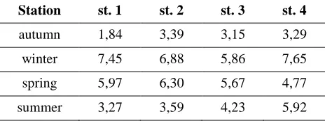
Fig. 2. Seasonal dynamics of bacteria MEM+ (live) in
each areas of the estuary (in percentage).
2.2. Marking of chloride ions concentration
Samples of the examined water were analy-zed chromatographically. Before injection to the chromatographic columns, samples were filtered by means of injection filters with the diameter of pores of 0.25 µm. Analysis of chloride anions in the examined waters was conducted using a ionic chromatograph by Metrohm (881 Compact IC pro), equipped with automatic sample feeder (863 Compact Autosampler). Separation of anions was carried out by means of column Metrosept A Supp 5-150/4.0 connected to protective column A Supp 4/5 Guard 4.0. The eluent was a mixture of 3.2 mM/L per2 CO3 and 1.0 mM/L of NaHCO3, flowing
with the intensity of 0.7 ml/min.
2.3. Marking organic matter
The organic matter was marked in terms of weight by marking out the difference between a dried sample, and a sample roasted for 4 hours in the temperature of 600°C. In order to remove carbonates the sample was previously treated with hydrochloric acid.
2.4. Marking the numbers of LIVE/DEAD bacteria
20 | Perliński et al. Participation of MEM+ bacteria in the bacterioplankton community in Ustka harbor
European Journal of Biological Research 2015; 5 (2): 17-24
(Millipore) type using Millipore filtration appa-ratus. After drying, the filters were observed under an epifluorescent microscope Olympus BX4 using dichronous filter B-2A (Ex. λ=480/490nm, Em. λ=500/635 nm) with the magnification of 1000x ("dry" lens, ocular 10x). From each preparation, fluorescent bacteria were counted from twenty ran-domly selected the fields of vision. Green stained cells were classified as MEM+, while cells revealing red fluorescence were qualified as MEM-. The results obtained during microscopic observations were converted according to the formula Zimmer-man, Mayer-Reil [20].
2.5. Statistical analysis
The statistical analysis of the obtained test results was done using STATISTICA 10 software. The variable distribution type was described by means of a Shapiro-Wilk normality test. If variable distribution met the condition of normality, for comparing the averages, ANOVA variance analysis was used. If variable distribution did not meet this condition, nonparametric test was used - the ANOVA Kruskal-Wallis rank test and the median test.
When testing the statistical association of two variables, the correlation coefficient was calculated. If at least one variable distribution did not meet the normality condition, Spearman's rank correlation
coefficient (r) was used. In order to determine the strength of association between correlated variables, appropriate tables with critical values of the corre-lation coefficient were used.
3. RESULTS
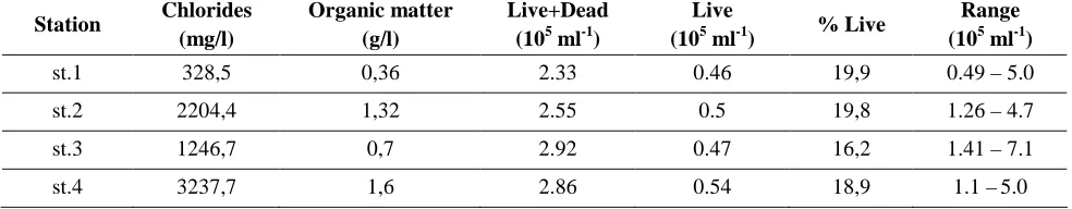
Data relating to concentration of chlorides and organic matter in particular zones of the port estuary have been presented in Table 1. They imply that the highest concentration of chlorides (3237.7 mg/l) was observed in the area of port breakwaters and the lowest (328.5 mg/l) in the area of the river mouth. A very similar situation was observed in concentration of organic matter, the most of which (1.6 g/l) was observed in the mouth of the estuary to the sea and the least (0.36 g/l) in the area of the river where water was least salty.
Table 1 also presents the results of the numbers of live and dead bacteria in particular zones of the Ustka estuary. It was stated that the total number of live and dead bacteria was highest in the coal basin (2.92·105 ml-1) and in the area of breakwaters (2.86·105 ml-1) whereas the smallest number of bacteria was recorded in the limnetic zone (2.33·105 ml-1). The share of live bacteria on three research posts in the mainstream remained at the level of approximately 19% only in the blind arm of the port, (st. 3) it was lower and amounted to 16.2%.
Table 1. The number of bacteria and the concentration of chloride ions and organic matter in the different zones
of the estuary.
Station Chlorides (mg/l)
Organic matter (g/l)
Live+Dead (105 ml-1)
Live
(105 ml-1) % Live
Range (105 ml-1)
st.1 328,5 0,36 2.33 0.46 19,9 0.49 – 5.0
st.2 2204,4 1,32 2.55 0.5 19,8 1.26 – 4.7
st.3 1246,7 0,7 2.92 0.47 16,2 1.41 – 7.1
st.4 3237,7 1,6 2.86 0.54 18,9 1.1 –5.0
Table 2 shows the dynamics of seasonal changes in the numbers of "live" bacteria on diffe-rent research posts. The presented data imply that at each post, bacteria were most numerous in the winter season, whereas the smallest number of bacteria populated all zones of the estuary in autumn. In addition, it was stated that in the limnetic