ONLINE FIRST
This is a provisional PDF only. Copyedited and fully formatted version will be made available soon.
ISSN: 0015-5659
e-ISSN: 1644-3284
Calvarial bone defects in ovariectomized rats treated with
mesenchymal stem cells and demineralized freeze-dried bone
allografts
Authors: E. T. Kadiroğlu, M. E. Akbalık, E. Karaöz, B. E. Kanay, A. Dağ, M. A.
Ketani, E. G. Eroğlu, E. Uysal, M. C. Tuncer
DOI: 10.5603/FM.a2020.0001
Article type: ORIGINAL ARTICLES
Submitted: 2019-11-06
Accepted: 2019-12-14
Published online: 2020-01-07
This article has been peer reviewed and published immediately upon acceptance.
It is an open access article, which means that it can be downloaded, printed, and distributed freely, provided the work is properly cited.
Articles in "Folia Morphologica" are listed in PubMed.
Calvarial bone defects in ovariectomized rats treated with mesenchymal stem cells and
demineralized freeze-dried bone allografts
Healing of calvarial bone defects with stem cells
E.T. Kadiroğlu1, M.E. Akbalık2, E. Karaöz3, B.E. Kanay4, A. Dağ5, M.A. Ketani6, E.G. Eroğlu7,
E. Uysal8, M.C. Tuncer9
1Department of Periodontology, Faculty of Dentistry, Dicle University, Diyarbakır, Turkey
2Department of Histology and Embryology, Faculty of Veterinary Medicine, Dicle University, Diyarbakır, Turkey
3Department of Histology and Embryology, Faculty of Medicine, University of İstinye, Istanbul, Turkey 4Department of Surgery, Faculty of Veterinary Medicine, Dicle University, Diyarbakır, Turkey
5Department of Periodontology, Faculty of Dentistry, Dicle University, Diyarbakır, Turkey
6Department of Histology and Embryology, Faculty of Veterinary Medicine, Dicle University, Diyarbakır, Turkey
7Oral and Dental Health Center, Van, Turkey
8Diyarbakır Vocational School of Technical Science, Dicle University, Diyarbakır, Turkey 9Department of Anatomy, Faculty of Medicine, Dicle University, Diyarbakır, Turkey
Address for correspondence: M.Cudi Tuncer, Professor, PhD (The Chief of Anatomy Department), Dicle University, Medical School, Department of Anatomy, 21280, Diyarbakır, Turkey, tel: +90 412 2488001 Ext. 4539 (Faculty room), fax: +90 412 2488440, Mobile phone: +90 532 2744926, e-mail: drcudi@hotmail.com
Abstract
The aim of the study was to investigate the ability of a combination of bone marrow
mesenchymal stem cells (BM-MSCs) with and without demineralized freeze-dried bone
allografts (DFDBAs) to induce bone regeneration in calvarial defects in ovariectomized rats.
Critical size defects were filled with a combination of demineralized freeze-dried bone
allografts and bone marrow mesenchymal stem cells (BM-MSCs) or BM-MSCs alone. Eight
weeks after calvarial surgery, the rats were sacrificed. The samples were analyzed
histologically and immunohistochemically. No difference was observed in vascularization
between groups C1 (animals with cranial defect only, control group) and O1 (animals with
cranial defect only, ovariectomy group). Intramembranous ossification was observed at a
limited level in groups C2 (animals with cranial defect with MSCs, control group) and O2
(animals with cranial defect with MSCs, ovariectomy group) compared to C1 and O1. In
group C3 (animals with demineralized freeze-dried bone allografts with MSCs, control
been placed in the cavity, but in group O3 (animals with demineralized freeze-dried bone
allografts with MSCs, ovariectomy group), the fibrous tissue was poorly distributed between
the bone grafts for the most parts. We conclude that the insertion of BM-MSCs enhances bone
healing; however, the DFDBA/BM-MSC combination has little effect on overcoming
impaired bone formation in ovariectomized rats.
Key words: bone healing, bone marrow mesenchymal stem cells (BM-MSCs),
demineralized freeze-dried bone allografts (DFDBAs), ovariectomy, calvarial defect
INTRODUCTION
Estrogen deficiency is an important cause of postmenopausal bone loss. It leads to an
imbalance in osteoblast and osteoclast number. The effect of estrogen on bone metabolism is
mediated by proinflammatory cytokines. In estrogen deficiency conditions, monocytes and
macrophages produce large amounts of the cytokines IL-1, IL-6, TNF-α,
granulocyte-macrophage colony-stimulating factor, granulocyte-macrophage colony-stimulating factor, and
prostaglandin-E2 that stimulate mature osteoclasts, and consequently induce bone resorption
(1).
Several studies have suggested that there is a relationship between systemic low bone
density and the onset of periodontitis, which is characterized by the loss of connective tissue
and alveolar bone, and risk factors, such as genetics, environmental factors, hormone levels,
smoking and diabetes, associated with osteoporosis. Authors have also remarked that
osteoporosis may have an effect on the progression of periodontitis via the loss of bone
mineral density in the maxilla and mandible in postmenopausal women (2,3).
Diminished bone density enhances the destruction of alveolar bone, which complicates bone
regenerative procedures (4).
Thus, immune cells directly contribute to bone remodeling, and the bone healing
process is known to be negatively affected by estrogen deficiency in elderly women, as
estrogen promotes osteoclastic activity. Many experimental studies have also shown delayed
wound healing (impaired bone healing) in ovariectomized rats (5-8).
The treatment of bone defects is particularly controversial in the case of osteoporosis.
The bone grafting procedure is thought to be the most widely used method to enhance bone
regeneration and repair bone defects, but it has certain drawbacks, including risks during
collection, hemorrhage, infection, chronic pain, sterilization, storage, especially foreign body
improve bony defects in osteoporosis. Mesenchymal stem cells (MSCs) have the ability to
differentiate into osteoblasts and are available from a wide variety of sources. Tissue
regeneration using autologous stem cells to form a suitable scaffold is an alternative to using
autografts and allografts (11). The bone-regeneration potential of mesenchymal stem cells has
been evaluated in bone defects in animals with or without scaffolds (12,13). Currently,
different methods for efficient tissue regeneration are being developed with various
combinations of stem cells and scaffolds (14,15).
In light of this information, we hypothesized that treatment with BM-MSCs combined
with bone grafts would facilitate bone repair in osteoporotic bone damage conditions. There is
little information available about the healing capacity of BM-MSCs used in combination with
demineralized freeze-dried bone allografts (DFDBAs) for the treatment of calvarial bone
defects. For this reason, we aimed to evaluate the effects of BM-MSCs and allografts on bone
healing in ovariectomized rats via the expression of immunohistochemical markers.
MATERIALS AND METHODS
Animals
A total of 48 female Wistar rats (250-300 g) provided by Scientific Application and
Research Centre of Dicle University (Protocol No: 12-DH-53) were used. All of the
procedures involved in the experimental protocols were approved by the Animal Ethics
Committee of Dicle University (Protocol No. 2011/15).The study was performed in
accordance with the Helsinki Declaration and with the permission of the Governmental
Animal Protection Committee. Because six animals died, the study was conducted with 42
rats. Whole animals were provided with commercial rat chow and water ad libitum and were
maintained on a 12 h light/12 h dark cycle at a temperature of 22 ±1 °C.
The animals were anesthetized by the intraperitoneal administration of xylazine and
ketamine and then subjected to ovariectomy. Ovariectomy was preceded by a 3 cm long
midline dorsal skin incision, approximately halfway between the middle of the back and the
base of the tail, according to the method described by Pires-Oliveira et al. (16). The animals
were monitored for infection.
Thirty days following ovariectomy, all animals were anesthetized for the introduction
of calvarial bone defects. After the head hair was shaved, a longitudinal midsagittal skin
incision was made to expose the parietal bones, and flaps were retracted in a subperiosteal
was made unilaterally in the parietal bone using trephine dental drills with saline water
irrigation. Care was taken to avoid injury to the dura in all animals (17).
The animals were randomized. The two groups were separated into three subgroups
(n=7). Group 1 (control group) was divided into C1; animals with cranial defect only, C2;
animals with cranial defectwith MSCs, and C3; animals with demineralized freeze-dried
bone allografts with MSCs. Group 2 (ovariectomy group) was divided into O1; animals with
cranial defect only, O2; animals with cranial defectwith MSCs, and O3; animals with
demineralized freeze-dried bone allografts with MSCs. Eight weeks after the calvarial
surgery, all of the rats were euthanized with an intraperitoneal overdose of ketamine
hydrochloride for histological evaluation.
Isolation and culture of rBM-MSCs
The isolation and culturing of rBM-MSCs (rat bone marrow mesenchymal stem cells)
were performed in vitro according to a published protocol (18). MSCs were isolated from the
bone marrow of rats. Under sterile conditions, femurs and tibias were excised from each rat,
bone marrow cells were isolated by flushing the bone marrow cavity with complete medium
(L-DMEM supplemented with 10 % fetal bovine serum [FBS, Gibco/Life Technologies] and
1% penicillin/streptomycin) delivered through a 21 gauge needle. After washing, the isolated
bone marrow cells were cultured in complete medium at 37°C in a humidified atmosphere of
5% CO2 for three days. The unattached cells were removed, and the adhered cells were
continually cultured until reaching 70-80%confluence. The cells were trypsinized and
passaged at a ratio of 1:2 or 1:3. The third-passage rBM-MSCs were pooled and used for
characterization and treatment.
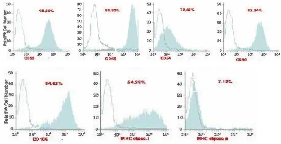
Characterization of rBM-MSCs
Undifferentiated rBM-MSCs were subjected to flow cytometry analysis (FACS
Calibur [BD Biosciences, San Jose, CA]). The cell suspension was spun at 1000 RPM for 5
minutes and the supernatant was decanted. The pellet was resuspended in 1X PBS. The cells
were counted with a hemocytometer.The desired total number of cells was added to a flow
tube (0.5-1 x 10e6 per sample). Wash the cells by adding ~ 1 ml 1X PBS to the flow tube. The
cell suspension was spun at 1000 RPM for 5 minutes and the supernatant was decanted.
Gently tap the tube to loosen the cell pellet.An appropriate amount of staining buffer (50 ul
of flow tubes. Finally, immunophenotyping analysis was performed for the antigens CD29,
CD45, CD54, CD90, CD106, MHC class-I and MHC class-II (BD Biosciences).
In vitro differentiation of rBM-MSCs
Adipogenic and osteogenic differentiation was performed in vitro according to a
published protocol (19). Adipogenic differentiation was performed by incubating rBM-MSCs
with L-DMEM supplemented with 0.5 mM isobutyl-methylxanthine, 10-6M dexamethasone, 10 μg/ml insulin and 200 μM indomethacin for two weeks. The medium was refreshed every
3–4 days. The formation of intracellular lipid droplets, which indicates adipogenic
differentiation, was confirmed by staining with 0.5% oil red O (Sigma-Aldrich, St. Louis,
MO).For osteogenic differentiation, the cells were cultured with L-DMEM supplemented with 100 nM dexamethasone, 0.05 μM ascorbate-2-phosphate, and 10 mM β-glycerophosphate for
four weeks. After four weeks, osteogenic differentiation was assessed via staining with 2%
alizarin red S (pH 4.1–4.3; Fluka, Buchs, Switzerland).
Green fluorescent protein (GFP) labeling of rBM-MSCs
Mesenchymal stem cells were transfected with pGFP-N (Clontech, Palo Alto, CA) by
electroporation (Neon Transfection System, Invitrogen, Carlsbad, CA) following the
instructions provided by the manufacturer. After transfection, the cells were cultured with
L-DMEM (supplemented with 10% FBS), and the transformed cells were selected with G418 (Gibco/Life Sciences; 200 μg/ml) under standard culture conditions for 48 h. GFP-positive cells were maintained in the same medium supplemented with G418 (200 μg/ml) for three
passages. The numberof GFP+ cells was monitored by flow cytometry;> 90% of the cells
used in the treatment were GFP positive.
Immunohistochemical analysis of GFP+rBM-MSCs in the tissue
Consecutive sections, each 4 µm thick, were taken from each paraffin-embedded
tissue. To detect GFP+rBM-MSCs, an immunofluorescence staining protocol was performed.
Slides were deparaffinized with xylene for 5 min twice and rehydrated in a series of graded
alcohol solutions (70 to 100%). Endogenous peroxidases were inhibited by incubation with
3% H2O2 in PBS buffer. For antigen retrieval, the samples were heated to 98–99 °C in
antigen retrieval buffer (10 mM sodium citrate, 0.05% Tween 20, pH 6.0) and incubated for
30 min in a pressurized vessel. Nonspecific staining was blocked with a mixture of 1.5%
monoclonal anti-GFP antibody (SC-9996) at a 1:50 dilution for 1 h at room temperature. After
incubation with appropriate fluorophore-conjugated secondary antibodies, the sections were
covered with mounting medium containing DAPI (Santa Cruz, Heidelberg, Germany). The
cells were investigated under a fluorescence microscope (Leica DMI 4000B, Wetzlar,
Germany).
Light microscopy
Calvarial tissues were removed and fixed in 10% neutral buffered formaldehyde
solution for 48 h and then decalcified in 10% ethylene diaminetetraacetic acid (EDTA)
prepared in 0.1 M Tris-HCl buffer (pH 7.4) for 14 days. The tissues were subsequently
dehydrated, cleared and embedded in paraffin blocks. Five-micrometer-thick sections were cut from these blocks and stained using Gomori’s method to determine ossification.
Immunohistochemical staining
Immunohistochemical investigations were performed on tissue preparations embedded
in paraffin and by using a Zymed Histostain Plus Bulk kit (code: 85-9043, Histostain Plus
Bulk Kit, Zymed, South San Francisco, CA, USA) and streptavidin-peroxidase (Akbalik and
Ketani 2013) (19). Briefly, sections were deparaffinized, rehydrated and incubated for 15 min
in 3% H2O2 in methanol. After the sections were washed in phosphate-buffered saline (PBS),
antigen retrieval was performed by boiling in 0.01 M citric buffer pH 6.0 for 30 min at 95 ºC
using a water bath and by cooling for 20 min prior to immunostaining. Sections were then
washed in PBS and incubated in protein blocking solution (Ultra V Block) for 10 min at room
temperature to prevent nonspecific binding. Subsequently, the preparations were incubated
with primary antibodies for 20 h at +4ºC (Table 1)
Table 1. Details of antibodies used.
Antibody Clone, isotype Host Cellular
localization
Dilution Supplier
(Catalog no.)
Osteopontin/OPN
(human)
AKm2A1, monoclonal
IgG
Mouse Cytoplasmic 1:200 Santa Cruz (sc-21742)
Osteonectin/Sparc
(human)
H-90, polyclonal IgG Rabbit Cytoplasmic 1:200 Santa Cruz (sc-25574)
After being washed in PBS, the sections were incubated with biotinylated secondary
antibodies for 20 min at room temperature and washed in PBS. Subsequently, the preparations
were incubated in streptavidin peroxidase conjugate for 20 min at room temperature and were
then washed with PBS. To visualize the reaction, the sections were treated with
3’3-diaminobenzidine (DAB) for 5-15 min. After the reaction developed, the sections were counterstained with Gill’s hematoxylin, dehydrated through an alcohol series, cleared in
xylene, and finally mounted in entellan. Negative controls were used for the confirmation of
the staining. As a negative control, the primary antibodies used for staining were replaced
with PBS. OPN, OC and ON expression in bone tissue was examined microscopically at x200
magnification. Immunohistochemical staining results were evaluated semi quantitatively. The
intensity of positive staining was defined as + weak, ++ medium, +++ strong, + / ++ weak to
moderate, and ++ / +++ moderate to strong. The slides were examined and photographed
using a Nikon Eclipse E400 (Nikon, Tokyo, Japan) microscope equipped with a digital
camera (Nikon Coolpix-4500).
Statistical analysis
Kruskal Wallis and Mann-Whitney U tests were used as nonparametric statistical
analyses. Values of p<0.05 were considered statistically significant.
RESULTS
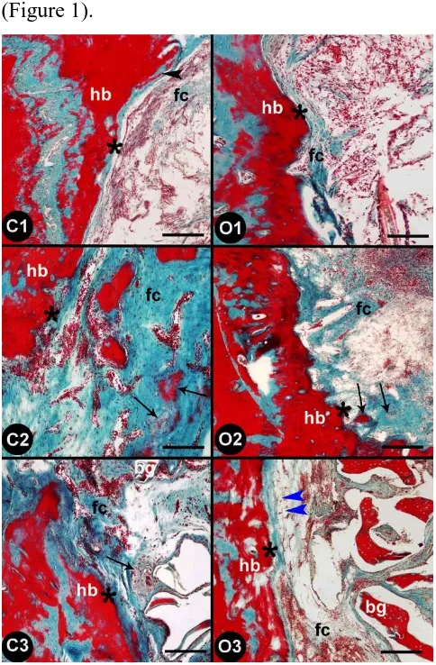
Histological findings in calvarial bone tissue
In C1 group (animals with cranial defect only, control group), structures (bone
spicules) characterized by intramembranous ossification were observed along the border of
the cavity. Furthermore, fibrous tissue was abundant in the cavity and vascularization
(angiogenesis) had occurred in some areas. In O1 group (animals with cranial defect only,
ovariectomy group), intramembranous ossification was observed neither in the cavity nor
along the border of the cavity, and fibrous tissue was not diffusely distributed, but cellular
structures were markedly abundant. No significant revascularization was observed between
control (animals with cranial defect only) and overectomy group rats (animals with cranial
defect only).
O1 (animals with cranial defect only, ovariectomy group), intramembranous
ossification and osteogenesis had occurred locally, and fibrous tissue was diffuse and
regularly distributed. Furthermore, vascularization was significantly increased. In group O2
was observed at a limited level and was localized to the border of the cavity. Furthermore,
fibrous tissue was irregularly distributed in only some parts of the cavity. The vascularization
in groups C2 (animals with cranial defectwith MSCs, control group) and O2 (animals with
cranial defectwith MSCs, ovariectomy group) did not differ.
When compared to those in the other control groups, the fibrous structures of the
matrix in group C3 (animals with demineralized freeze-dried bone allografts with MSCs,
control group) displayed a compact structure as a result of a bone graft having been placed in
the cavity, and these structures were also tightly adhered to the border of the cavity.
Intramembranous ossification was observed between the portions of the bone graft. In group
O3 (animals with demineralized freeze-dried bone allografts with MSCs, ovariectomy group),
ossification was not observed. The fibrous tissue showed a weak distribution between the
bone grafts and along the border of the cavity, in a strip-like formation along the latter.
Furthermore, in contrast to those in the other groups, the stem cells had differentiated into
adipose cells along the border of the cavity and were present in the form of infiltrating cells
(Figure 1).
and O1, and the defect areas were treated with stem cells (C2, O2) and stem cells+bone grafts (C3, O3); hb: host bone, fc: fibrous connective tissue, bg: bone graft, asterisk: interface between host bone and defect, arrow:
ossification areas, black arrowhead: bone spicule, blue arrowhead: adipocytes. Gomori’s staining method. Scale bars: 100 μm (C1-C2 and O1-O2) and 250 μm (C3-O3).
Immunohistochemistry for osteopontin (OPN)
In C1 group (animals with cranial defect only, control group), the osteoprogenitor cells
along the border of the cavity were not immunoreactive for OPN. However, while the
osteoblasts and extracellular matrix in the areas of intramembranous ossification along the
border of the cavity showed weak OPN immunoreactivity, the newly formed blood vessels in
the cavity were positively stained for OPN (Figure 2-C1). In O1 group (animals with cranial
defect only, ovariectomy group), the normal bone tissue showed positive immunoreactivity
for OPN, but OPN immunoreactivity was limited to the blood vessels in the newly formed
tissue in the cavity (Figure 2-O1).
In group C2 (animals with cranial defectwith MSCs, control group), similar to group
C1, the osteoprogenitor cells along the border of the cavity did not show OPN
immunoreactivity. Although the osteoblasts in the cavity presented weak OPN staining, OPN
expression in the blood vessels was increased (Figure 2-C2). In group O2 (animals with
cranial defectwith MSCs, ovariectomy group), staining for OPN was observed neither in the
osteoprogenitor cells along the border of the cavity nor in the osteoblasts in the cavity, but the
extracellular matrix and blood vessels were positive for OPN (Figure 2-O2).
In group C3 (animals with demineralized freeze-dried bone allografts with MSCs,
control group), the osteoprogenitor cells along the border of the cavity and the newly formed
osteoblasts and extracellular matrix in the periphery of the bone graft demonstrated moderate
OPN immunoreactivity, whereas the blood vessels showed strong OPN immunoreactivity
(Figure 2-C3). In group O3 (animals with demineralized freeze-dried bone allografts with
MSCs, ovariectomy group), the newly formed osteoblasts in the periphery of the bone graft
presented moderate immunoreactivity for OPN, whereas the staining of the blood vessels was
Figure 2. Osteopontin expression in the rat calvaria bone defect areas in the control (C1, C2, and C3) and ovariectomy (O1, O2, and O3) groups. The formation of the groups is shown in figure 1; hb: host bone, fc:
fibrous connective tissue, bv: blood vessel, bg: bone graft, asterisk: interface between host bone and defect,
arrow: osteoprogenitor cell, arrowhead: osteoblast. Scale bars: 25 μm (C1), 50 μm (C2-C3-O3), and 100 μm (O1-O2).
Immunohistochemistry for osteocalcin (OC)
In C1 group (animals with cranial defect only, control group), the osteoprogenitor cells
along the border of the cavity did not show immunoreactivity for OC, but the osteoblasts
presented weak OC immunoreactivity. The extracellular matrix in the border of the cavity
showed moderate staining, and the blood vessels stained positively for OC (Figure 3-C1). In
O1 group (animals with cranial defect only, ovariectomy group), the osteoprogenitor cells
also showed OC immunoreactivity, and when compared to that of group C1, the OC
immunoreactivity of the osteoblasts and extracellular matrix was weaker (Figure 3-O1).
Compared with that in the control group, the OC immunoreactivity of the
osteoprogenitor cells along the border of the cavity in group C2 (animals with cranial defect
matrix and the blood vessels localized to the border of the cavity and the cavity was more
intense. Furthermore, the osteoblasts in the cavity displayed weak immunoreactivity for OC
(Figure 3-C2). In group O2 (animals with cranial defectwith MSCs, ovariectomy group), the
osteoprogenitor cells did not show any OC immunoreactivity, and the osteoblasts displayed
weak OC immunoreactivity. The extracellular matrix of the border of the cavity stained
strongly for OC and the staining in the cavity was observed to be weak. The OC
immunoreactivity of the blood vessels was also determined to be strong (Figure 3-O2).
In group C3 (animals with demineralized freeze-dried bone allografts with MSCs,
control group), the newly formed osteoblasts in the periphery of the bone graft displayed OC
immunoreactivity ranging from moderate to strong, whereas the osteocytes in the ossified
areas displayed weak OC immunoreactivity. The OC immunoreactivity of the extracellular
matrix was more homogenous than that in the other groups. Blood vessels also stained
positively for OC (Figure 3-C3). In group O3 (animals with demineralized freeze-dried bone
allografts with MSCs, ovariectomy group), the osteoprogenitor cells displayed negative
staining and the osteoblasts and extracellular matrix were stained moderately. However, the
Figure 3. Osteocalcin expression in the rat calvaria bone defect areas in the control (C1, C2, and C3) and ovariectomy (O1, O2, and O3) groups. The formation of the groups is shown in Figure 1; hb: host bone, fc:
fibrous connective tissue, bv: blood vessel, bg: bone graft, asterisk: interface between host bone and defect,
arrow: osteoprogenitor cell, black arrowhead: osteoblast, blue arrowhead: osteocyte. Scale bars: 50 μm (C1-C2-O2), and 100 μm (C3-O1-O3).
Immunohistochemistry for osteonectin (ON)
In C1 group (animals with cranial defect only, control group), while the
osteoprogenitor cells along the border of the cavity and the osteoblasts in the cavity stained
negatively for ON, the extracellular matrix showed weak immunoreactivity, and the blood
vessels showed moderate immunoreactivity for ON (Figure 4-C1). Group O1(animals with
cranial defect only, ovariectomy group) showed staining results similar to those of group C1,
but the extracellular matrix stained weakly (Figure 4-O1).
In group C2 (animals with cranial defectwith MSCs, control group), the
osteoprogenitor cells along the border of the cavity were stained negatively, and the
osteoblasts in the cavity showed weak ON expression. In contrast, the extracellular matrix and
blood vessels were strongly stained (Figure 4-C2). In group O2 (animals with cranial defect
with MSCs, ovariectomy group), the osteoprogenitor cells along the border of the cavity did
not stain, the osteoblasts in the cavity showed weak immunoreactivity for ON. The
extracellular matrix and blood vessel findings were similar to those of group C2 (Figure
4-O2).
In group C3 (animals with demineralized freeze-dried bone allografts with MSCs,
control group), the osteoprogenitor cells along the border of the cavity did not express ON,
but the newly formed osteoblasts in the periphery of the bone graft displayed moderate
staining. In contrast, the extracellular matrix and blood vessels displayed strong ON
expression (Figure 4-C3). In group O3 (animals with demineralized freeze-dried bone
allografts with MSCs, ovariectomy group), the osteoprogenitor cells showed no staining and
the osteoblasts in the periphery of the bone graft displayed moderate ON expression. It was
observed that adipose cells had formed in the cavity, yet these cells displayed no
immunoreactivity. However, the extracellular matrix and blood vessels showed strong ON
Figure 4. Osteonectin expression in the rat calvaria defect areas in the control (C1, C2, and C3) and ovariectomy (O1, O2, and O3) groups. The formation of the groups is shown in Figure 1; hb: host bone, fc: fibrous
connective tissue, bv: blood vessel, bg: bone graft, asterisk: interface between host bone and defect,
arrowhead: osteoblast. Scale bars: 50 μm (C1-C2-O2) and 100 μm (C3-O1-O3).
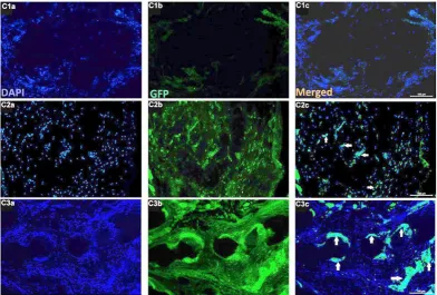
Findings of green fluorescent protein (GFP) labeling of RBM-MSCs
In the control group, no immune reaction was observed for GFP in animals with
cranial defect only, without MSCs, whereas GFP and cells (arrows) were observed in C2
(animals with cranial defectwith MSCs, control group) and C3 (animals with demineralized
dried bone allografts with MSCs, control group). Animals with demineralized
freeze-dried bone allografts in the control group, the MSCs transplanted with the graft show the new
tissue formation (Figure 5).In the overectomy group, no immune reaction was observed for
GFP in the non- MSCs group (O1), whereas GFP and cells (arrows) were observed in O2
(animals with cranial defectwith MSCs, ovariectomy group) and O3 (animals with
demineralized freeze-dried bone allografts with MSCs, ovariectomy group). MSCs
Figure 5. GFP fluorescence after immunostaining of the tissue sections from the control group. C1; animals with cranial defect only, C2; animals with cranial defectwith MSCs, C3; animals with demineralized freeze-dried bone allografts with MSCs. Immune reaction (white arrows) was observed in GFP and cells belonging to C2 and C3 groups. MSCs transplanted in C3 with the graft showed new bone tissue formation. Scale bars:100 µm.
In addition, the cell expression of each marker is given in Table 2 for fluorescence intensity.
Table 2. Fluorescence intensity. Numbers in panels represent mean fluorescent intensity of
the cells expressing each marker.
Statistical findings
Groups p Parameter n Average Rank Different (p<0.05) from factor nr
(1) C1
0,0001 1
7 7,50 (3)
(2) C2 7 7,50 (3)
(3) C3 7 18,00 (1)(2)
(1) C1
0,0109 2
7 7,36 (3)
(2) C2 7 9,71 (3)
(3) C3 7 15,93 (1)(2)
(1) C1
ns 3
7 11,00
ns
(2) C2 7 11,00
(3) C3 7 11,00
(1) C1
0,0007 4
7 4,86 (2)(3)
(2) C2 7 16,57 (1)(3)
(3) C3 7 11,57 (1)(2)
(1) O1
ns 1
7 11,00
ns
(2) O2 7 11,00
(3) O3 7 11,00
(1) O1
0,001 2
7 7,50 (3)
(3) O3 7 18,00 (1)(2)
(1) O1
ns 3
7 11,00
ns
(2) O2 7 11,00
(3) O3 7 11,00
(1) O1
0,0004 4
7 4,00 (2)(3)
(2) O2 7 15,00 (1)
(3) O3 7 14,00 (1)
Table 3. Comparisons of the parameters within the groups (ns; non-significant ; p>0,05)
1; osteopregenitor cells, 2; osteoblasts, 3;osteocytes, 4;extracelluler matrix
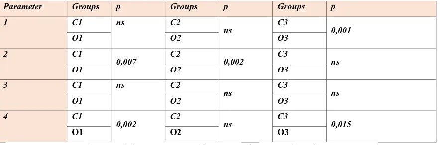
Parameter Groups p Groups p Groups p
1 C1 ns C2
ns C3 0,001
O1 O2 O3
2 C1
0,007 C2 0,002 C3 ns
O1 O2 O3
3 C1 ns C2
ns C3 ns
O1 O2 O3
4 C1
0,002 C2 ns C3 0,015
O1 O2 O3
Table 4. Comparisons of the parameters between the control and test groups (ns;
non-significant ; p>0,05) 1; osteopregenitor cells, 2; osteoblasts, 3; osteocytes, 4; extracelluler
matrix
When osteopregenitor cells were evaluated between control (animals with
demineralized freeze-dried bone allografts) and overectomy groups (animals with
demineralized freeze-dried bone allografts), a significant statistical difference was found
(Table 3 and 4).When osteoblast cells were evaluated between control and overectomy
groups,the difference between C1 (animals with cranial defect only, control group)-O1
(animals with cranial defect only, ovariectomy group) and C2 (animals with cranial defect
with MSCs, control group) -O2 (animals with cranial defect with MSCs, ovariectomy group)
groups was found to be statistically significant (Table 3 and 4). And,when the extracellular
matrix was evaluated, the difference between C1(animals with cranial defect only, control
group)-O1 (animals with cranial defect only, ovariectomy group) and C3 (animals with
demineralized freeze-dried bone allografts with MSCs, control group)-O3 (animals with
demineralized freeze-dried bone allografts with MSCs, ovariectomy group) groups was found
DISCUSSION
We aimed to investigate the healing capacity of BM-MSCs used with DFDBAs for the
treatment of Calvarial bone defects, assuming that BM-MSC combined with bone grafts
would facilitate bone repair under conditions of osteoporotic bone injury.Therefore, the
effects of BM-MSCs and allografts on bone healing in ovariectomized rats were concluded by
evaluating immunohistochemical results.
Sethi et al. (20) evaluated and interpreted both clinically and radiographically by
studying the changes post 1 week, 1 month, 3 months, and 6 months, respectively. It was
stated that there was evidence of trabecular formation and calcification. They concluded that
platelet-rich plasma-enriched DFDBA was a superior inoculant in terms of other available
inoculants in patients (20).In this experimental study, we observed the successful results of
demineralized freeze-dried bone allografts. Called ‘‘Mesenchymal Stem Cell Chondrocytes
technique’’ was used to reconstruct a 15 mm massive femoral defect (approximately 50% of
rat femur shaft length) in an experimental study.According to their results, considering the
high repairability and the excellent biomechanical forces of the repaired femora, they
concluded that the reconstruction of the large bone defect may be possible (21).
In an experimental study in which 8 mm defect was applied to one of the groups in
which decalcified freeze-dried bone allograft was applied, statistically significant results were
obtained when compared with the other groups (22).In an experimental study, ovarectomy
was performed methodologically as in our study.It has been stated that limited proteins are
known to be involved in the subsequent stages of bone formation and maturation in the
proteomes of animals (23)
As a result of these cellular changes ,osteogenesis becomes impaired and the bone
formation period is insufficient to repair the increased bone destruction in estrogen
deficiency-related osteoporosis (24,25). He et al. (8) also found that the femurs of mice with
OVX-induced osteoporosis showed impaired angiogenesis, osteogenesis, and remodeling in
their study (8). Calciolari et al. (23) observed immature bone formation both in OVX and
control calvarial CSDs over a 30-day period (23). Stockmann et al. (26) also observed a
similar result during the early stage of bone healing on the 30th day. There were no
differences between the test and control groups, but at 60 days, new bone formation was
achieved in the MSC group; however, significant pig calvarial bone regeneration was
measured at day 90 (26). In our study, we observed intramembranous ossification in animals
with cranial defect only of the healthy control rats , after 60 days. Consistent with other
either in the cavity or along the border of the cavity of animals with cranial defect only with
ovariectomy group after 8 weeks. However, in cortical bones, such as the calvarial bone, the
healing process is slower than that in cancellous bone, with a poorer blood supply and less
bone marrow; thus, much more than 60 days might be necessary for mature bone formation.
Recently, it has been stated that the advantages and disadvantages of autogenous,
allogeneic, xenogenic and alloplastic materials have gained meaning in periodontal treatment.
(27). Kurkalli et al. (28) concluded that the placement of the osteogenic composite in a large
deficient area of the parietal bone of the skull of rats resulted in a large demineralized bone
matrix particle structure, fully reconstituted hematopoietic microenvironment within thirty
days, and a well-integrated normal smooth bone. (28). Intini et al. (29) found that
demineralized freeze-dried bone was not effective enough to induce bone formation in rat
calvaria 8 weeks after surgery (29). Caplanis et al. (30) did not find any histological effects on
bone formationin canine defects treated with demineralized freeze-dried bone allografts after
4 weeks (30). Bertolai et al. (31) have successfully used freeze-dried bone as a graft material
in the treatment of maxillary atrophy, as in our study. (31).
In the meta-analysis, the authors concluded that MSCs improved bone regeneration,
and it is preferable to use MSCs with an appropriate scaffold. Koob S et al. mentioned that
mesenchymal stem cells (MSCs) enhance bone formation in calvarial defects (32). Moreover,
in large animal studies, autologous MSCs transplanted alone or in combination with different
bone substitutes were found to significantly increase bone formation in critical-sized defects
(9). Kandal et al. suggested that the combined use of demineralized bone matrix with MSCs
increases the osteoinductive responses in the frontal bone of rats. They suggested that this
combination can provide enhanced craniofacial bone reconstruction results at the end of 12
weeks (33). In another experimental study, the utilization of mesenchymal stem cells with
platelet-rich plasma and synthetic bone substitutes was found to enhance new bone formation
(34). Semyari et al. observed the overall recovery of a bony defect treated with mesenchymal
stem cells on different scaffolds with membranes after 8 weeks of calvarial surgery in rabbits
(35).
Osteopontin has been implicated as being an important factor in bone
remodeling. Research suggests it plays a role in attaching osteoclasts to the mineral matrix of
bones and in the regulation of normal mineralization within the extracellular matrices of
bones and teeth. Osteocalcin and osteonectin are not observed during initial crystal formation
but are seen in the later stages of bone formation (36). Therefore, we chose these three bone
deficiency on bone healing via the expression of these bone markers, as there are few studies
on this issue. In our study, the osteoblasts and extracellular matrix staining for these proteins
was weak in animals with cranial defect only in the control and ovariectomy groups without
MSCs. Histological and immunohistochemical evaluation revealed that the findings obtained
for the extracellular matrix, ossification and blood vessels were similar between groups O2
(animals with cranial defect with MSCs, ovariectomy group) and C2 (animals with cranial
defectwith MSCs, control group). Thus, estrogen deficiency may not influence the expression
of bone markers, which is consistent with the findings of Tera Tde et al. (36). According to
the results of this study, more new bone formation was observed in defects treated with MSCs
alone than was observed in animals with cranial defect only. However, the combination of
DFDBA/MSCs in animals with demineralized freeze-dried bone allografts of ovariectomy
group was not as effective on compact intramembranous ossification at the end of the 8 weeks
as expected.
Akita et al. found that there were no significant differences in 4 mm cranial rat defects
among groups treated with MSCs only or MSCs with FGF-BMP at 8 weeks after
transplantation (17). Similar to our study, Wang et al. created bony defects in ovariectomized
rabbits and treated the defects with mesenchymal stem cells/decalcified bone matrix. Three
months later, the authors concluded that the defect treatment was ineffective for the
osteoporotic state and that the bone formation was significantly worse than that of the control
group (37).
The properties of scaffolds are important for the migration, proliferation and
differentiation of living cells during bone regeneration. In this study, the combination of
human (DFDBAs) and animal (rBM-MSCs) scaffolds may be biologically incompatible.
However, the osteogenic potential of the DFDBA may be diminished during the production
process. Additionally, bone healing may have been negatively affected by the absence of a
collagen membrane in the scaffold.
There are some limitations to the current study. It would be better to evaluate bone
formation with histomorphometric parameters than histochemical staining. In addition, a
collagen membrane may be used with the scaffold for 12 weeks to achieve complete bone
regeneration. In conclusion, stem cell therapy could be an option to manage impaired bone
formation. However, to achieve compact bone formation it is preferential to use proper
scaffolds loaded with BM-MSCs for the appropriate healing time. Because there are limited
differentiation of MSCs in different scaffolds for the enhancement of impaired bone
formation.
Acknowledgments
This work was supported by the Scientific Application and Research Centre of Dicle
University.(Protocol No: 12-DH-53).
REFERENCES
1. Riggs BL. The mechanisms of estrogen regulation of bone resorption.J Clin Invest. 2000;106(10):1203-4. doi: 10.1172/JCI11468.
2. Juluri R, Prashanth E, Gopalakrishnan D, Kathariya R, Devanoorkar A, Viswanathan V, et al. Association of Postmenopausal Osteoporosis and Periodontal Disease: A Double-Blind Case-Control Study. J Int Oral Health. 2015;7(9):119-23.
3.Richa, R Y, Puranik MP, Shrivastava A. Association between osteoporosis and periodontal disease among postmenopausal Indian women. J Investig Clin Dent. 2017;8(3). doi: 10.1111/jicd.12223.
4. Miron RJ, Wei L, Yang S, Caluseru OM, Sculean A, Zhang Y. Effect of enamel matrix derivative on periodontal wound healing and regeneration in an osteoporotic model. J Periodontol. 2014;85(11):1603-11. doi: 10.1902/jop.2014.130745.
5. Palomo L, Williams K, Thacker H. Periodontal Healing and Osteoporosis in Postmenopausal Women. 2016;Ann Gerontol Geriatric Res. 3(3):1043.
6. Tarantino U, Cerocchi I, Scialdoni A, Saturnino L, Feola M, Celi M, et al.Bone healing and osteoporosis. Aging Clin Exp Res. 2011;23(2 Suppl):62-4.
7. Weitzmann MN, Pacifici R.Estrogen deficiency and bone loss: an inflammatory tale.J Clin Invest. 2006;116(5):1186-94.doi: 10.1172/JCI28550.
8. He YX, Zhang G, Pan XH, Liu Z, Zheng LZ, Chan CW, et al.Impaired bone healing pattern in mice with ovariectomy-induced osteoporosis: A drill-hole defect model.Bone. 2011;48(6):1388-400. doi:
10.1016/j.bone.2011.03.720.
9. Im JY, Min WK, You C, Kim HO, Jin HK, Bae JS. Bone regeneration of mouse critical-sized calvarial defects with human mesenchymal stem cells in scaffold.Lab Anim Res. 2013;29(4):196-203. doi:
10.5625/lar.2013.29.4.196.
10. Saad KA, Abu-Shahba AG, El-Drieny EA, Khedr MS. Evaluation of the role of autogenous bone-marrow-derived mesenchymal stem cell transplantation for the repair of mandibular bone defects in rabbits. J
Craniomaxillofac Surg. 2015;43(7):1151-60. doi: 10.1016/j.jcms.2015.04.013.
11. Lu W, Ji K, Kirkham J, Yan Y, Boccaccini AR, Kellett M, et al.Bone tissue engineering by using a combination of polymer/Bioglass composites with human adipose-derived stem cells.Cell Tissue Res. 2014;356(1):97-107. doi: 10.1007/s00441-013-1770-z.
13. Gamie Z, Tran GT, Vyzas G, Korres N, Heliotis M, Mantalaris A, et al.Stem cells combined with bone graft substitutes in skeletal tissue engineering.Expert Opin Biol Ther. 2012;12(6):713-29. doi:
10.1517/14712598.2012.679652.
14. Alfotawei R, Naudi KB, Lappin D, Barbenel J, Di Silvio L, Hunter K, et al.The use of TriCalcium
Phosphate (TCP) and stem cells for the regeneration of osteoperiosteal critical-size mandibular bony defects, an in vitro and preclinical study.J Craniomaxillofac Surg. 2014;42(6):863-9. doi: 10.1016/j.jcms.2013.12.006. 15. Vahabi S, Amirizadeh N, Shokrgozar MA, Mofeed R, Mashhadi A, Aghaloo M, et al. A comparison between the efficacy of Bio-Oss, hydroxyapatite tricalcium phosphate and combination of mesenchymal stem cells in inducing bone regeneration.Chang Gung Med J. 2012;35(1):28-37.
16. Pires-Oliveira DA, Oliveira RF, Amadei SU, Pacheco-Soares C, Rocha RF.Laser 904 nm action on bone repair in rats with osteoporosis. Osteoporos Int. 2010;21(12):2109-14. doi: 10.1007/s00198-010-1183-8. 17. Akita S, Fukui M, Nakagawa H, Fujii T, Akino K. Cranial bone defect healing is accelerated by mesenchymal stem cells induced by coadministration of bone morphogenetic protein-2 and basic fibroblast growth factor.Wound Repair Regen. 2004;12(2):252-9.doi: 10.1111/j.1067-1927.2004.012118.x
18. Karaoz E, Aksoy A, Ayhan S, Sariboyaci AE, Kaymaz F, Kasap M.Characterization of mesenchymal stem cells from rat bone marrow: ultrastructural properties, differentiation potential and immunophenotypic markers. Histochem Cell Biol. 2009;132(5):533-46. doi: 10.1007/s00418-009-0629-6.
19. Akbalik ME, Ketani MA.Expression of epidermal growth factor receptors and epidermal growth factor, amphiregulin and neuregulin in bovine uteroplacental tissues during gestation.Placenta. 2013;34(12):1232-42. doi: 10.1016/j.placenta.2013.09.019.
20. Sethi AK, Kar IB, Mohanty T, Mishra N, Singh AK.Use of plasma-enriched demineralized freeze-dried bone matrix in postsurgical jaw defects.Natl J Maxillofac Surg. 2018;9(2):174-183. doi:
10.4103/njms.NJMS_33_18.
21.Watanabe Y, Harada N, Sato K, Abe S, Yamanaka K, Matushita T.Stem cell therapy: is there a future for reconstruction of large bone defects? Injury. 2016;47(Suppl 1):S47-51. doi: 10.1016/S0020-1383(16)30012-2. 22. Mokbel N, Bou Serhal C, Matni G, Naaman N.Healing patterns of critical size bony defects in rat following bone graft.Oral Maxillofac Surg. 2008;12(2):73-8. doi: 10.1007/s10006-008-0107-7.
23. Calciolari E, Mardas N, Dereka X, Anagnostopoulos AK, Tsangaris GT, Donos N.The effect of experimental osteoporosis on bone regeneration: part 2, proteomics results.Clin Oral Implants Res. 2017;28(9):e135-e145. doi: 10.1111/clr.12950.
24. McCann RM, Colleary G, Geddis C, Clarke SA, Jordan GR, Dickson GR, et al.Effect of osteoporosis on bone mineral density and fracture repair in a rat femoral fracture model.J Orthop Res. 2008;26(3):384-93.doi: 10.1002/jor.20505.
25. Namkung-Matthai H, Appleyard R, Jansen J, Hao Lin J, Maastricht S, Swain M, et al.Osteoporosis influences the early period of fracture healing in a rat osteoporotic model. Bone. 2001;28(1):80-6.doi: 10.1016/s8756-3282(00)00414-2.
26. Stockmann P, Park J, von Wilmowsky C, Nkenke E, Felszeghy E, Dehner JF, et al.Guided bone
regeneration in pig calvarial bone defects using autologous mesenchymal stem/progenitor cells - a comparison of different tissue sources.J Craniomaxillofac Surg. 2012;40(4):310-20. doi: 10.1016/j.jcms.2011.05.004.
28. Kurkalli BG, Gurevitch O, Sosnik A, Cohn D, Slavin S.Repair of bone defect using bone marrow cells and demineralized bone matrix supplemented with polymeric materials.Curr Stem Cell Res Ther. 2010;5(1):49-56. 29. Intini G, Andreana S, Buhite RJ, Bobek LA.A comparative analysis of bone formation induced by human demineralized freeze-dried bone and enamel matrix derivative in rat calvaria critical-size bone defects.J Periodontol. 2008;79(7):1217-24. doi: 10.1902/jop.2008.070435.
30. Caplanis N, Lee MB, Zimmerman GJ, Selvig KA, Wikesjö UM.Effect of allogeneic freeze-dried demineralized bone matrix on regeneration of alveolar bone and periodontal attachment in dogs.J Clin Periodontol. 1998;25(10):801-6.doi: 10.1111/j.1600-051x.1998.tb02373.x.
31. Bertolai R, Catelani C, Aversa A, Rossi A, Giannini D, Bani D.Bone graft and mesenchimal stem cells: clinical observations and histological analysis.Clin Cases Miner Bone Metab. 2015;12(2):183-7. doi: 10.11138/ccmbm/2015.12.2.183.
32. Koob S, Torio-Padron N, Stark GB, Hannig C, Stankovic Z, Finkenzeller G.Bone formation and
neovascularization mediated by mesenchymal stem cells and endothelial cells in critical-sized calvarial defects. Tissue Eng Part A. 2011;17(3-4):311-21. doi: 10.1089/ten.TEA.2010.0338.
33. Kandal S, Özmen S, Uygur S, Yagci M, Kayhan H, Elmas C, et al. Effects of Rat Bone Marrow-Derived Mesenchymal Stem Cells and Demineralized Bone Matrix on Cranial Bone Healing.Ann Plast Surg. 2016;77(2):249-54. doi: 10.1097/SAP.0000000000000274.
34. Agacayak S, Gulsun B, Ucan MC, Karaoz E, Nergiz Y.Effects of mesenchymal stem cells in critical size bone defect.Eur Rev Med Pharmacol Sci. 2012;16(5):679-86.
35. Semyari H, Rajipour M, Sabetkish S, Sabetkish N, Abbas FM, Kajbafzadeh AM.Evaluating the bone regeneration in calvarial defect using osteoblasts differentiated from adipose-derived mesenchymal stem cells on three different scaffolds: an animal study.Cell Tissue Bank. 2016;17(1):69-83. doi: 10.1007/s10561-015-9518-5. 36. Tera Tde M, Nascimento RD, Prado RF, Santamaria MP, Jardini MA.Immunolocalization of markers for bone formation during guided bone regeneration in osteopenic rats.J Appl Oral Sci. 2014;22(6):541-53. doi: 10.1590/1678-775720140190.