IJBCP
International Journal of Basic & Clinical Pharmacology
Review Article
Treatment and prognostic assessment of acute myeloid leukemia
Bannur Ramanna Nandeesh
1*, Subramanya S. Mallikarjunappa
2INTRODUCTION
Acute myeloid leukemia (AML) is a clonal malignant hematopoietic disease characterized by accumulation of abnormal leukemic blasts primarily in the bone marrow leading to impaired production of normal blood cells.1 The incidence of AML has been nearly stable over years with two peaks in occurrence, one in early childhood and the other in later adulthood. AML has an incidence of 3.7 per 100,000 persons and an age-dependent mortality of 2.7 to nearly 18 per 100,000 persons.2 The exact etiology of AML is not known in most cases, however some risk factors like defect in DNA repair mechanism, radiation exposure, exposure to benzene, prior treatment with alkylating agents such as etoposide, cisplatin, procarbazine, etc, exposure to drugs like phenylbutazone and chloramphenicol and myelodysplastic disorders all have been identified.3
Classification
Acute myeloid leukemia is not a single disease but a group of heterogeneous myeloid neoplasms. The world health organization defines AML as blasts more than or equal to 20% in bone marrow or in the peripheral blood.4 The diagnosis of AML is specifically confirmed by identifying myeloperoxidase activity and characteristic expression of cluster differentiating antigens CD34, CD13 and CD33 on the blasts.5,6 With improved understanding of tumor biology WHO now classifies AML based on cytogenetics, molecular lesions, disease biology, immune-phenotype and clinical features. Based on cell morphology, AML was earlier classified into 8 subtypes by the French-American-British (FAB) co-operative group (Table 1).5 Due to biologic heterogeneity, based on cytogenetic abnormalities AML is divided into different sub categories that have different prognosis.
ABSTRACT
Acute myeloid leukemia (AML) is a heterogeneous group of clonal malignant myeloid neoplasms. Malignant transformation of hematopoietic progenitor cell leads to clonal expansion and replacement of normal bone marrow cells with malignant cells leading to suppression of normal haematopoiesis. Advancements in our understanding of disease biology have allowed AML to be classified based on its gene expression profile, which includes previously identified cytogenetic subgroups, and distinct novel subgroups which have prognostic significance. Identification of mutations in DNMT3A and IDH 1 genes in cytogenetically normal AML (by gene sequencing) helps to identify patients with poor prognosis. Redesigning the treatment regimen consisting of cytarabine and daunorubicin has improved the treatment outcomes without increase in the treatment-related mortality. Increasing the dose of daunorubicin to 90 mg/m2 improves complete remission rates without increasing treatment-related complications both in young and elderly patients. Cytarabine (200 mg/m2 in cycle I and 2 g/m2 in cycle 2) is shown to be as effective as high dose cytarabine (1000 mg/m2 twice daily in cycle 1and 2 g/m2 twice daily in cycle 2) and is associated with less treatment-related toxicities.
Keywords: AML, Daunorubicin, Cytarabine, FLT3 inhibitors DOI: http://dx.doi.org/10.18203/2319-2003.ijbcp20161494
1Department of Pharmacology,
All India Institute of Medical Sciences, New Delhi, India
2Department of Pharmacology,
Mandya Medical College Mandya, Karnataka, India
Received: 29 March 2016
Accepted: 27 April 2016
*Correspondence to:
Dr. Bannur Ramanna Nandeesh, Email: bannurnandeesh
@gmail.com
Table 1: Classification of AML based on gene expression.5,9,11,16,19,20
Class Incidence Morphology`
M0 - Minimally differentiated AML 4-6% Myeloperoxidase negative-resembles myeloblasts ultra structurally.
M1 - AML without differentiation 17-27% Blasts 3% are myeloperoxidase positive. Few auer rods.
M2 - AML with maturation 15-29% Full range of myeloid maturation. Auer rods present in most cells.
M3 - Acute promyelocytic leukemia Considered separately Hyper granular promyelocytes with many auer rods per cells.
M4 - Acute myelomonocytic leukemia 11-31% Myelocytic and monocytic differentiation evident. Monoblasts are positive for nonspecific esterase.
M5 - Acute monocytic leukemia 6-14% Monoblasts and promonocytes, organomegaly and tissue infiltration present.
M6 - Acute erythro leukemia 4 % Dysplastic erythroid precursors.
M7 - Acute megakaryocytic leukemia 1 % Megakaryocytic blasts predominate, glycoprotein 2b/3a positive.
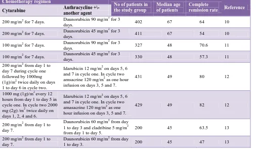
Table 2: Results of using cytarabine with different treatment regimens and the results.
Chemotherapy regimen
No of patients in the study group
Median age of patients
Complete
remission rate Reference Cytarabine Anthracycline +\-
another agent
200 mg/m2 for 7 days. Daunorubicin 90 mg/m
2
for 3
days. 402 67 64 10
200 mg/m2 for 7 days. Daunorubicin 45 mg/m
2
for 3
days. 411 67 54 10
100 mg/m2 for 7 days. Daunorubicin 90 mg/m
2
for 3
days. 327 48 70.6 11
100 mg/m2 for 7 days. Daunorubicin 45 mg/m
2
for 3
days. 330 48 57.3 11
200 mg/m2 from day 1 to day 7 during cycle one followed by 1000mg (1g)/m2 twice daily on days 1 to day 6 in cycle two.
Idarubicin 12 mg/m2 on days 5, 6 and 7 in cycle one. In cycle two amsacrine 120 mg/m2 as one hour infusion on days 3, 5 and 7.
431 49 80 12
1000 mg (1g)/m2 every 12 hours from day 1 to day 5 in cycle one. In cycle two 2000 mg (2g) /m2 twice daily on days 1, 2, 4 and 6.
Idarubicin 12 mg/m2 on days 5, 6 and 7 in cycle one. In cycle two amasacrine 120 mg/m2 as one hour infusion on days 3, 5 and 7.
429 49 82 12
200 mg/m2 from day 1 to day 7.
Daunorubicin 60 mg/m2 from day 1 to day 3 and cladribine 5 mg/m2 from day 1 to day 5.
200 45 63.5 13
200 mg/m2 from day 1 to day 7.
Daunorubicin 60 mg/m2 from day
1 to day 3. 200 45 47 13
Classification based on gene expression: Based on gene expression profile, genetically distinct groups have been identified.7-9 Each group has a distinct gene expression profile (eg: CBFβ-MYH11, PML-RARα or AML1-ETO) or a particular chromosomal lesion [eg: inv (16), t (15;17) or t (8;21)] or an abnormal oncogene expression (eg: EVI 1]). Gene expression characteristics of each of these groups have been identified, which may reveal critical functional pathways involved in the development of AML.
Therapy
[image:2.595.52.558.327.621.2]Remission-induction therapy
Administration of cytarabine (Ara-C) in combination with anthracyclines has been the cornerstone in the management of AML for more than 30 years.14 Various dosage regimens have been followed with varying results (Table 2). The variation in the remission rate is not only because of the use of different chemotherapy regimens but also due to variation in the supportive care the patients receive in different treatment centers.
Bone marrow evaluation is usually done between day 17 to day 21 to assess the response to chemotherapy. Complete remission is defined as normalization of neutrophil count (at least 1.5 X10⁹ \L) and platelet count (more than 100X109/L) with bone marrow aspirate or biopsy having at least 20% cellularity with less than 5% blasts with no auer rods and absence of extra medullary leukemia.15 About 60-70% of patients usually achieve complete remission after the first cycle of chemotherapy. For patients who do not attain complete remission after the first cycle of chemotherapy, a second cycle of chemotherapy is given. For the second cycle either the regimen used during the first cycle or a new chemotherapy regimen can be used. Of patients with persistent AML after the first cycle of chemotherapy approximately 20% will achieve complete remission after receiving the second cycle of chemotherapy. For patients not responding to the second cycle of chemotherapy a salvage treatment can be suggested.
Post remission therapy
It is given after achieving complete remission by induction chemotherapy. The goal of post remission chemotherapy is to prolong the duration of complete remission and to improve overall survival by eradicating residual leukemic cells.
Post-remission treatment options are.10-12
Bone marrow transplantation (allogenic or autologous) after preconditioning. Allogenic bone marrow transplantation is preferred because of graft versus leukemia effect.
High dose cytarabine regimen.
Conventional consolidation chemotherapy, where a regimen same or different from that used for obtaining complete remission is given.
Maintenance therapy which is less myelosuppressive than that used for obtaining complete remission (Mostly used for acute promyelocytic leukemia).
Prognosis
There is no single factor that can reliably predict the complete remission or the long-term survival. However, there are a few factors that are associated with the probability of obtaining complete response to
chemotherapy. They are age, cytogenetic and antecedent hematological disorder.
Age
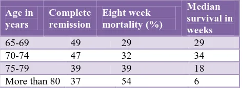
[image:3.595.312.548.291.377.2]It is the single most significant patient variable that correlates inversely with prognosis. This is because a greater proportion of older people die of complications related to myelosuppressive effects of chemotherapy like infections and bleeding before achieving a complete remission. Overall, patients less than 65 year age have a five year survival rate of 32.7 % and patients above 65 year age have a 3.8% five-year survival rate. The mortality rate for elderly people in different age groups is shown in (Table 3).
Table 3: Mortality rate for elderly people in different age groups.16-19
Age in years
Complete remission
Eight week mortality (%)
Median survival in weeks
65-69 49 29 29
70-74 47 32 34
75-79 39 39 18
More than 80 37 54 6
Antecedent hematological disorder
Patients with prior myeloproliferative disorder, refractory anemia with excess blasts, prior cytotoxic drug therapy, etc are less likely to respond to the treatment.17,19,20 Performance status of the patient, high leukocyte count and splenomegaly (especially in elderly patients) also have prognostic significance.17
Cytogenetics
A large number of recurrent balanced and unbalanced chromosomal aberrations have been described in AML.19,20 A strong association has been described between cytogenetic abnormality and prognosis. Currently pre-treatment cytogenetics has been divided into three groups-favourable, intermediate and unfavourable.
Table 4: Pretreatment cytogenetics and prognosis.16,17,19
Cytogenetic subtype
Karyotype abnormality
Associated subtype
Five year Survival rate
Favorable
t (15:17) M3 63 t (8:21) M2 69 Inv (16) M4E0 61
Intermediate
Trisomy 8 M2, M4, and M5 48 Normal No association 45 Trisomy 21 No association 47
Unfavorable
Deletion 7q No association 10 Deletion 5q No association 11 Complex No association 21
Other gene mutations
Assessment of mutational status of several genes and microRNA expression has improved the risk stratification of the patients, especially in cytogenetically normal AML patients who would be otherwise classified to have intermediate prognosis.9,23 Somatic mutations in the DNMT3A gene which encodes for DNA methyl transferase have been identified in patients with normal karyotype and are independently associated with poor outcomes. Nucleophosmin (NPM1) mutations are associated with good response to induction chemotherapy in patients younger than 60 years. Based on microRNA expression cytogenetically normal patients with FLT3-ITD, wild type NPM1 or both are considered as high risk groups and have poor prognosis.24 Genome sequencing has identified recurrent mutations in IDH 1 gene in cytogenetically normal AML patients MLL gene translocations are seen in patients with therapy related leukemias and are associated with improved response to high dose daunorubicin treatment.24,25
Drugs used in the treatment of acute myeloid leukemia
Cytarabine (cytosine arabinoside, aslo called as Ara-C)
Cytarabine has been a cornerstone in the treatment of acute myeloid leukemia for over 30 years. Cytarabine is an important component of both remission induction regimens and in remission maintenance regimens. As less than 20% of cytarabine is absorbed orally, it is always given parenterally. It is usually administered as a continuous intravenous infusion or subcutaneously. It can also be given intramuscularly and intrathecally (to treat leukemic infiltration of brain). Intravenous administration exhibits biphasic elimination. It is metabolized in the liver into an inactive metabolite (uracil arabinoside) by the enzyme cytidine deaminase. About 70-80% of the administered dose appears in urine within 24 hours, of which 90% is metabolite and 10% is active drug. The exact mechanism of anti-leukemic action is not yet
known. After transportation into the cell by a nucleoside carrier it is converted into cytosine triphosphate by deoxycytidine kinase. The deoxy cytarabine triphosphate (dCTP) when incorporated into DNA inhibits DNA synthesis. dCTP also inhibits the enzyme DNA polymerase, thus cytarabine is a S phase-specific anti-cancer drug. Since even a very low dose of cytarabine (20 mg/m2) evokes an appreciable response, the cytotoxicity of cytarabine cannot explain its anti-leukemic action. Hence, other effects like terminal differentiation of leukemic cells by decreasing c myc expression, induction of apoptosis by activating transcription factor AP-1, etc, have been suggested to be responsible for its remission-inducing properties.32 The sensitivity of cells to cytarabine is determined by the expression of the nucleoside carrier and the predominance of activity of the activating enzyme deoxycytidine kinase over the inactivating enzyme cytidine deaminase. The duration of cytarabine triphosphate retention has been correlated with remission.26 Cytarabine has a dose-dependent anti-leukemic effect, which reaches a ceiling.12,27,28 Although increasing the dose leads to an increase in remission rate and relapse-free survival, toxicity rate also increases with increase in dose.12,29 Treatment with an intermediate dose of cytarabine (200 mg/m2 in cycle one and 2 g/m2 in cycle two) achieves the same complete remission rate and relapse-free survival rate as that of high dose cytarabine (1000 mg/m2 twice daily in cycle one and 2 g/m2 twice daily in cycle two) with similar treatment related toxicity.12 Cytarabine is usually administered at a dose of 100 to 200 mg/m2 for 7 days for induction of remission along with daunorubicin 90 mg/m2 for initial 3 days. After achieving complete remission, three cycles of cytarabine at dose of 18 g/m2 is usually administered as post-remission maintenance chemotherapy.30 Three cycles of cytarabine is an acceptable post remission maintenance therapy.30,31 In some centers low dose of cytosine arabinoside (20 mg/m2) is used in elderly patients and in patients with poor performance status who are unable to tolerate a high myelosuppressive dose. Cytarabine causes myelosuppression (dose-limiting toxic effect), conjunctivitis, dermatitis (cytarabine hands), elevation of liver enzymes, cerebellar toxicity, stomatitis, nausea, vomiting and non-cardiogenic pulmonary edema as side effects.12,33
Anthracycline antibiotics
improving the complete remission rate and duration of overall survival in patients younger than 50 years of age. Improved complete remission rate is also seen in elderly patients (older than 60 years), but overall survival does not improve with increase in the dose in elderly patients.10 The increase in dose from 45 mg/m2 to 90 mg/m2 does not increase the hematologic toxic effects or 30 day mortality rate or moderate to severe life threatening side effects across the patient age group10,11. Increasing the dose of daunorubicin does not provide any additional benefit for patients with unfavorable cytogenetic profile.12 Idarubicin is a newer anthracycline which is at least as efficacious as daunorubicin. It is used in a dose of 12 mg/m2 for 3 days. Mitoxantrone has also been evaluated because of its less cardiotoxic potential but percentage of patients achieving complete remission is less than that of those treated with daunorubicin. Though P-glycoprotein (Pgp) (an efflux pump which actively transports cytotoxic drugs like anthracyclines out of the cell) has been implicated in resistance to chemotherapy, addition of Pgp inhibitors like cyclosporine or zosuquidar (highly selective Pgp inhibitor) have failed to improve the complete remission rate of the treatment.35,36 Acute effects on the myocardium like transient tachycardia and reversible ECG changes are seen, but chronic cadiotoxicity is not a common side effect in patients with AML, as chronic cardiotoxicity usually occurs in patients who received a total dose at or above 550 mg/m2. The total dose of anthracyclines used during induction therapy is far less than the chronic cardiotoxic dose. However, some patients may develop pericarditis-myocarditis syndrome which is characterized by congestive heart failure, conduction abnormalities and pericardial effusion even with low dose of daunorubicin. Myelosupression is the acute dose-limiting side effect of daunorubicin with drop in neutrophil count reaching the nadir within 10-14 days after the drug administration. A transient erythematic reaction can be seen along the intravenous line; this is sometimes called “adriamycin flare”, and is a benign lesion compared to extravasation of adriamycin from the intravenous line.
All trans-retinoic acid (Tretinoin, ATRA)
Tretinoin induces the differentiation of leukemic cells bearing the t (15;17) translocation. ATRA is used in the treatment of acute promyelocytic leukemia, which characteristically has a PML-RAR translocation due to the t (15;17) translocation. Oral dose of 45 mg/m2 per day, plus concurrent anthracycline chemotherapy is an effective treatment for acute promyelocytic leukemia. The first 3 weeks of treatment may be complicated by “retinoic acid syndrome” or “ATRA syndrome” which is characterized by fever, dyspnea, chest pain, pulmonary infiltrates, pleural and pericardial effusions, and hypoxia, with mortality around 10%. This may be due to adhesion of differentiated neoplastic cells to the pulmonary vasculature endothelium. The risk of differentiation
syndrome can be minimized by prior administration of dexamethasone.
Arsenic trioxide
Arsenic trioxide in combination with ATRA has shown to be as effective as ATRA and anthracycline combination in patients with low to intermediate risk APL with two year event free survival rate of 97%.37 This is because of synergy: while ATRA binds to the RAR alpha moiety of PML-RAR, arsenic binds to the PML moiety. This synergy leads to rapid degradation of PML-RAR oncoprotein and cell differentiation. The advantage of using arsenic trioxide and ATRA combination is that it is associated with lower hematological toxicity, hence it has a lower risk of severe cytopenias, mucositis, and infections, and thus less treatment-related deaths. However, arsenic trioxide causes more frequent elevation of liver enzymes and prolongation of QTc interval.37 Use of this combination cures APL without the use of cytotoxic chemotherapy. Incorporating arsenic trioxide into the ATRA and anthracycline combination improves outcomes while decreasing the exposure to anthracycline.38
Other conventional cytotoxic drugs
Various other drugs like etoposide, thioguanine, hydroxyurea, fludarabine, etc. have been evaluated either in combination with the standard regimen (3+7) or with other cytarabine-based regimens to increase complete remission rate, but results are either unsatisfactory or equivocal.
FLT3 inhibitors
Use of FLT3 receptor tyrosine kinase inhibitors like midostaurin, lestsutinib, sorafinib, etc. in patients with the FLT3-ITD mutation reduces the peripheral blast cells and bone marrow blast cells.21 The initial results are encouraging but in most patients the response was incomplete and of short duration. The development of resistance during therapy and also the lack of correlation between in vitro cytotoxicity with actual clinical outcomes have been a challenge in use of these drugs. Incorporating FLT3 inhibitors into the salvage regimen has shown to improve long term outcomes.39,40
Gemtuzumab ozogamicin
in the course of the disease.43 Recent phase III clinical trials have shown that Gemtuzumab ozogamicin does not improve either complete remission rate or overall survival, both in younger and elderly patients.44,45 Since later studies failed to show the survival benefit, on the advice of USFDA, the manufacturers withdrew the drug from the market.
Drugs in clinical trial
Clofarabine, a second generation of purine nucleoside analogues which acts by inhibiting the enzymes DNA polymerases and ribonucleotide reductase induces apoptosis in leukemic cells46. It has shown significant efficacy in acute myeloid leukemia.47,49 However, there are reports of fatal skin and liver toxicity.48
Cloretazine is a novel alkylating agent. A phase II study has revealed significant anti-leukemic activity with modest extra-medullary toxicity in elderly patients with AML.50 However it is reported to have minimal activity in very high risk patients with relapsed AML.48 Demethylating agents like decitabine, 5-azacytidine and zebularine and FLT3 inhibitors like SU11248 and MLN518 have been evaluated for anti-leukemic action especially in elderly. 51,52
CONCLUSION
With better understanding of the pathogenesis, it is now possible to assess and stratify patients with AML into different categories based on the gene expression profile. Conventional cytotoxic drugs still remain the mainstay of therapy. Redesigning the treatment regimen by modifying the dosage of cytarabine and anthracyclines has improved the complete remission rate and overall all survival. Increasing the dose of daunorubicin increases the complete remission rate without increase in the toxicity of the therapy. Newer treatments have been developed targeting the specific abnormality in the gene expression and are less toxic than conventional treatment. The advancement in the understanding of the disease biology of this heterogeneous disease may eventually lead to development of therapies directed against specific abnormalities.
Funding: No funding sources Conflict of interest: None declared Ethical approval: Not required
REFERENCES
1. Genovese G, Kähler AK, Handsaker RE, Lindberg J, Rose SA, Bakhoum SF, et al. Clonal hematopoiesis and blood-cancer risk inferred from blood dna sequence. N Engl J Med.2015;372:1071-2.
2. Deschler B, Lübbert M. Acute myeloid leukemia: epidemiology and etiology. Cancer. 2006;1;107(9):2099-107.
3. Walter MJ, Shen D, Ding L, Shao J, Koboldt DC, Chen K, et al. Clonal architecture of secondary acute myeloid leukemia. N Engl J Med. 2012;366(12):1090-8.
4. Vardiman JW, Thiele J, Arber DA, Brunning RD, Borowitz MJ, Porwit A, et al. The 2008 revision of the World Health Organization (WHO) classification of myeloid neoplasms and acute leukemia: rationale and important changes. Blood. 2009;114(5):937-51. 5. Eivazi-Ziaei J. Myeloperoxidase index and subtypes
of acute myeloid leukemia. J Pak Med Assoc. 2009;59(6):406-7.
6. Kaleem Z, Crawford E, Pathan MH, Jasper L, Covinsky MA, Johnson LR, et al. Flow cytometric analysis of acute leukemias. Diagnostic utility and critical analysis of data. Arch Pathol Lab Med. 2003;127(1):42-8.
7. Valk PJ, Verhaak RG, Beijen MA, Erpelinck CA, Barjesteh van Waalwijk van Doorn-Khosrovani S, Boer JM, et al. Prognostically useful gene-expression profiles in acute myeloid leukemia. N Engl J Med. 2004;350(16):1617-28.
8. Cancer Genome Atlas Research Network. Genomic and epigenomic landscapes of adult de novo acute myeloid leukemia. N Engl J Med. 2013;368(22):2059-74.
9. Schlenk RF, Döohner K, Krauter J, Fröhling S, Corbacioglu A, Bullinger L, et al. Mutations and treatment outcome in cytogenetically normal acute myeloid leukemia. N Engl J Med. 2008;358:1909-18. 10. Löwenberg B, Ossenkoppele GJ, van Putten W,
Schouten HC, Graux C, Ferrant A, et al. High-dose daunorubicin in older patients with acute myeloid leukemia. N Engl J Med. 2009;361(13):1235-48. 11. Fernandez HF, Sun Z, Yao X, Litzow MR, Luger
SM, Paietta EM, et al. Anthracycline dose intensification in acute myeloid leukemia. N Engl J Med. 2009;361(13):1249-59.
12. Löwenberg B, Pabst T, Vellenga E, van Putten W, Schouten HC, Graux C, et al. Cytarabine dose for acute myeloid leukemia. N Engl J Med. 2011;364(11):1027-36.
13. Holowiecki J, Grosicki S, Robak T, Kyrcz-Krzemien S, Giebel S, Hellmann A, et al. Polish adult leukemia group (PALG): addition of cladribine to daunomycin and cladribine increases remission rate after a single course of induction treatment in acute myeloid leukemia. Multi-center phase 3 study. Leukemia. 2004;18(5):989-97.
14. Yates J, Glidewell O, Wiernik P, Cooper MR, Steinberg D, Dosik H, et al. Cytosine arabinoside with daunorubicin or adriamycin for therapy of acute myelocytic leukemia: a CALGB study. Blood. 1982;60:454-62.
myeloid leukaemia revisited. An analysis of patients treated in HOVON-SAKK co-operative group studies. Br J Haematol. 2005;128(2):184-91.
16. Kantarjian HM, Keating MJ, Walters RS, Smith TL, Cork A, McCredie KB, et al. Therapy related leukemia and myelodysplastic syndrome: clinical, cytogenetic and prognostic features. J Clin Oncol. 1986;4(12):1748-57.
17. Kantarjian H, O’Brien S, Cortes J, Giles F, Faderl S, Jabbour E, et al. Results of intensive chemotherapy in 998 patients age 65 years or older with acute myeloid leukemia or high-risk myelodysplastic syndrome: predictive prognostic models for outcome. Cancer. 2006;106:1090-8.
18. Buchner T, Berdel WE, Haferlach C, Haferlach T, Schnittger S, Müller-Tidow C, et al. Age-related risk profile and chemotherapy dose response in acute myeloid leukemia: a study by the German Acute Myeloid Leukemia Cooperative Group. J Clin Oncol. 2009;27(1):61-9.
19. Frohling S, Schlenk RF, Kayser S, Morhardt M, Benner A, Döhner K, et al. Cytogenetics and age are major determinants of outcome in intensively treated acute myeloid leukemia patients older than 60 years: results from AMLSG trial AML HD98-B. Blood 2006;108:3280-8.
20. Schiffer CA, Lee EJ, Tomiyasu T, Wiernik PH, Testa JR. Prognostic impact of cytogenetic abnormalities in patients with de novo acute non lymphocytic leukemia. Blood. 1989;73(1):263-70.
21. Kindler T, Lipka DB, Fischer T. FLT3 as a therapeutic target in AML: still challenging after all these years. Blood. 2010;116(24):5089-102.
22. Linch DC, Hills RK, Burnett AK, Khwaja A, Gale RE. Impact of FLT3(ITD) mutant allele level on relapse risk in intermediate-risk acute myeloid leukemia. Blood. 2014;124(2):273-6.
23. Marcucci G, Radmacher MD, Maharry K, Mrózek K, Ruppert AS, Paschka P, et al. MicroRNA expression in cytogenetically normal acute myeloid leukemia. N Engl J Med. 2008;358(18):1919-28.
24. Ley TJ, Ding L, Walter MJ, McLellan MD, Lamprecht T, Larson DE, et al. DNMT3A mutations in acute myeloid leukemia. N Engl J Med. 2010;363(25):2424-33.
25. Patel JP, Gönen M, Figueroa ME, Fernandez H, Sun Z, Racevskis J, et al. Prognostic relevance of integrated genetic profiling in acute myeloid leukemia. N Engl J Med. 2012;366(12):1079-89. 26. Plunkett W, Liliemark JO, Adams TM, Nowak B,
Estey E, Kantarjian H, et al. Saturation of 1-β-D-arabinofuranosylcytosine 5¢-triphosphate accumulation in leukemia cells during high-dose 1-β-Darabinofuranosylcytosine therapy. Cancer Res. 1987;47:3005-11.
27. Bishop JF, Matthews JP, Young GA, Szer J, Gillett A, Joshua D, et al. A randomized study of high-dose cytarabine in induction in acute myeloid leukemia. Blood. 1996;87:1710-7.
28. Weick JK, Kopecky KJ, Appelbaum FR, Head DR, Kingsbury LL, Balcerzak SP, et al. A randomized investigation of highdose versus standard-dose cytosine arabinoside with daunorubicin in patients with previously untreated acute myeloid leukemia: a Southwest Oncology Group study. Blood. 1996;88:2841-51.
29. Mayer RJ, Davis RB, Schiffer CA, Berg DT, Powell BL, Schulman P, et al. Intensive postremission chemotherapy in adults with acute myeloid leukemia. N Engl J Med. 1994;331:896-903.
30. Döhner H, Estey EH, Amadori S, Appelbaum FR, Büchner T, Burnett AK, et al. Diagnosis and management of acute myeloid leukemia in adults: recommendations from an international expert panel, on behalf of the European Leukemia Net. Blood. 2010;115:453-74.
31. JC Byrd, AS Ruppert, K Mrózek , Carroll AJ, Edwards CG, Arthur DC, et al. Repetitive cycles of high-dose cytarabine benefit patients with acute myeloid leukemia and inv (16) (p13q22) or t (16; 16) (p13;q22): results from CALGB 8461. J Clin Oncol. 2004;22(6):1087-94.
32. Bianchi Scarrà GL, Romani M, Coviello DA, Garrè C, Ravazzolo R, Vidali G, et al. Terminal erythroid differentiation in the K-562 cell line by 1-beta-D-arabinofuranosylcytosine: accompaniment by c-myc messenger RNA decrease. Cancer Res. 1986;46:6327-32.
33. Forghieri F, Luppi M, Morselli M, Potenza L. Cytarabine-related lung infiltrates on high resolution computerized tomography: a possible complication with benign outcome in leukemic patients. Haematologica: 2007;92:e85-e90.
34. Langer SW, Sehested M, Jensen PB. Treatment of anthracycline extravasation with dexrazoxane. Clin Cancer Res. 2000;6:3680-6.
35. List AF, Kopecky KJ, Willman CL, Head DR, Persons DL, Slovak ML. Benefit of cyclosporine modulation of drug resistance in patients with poor-risk acute myeloid leukemia: a Southwest Oncology Group study. Blood. 2001;98:3212-20.
36. Baer MR, George SL, Dodge RK, O'Loughlin KL, Minderman H, Caligiuri MA, et al. Phase 3 study of the multidrug resistance modulator PSC-833 in previously untreated patients 60 years of age and older with acute myeloid leukemia: Cancer and Leukemia Group B Study 9720. Blood. 2002;100(4):1224-32.
37. Lo-Coco F, Avvisati G, Vignetti M, Thiede C, Orlando SM, Iacobelli S, et al. Retinoic acid and arsenic trioxide for acute promyelocytic leukemia. N Engl J Med. 2013;369(2):111-21.
38. Iland HJ, Bradstock K, Supple SG, Catalano A, Collins M, Hertzberg M, et al. All-trans-retinoic acid, idarubicin, and IV arsenic trioxide as initial therapy in acute promyelocytic leukemia (APML4). Blood. 2012;120(8):1570-80.
mechanisms of resistance. Int J Hematol. 2013;97(6):683-94.
40. Takahashi K, Kantarjian H, Pemmaraju N, Andreeff M, Borthakur G, Faderl S, et al. Salvage therapy using FLT3 inhibitors may improve long-term outcome of relapsed or refractory AML in patients with FLT3-ITD. Br J Haematol. 2013;161(5):659-66. 41. Stasi R, Evangelista ML, Buccisano F, Venditti A,
Amadori S. Gemtuzumab ozogamicin in the treatment of acute myeloid leukemia. Cancer Treat Rev. 2008;34(1):49-60.
42. Larson RA, Sievers EL, Stadtmauer EA, Löwenberg B, Estey EH. Final report of the efficacy and safety of gemtuzumab ozogamicin (Mylotarg) in patients with CD33-positive acute myeloid leukemia in first recurrence. Cancer. 2005;104(7):1442-52.
43. Chevallier P, Robillard N, Ayari S, Guillaume T, Delaunay J, Mechinaud F, et al. Persistence of CD33 expression at relapse in CD33(+) acute myeloid leukaemia patients after receiving Gemtuzumab in the course of the disease. Br J Haematol. 2008;143(5):744-6.
44. Petersdorf SH, Kopecky KJ, Slovak M, Willman C, Nevill T, Brandwein J, et al. A phase 3 study of gemtuzumab ozogamicin during induction and postconsolidation therapy in younger patients with acute myeloid leukemia. Blood. 2013;121(24):4854-60.
45. Amadori S, Suciu S, Stasi R, Salih HR, Selleslag D, Muus P, et al. Sequential combination of gemtuzumab ozogamicin and standard chemotherapy
in older patients with newly diagnosed acute myeloid leukemia: results of a randomized phase III trial by the EORTC and GIMEMA consortium (AML-17). J Clin Oncol. 2013;31(35):4424-30.
46. Faderl S, Ferrajoli A, Wierda W, Huang X, Verstovsek S, Ravandi F, et al. Clofarabine combinations as acute myeloid leukemia salvage therapy. Cancer. 2008;113(8):2090-6.
47. Kantarjian HM, Jeha S, Gandhi V, Wess M, Faderl S. Clofarabine: past, present, and future. Leuk Lymphoma. 2007;48(10):1922-30.
48. Johnston DL, Mandel KM. Fatal skin and liver toxicity in a patient treated with clofarabine. Pediatr Blood Cancer. 2008;50(5):1082.
49. Larson ML, Venugopal P. Clofarabine: a new treatment option for patients with acute myeloid leukemia. Expert Opin Pharmacother. 2009;10(8):1353-7.
50. Giles F, Verstovsek S, Faderl S, Vey N, Karp J, Roboz G, et al. A phase II study of cloretazine (VNP40101M), a novel sulfonylhydrazine alkylating agent, in patients with very high risk relapsed acute myeloid leukemia. Leuk Res. 2006;30:1591-5. 51. Pratz KW, Cortes J, Roboz GJ, Rao N, Arowojolu O,
Stine A, et al. A pharmacodynamic study of the FLT3 inhibitor KW-2449 yields insight into the basis for clinical response. Blood. 2009:23:3938-46.
52. Stubbs MC, Armstrong SA. FLT3 as a therapeutic target in childhood acute leukemia. Curr Drug Targets. 2007;8(6):703-14.
Cite this article as: Nandeesh BR,