R E S E A R C H
Open Access
Evaluation of the expression of Bmi-1 stem
cell marker in sinonasal melanomas and its
correlation with the expression of cell cycle
proteins
Harim Tavares dos Santos
1, Juliana de Souza do Nascimento
3, Fernanda Meireles
2, João Figueira Scarini
3,
Erika Said Egal
2, Victor Angelo Montalli
4, Felipe Paiva Fonseca
5, Fernanda Viviane Mariano
2and Albina Altemani
2*Abstract
Background:Sinonasal melanomas (SNM) are aggressive neoplasms, which present distinct clinicopathological and
molecular aspects when compared to cutaneous melanomas (CM). B-cell-specific moloney murine leukemia virus integration site-1 (Bmi-1) is a stem cell marker involved in the regulation of the cell cycle and has been found to be expressed in 70% of CM and 100% of benign nevi. Regarding the cell cycle, Bmi-1 is known to be an upstream repressor of p16, which is a tumor suppressor encoded by the INK4a/Arf locus. Considering this, the aim of this study is to evaluate the immunohistochemical expression of Bmi-1 in a series of SNM and its correlation with the expression of cell cycle proteins (p16 and Ki-67, a nuclear antigen of proliferating cells).
Methods:In 16 cases of SNM, nuclear expression of Bmi-1 and nuclear and cytoplasmic of p16 was classified as: absent, low (> 5 to < 50% of cells) and high (≥50%). Ki-67 proliferation index was represented by the ratio positive cells/ total cells.
Results:Histologically, all cases presented varying amount of necrosis and 75% contained undifferentiated cells. Bmi-1 was detected in 6 cases (37.5%) with high level of expression in 2; p16 expression was seen in 10 cases (62.5%) with high level in 7. The frequency of p16 expression did not differ significantly between tumors with or without Bmi-1 expression. Ki-67 index ranged from 8 to 22%. Neither Bmi-1 nor p16 expression showed correlation with Ki-67 index. Bmi-1 negative tumors presented more extensive necrosis (71.4%); no association between Bmi-1 expression and undifferentiated phenotype was observed.
Conclusions:In our SNM series, low immunohistochemical expression of Bmi-1 was a common phenomenon
favoring the hypothesis that mucosal melanoma possibly presents molecular pathways different from the
cutaneous counterpart. In SNM, Bmi-1 and p16 expression levels did not correlate with each other or with the cell proliferative index.
Keywords:Sinonasal melanoma, Bmi-1, p16, Immunohistochemical expression
* Correspondence:[email protected]
2Universidade Estadual de Campinas, Faculdade de Ciências Médicas,
Departamento de Anatomia Patológica, Campinas, SP, Brazil Full list of author information is available at the end of the article
Background
The sinonasal region is the major site of mucosal melan-oma of the head and neck (Williams, 2017). Sinonasal melanomas (SNM) are rare, their etiology is poorly understood, and they present distinct clinicopathological and molecular aspects when compared to their cutane-ous counterpart, which is etiologically related to ultra-violet radiation (Franchi et al., 2006). SNM are associated with poor overall survival at 5 years, frequent undifferentiated phenotype and higher rates of muta-tions involving C-Kit (CD 117) pathway (López et al.,
2016). These particularities show the need to deepen knowledge about SNM, in order to better understand its pathogenesis, which is important for the development of therapeutic strategies.
It is known that in malignant tumors a subgroup of neoplastic cells expresses proteins that are considered stem cell (SC) markers. Neoplastic SCs are capable of self-renewal and differentiation; in addition, they have been considered to play an important role in tumor initi-ation and progression as well as in therapeutic resistance (Siclari & Qin, 2010; Rangwala et al., 2011; Schatton et al., 2008). Among the SC markers, B-cell-specific moloney murine leukemia virus integration site-1 (Bmi-1) is a transcription factor involved in the regula-tion of the cell cycle and apoptosis. Deregularegula-tion of Bmi-1 expression has been described in several types of cancer and associated with histological features of tumor aggressiveness (increased number of mitotic figures and necrosis) as well as with tumor biological behavior (metastatic capacity, predictor of prognosis) (Bachmann et al.,2006; Vrzalikova et al.,2008; Song et al.,2006; Liu et al., 2008; Mihic-Probst et al.,2007; Silva et al.,2007; Bonora et al., 2015) P16 is a tumor suppressor protein, cyclin dependent kinase inhibitor involved in cell prolif-eration pathways.(Li et al.,2006). As Bmi-1 is a suppres-sor of the Ink4a / Arf locus, which encodes p16, it has been proposed that this interaction could lead to in-creased cell proliferation, affecting the biological behav-ior of the tumor (Song et al., 2006; Mihic-Probst et al.,
2007; Allegra et al., 2012; Vormittag et al., 2009; Chen et al.,2011).
Drawing from this background, the aim of our study was to evaluate the immunohistochemical expression of Bmi-1 in a series of SNM and its correlation with the expression of cell cycle proteins (p16 and Ki-67, a nuclear antigen of proliferating cells). To the best of our knowledge, expres-sion of Bmi-1 in SNM has yet to be determined.
Material and methods
This study was approved by the Institutional Ethics Committee. The surgical pathology archives of the Hospital of the University of Campinas (UNICAMP), São Paulo-Brazil, were reviewed between 1990 and 2016
and contained 16 tumors which had been diagnosed as SNM and had available slides and/ or blocks. All cases were reviewed to confirm the diagnosis and clinical de-tails were obtained from medical records.
Immunohistochemistry
Immunohistochemical studies were performed on sec-tions from representative formalin fixed paraffin embed-ded blocks. All cases were stained with antibodies showed in the Table 1. Immunoreactivity for Bmi-1 and p16 was assessed and classified as absent (0 to 5%), low (> 5 to < 50% of cells) and high (≥50%) according to Mihic-Probst et al.(Mihic-Probst et al.,2007). For Bmi-1, only nuclear staining was considered positive whereas for p16, both nuclear and cytoplasmic reactivity was used; high p16 expression could be nuclear and cytoplas-mic or exclusively cytoplascytoplas-mic in ≥50% of tumor cells. Ki-67 nuclear immunohistochemical assessment was performed with the help of Aperio ImageScope nuclear algorithm (Aperio ScanScope; Aperio Technologies, Vista, Calif ). Five high-power fields were randomly se-lected (original magnification × 200), and 3000 tumor cells per slide were counted approximately. The labeling index for Ki-67 in each case of nasal melanoma was expressed as a percentage of positive tumor cells.
Statistical analysis
A Student’s T test was used for comparison of the quan-titative variables. Mann-Whitney U test was used for comparison of the numeric variables between the groups as appropriate. Data were presented as mean ± SD (standard deviation), and the results with p< 0.05 were considered significant. All the statistical procedures were performed using Graph Prism version 6.0 for Mac (GraphPad Software® La Jolla, USA).
Results
Clinicopathologic findings
Table 2 shows the clinicopathological findings of 16 cases of SNM. In all cases but one, the nasal cavity was the site of origin of the tumor and all were advanced le-sions, i.e., they invaded submucosa and deep soft tissue (T3/ T4 in the TNM classification). The median age of the patients was 62.2 years (range, 24–78 years) and 60% were women. Histologically, growth pattern of the solid type was observed in almost all cases and most of them presented areas with peritheliomatous arrangement of
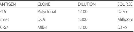
Table 1Antibodies used in this study
ANTIGEN CLONE DILUTION SOURCE
P16 Polyclonal 1:100 Dako
Bmi-1 DC9 1:300 Millipore
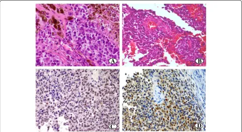
tumor cells (Fig. 1). Tumor cellular composition varied but 75% of cases contained undifferentiated cells (Fig.1). Neoplastic cells with melanin were seen in 43% of cases. All tumors exhibited areas of necrosis, which was exten-sive (> 50% of tumor area) in 56.2% (9/16) of cases. Vas-cular invasion was observed in two cases, but neural infiltration was not seen. However, it should be noted that most of cases were incisional biopsies.
Immunohistochemical findings
In SNM, the nuclear expression of Bmi-1 was detected in 6 cases (6/16–37.5%), with high level of expression in 2 of them (Fig.1). Nuclear and cytoplasmic p16 expres-sion was seen in 11 cases (11/16–68.7%), which pre-sented high level of expression in 7 (7/16–43.7%) and low in 4 (4/16–25%). P16 expression was absent in 5 cases (5/16–31.2%) (Fig. 1). The frequency of p16 ex-pression did not differ significantly between tumors with or without Bmi-1 expression (66.6 and 60% of cases were p16 positive, respectively) (p= 0.84). The combin-ation high Bmi-1 and low expression ratio of p16 was noted in only 1 of 16 cases. Regarding the morphological aspects associated with tumor aggressiveness (Table 3), most tumors with extensive necrosis were Bmi-1 nega-tive (71.4%). However, Bmi-1 expression was not related to the undifferentiated cellular pattern; Bmi-1 positive and negative tumors showed a similar frequency of le-sions composed predominantly of undifferentiated cells. The Ki-67 proliferation index ranged from 8 to 22% and no significant difference was detected between tumors with and without Bmi-1 expression (means 17.3% versus
15%) (p= 0.33) and the ones with or without p16 expres-sion (means 16.3% versus 14.7%, respectively) (p= 0.83).
Discussion
Overexpression of Bmi-1 has been described in several malignant tumors and related to tumorigenesis, metasta-sis, and increased resistance to ionizing radiation (Song et al., 2006; Allegra et al., 2012; Vormittag et al., 2009; Chen et al., 2011). Furthermore, Bmi-1 has been found as a predictor of prognosis in breast, gastric, nasopha-ryngeal and salivary adenoid cystic carcinomas (Bachmann et al., 2006; Vrzalikova et al., 2008; Song et al., 2006; Liu et al., 2008; Mihic-Probst et al., 2007; Silva et al., 2007). However, in melanoma, studies on Bmi-1 expression have shown conflicting results. Bachmann et al .(Bachmann et al.,2008) reported that in invasive melanomas, the loss of Bmi-1 expression was associated with increased cell proliferation, necrosis, and decreased patient survival. In contrast, Mihic-Probst et al.(Mihic-Probst et al., 2007) suggested that the in-crease of Bmi-1 expression could induce a metastatic tendency in cutaneous melanoma. Interestingly, recently, experimental studies have reinforced Mihic–Probst et al.(Mihic-Probst et al., 2007) findings in human mel-anoma. In these, downregulation of Bmi-1 was shown to inhibit the aggressive behavior of melanoma cells by re-versing epithelial-mesenchymal transition,(Liu et al.,
2017) whereas Bmi-1 levels increased with tumor pro-gression, promoting all the steps of the metastatic cas-cade (Ferretti et al.,2016).
Table 2Clinicopathological findings of 16 cases of sinonasal melanoma
Cases Clinical findings Predominant cellular composition
Melanogenesis Necrosis Vascular invasion Neural invasion
Gender Age Origin
1 – – NA- undifferentiated 0 < 50% Negative Negative
2 male 69 Nasal cavity Epithelioid < 50% < 50% Negative Negative
3 female 71 Nasal cavity undifferentiated < 50% < 50% Positive Negative
4 female 24 Maxillary sinus Spindle cell > 50% < 50% Negative Negative
5 male 65 Nasal cavity Epithelioid 0 > 50% Negative Negative
6 female 67 Nasal cavity undifferentiated < 50% < 50% Negative Negative
7 female 60 Nasal cavity undifferentiated 0 > 50% Negative Negative
8 male 66 Nasal cavity undifferentiated 0 > 50% Negative Negative
9 male 55 Nasal cavity Spindle cell 0 < 50% Positive Negative
10 female 58 Nasal cavity undifferentiated 0 > 50% Negative Negative
11 female 67 Nasal cavity undifferentiated < 50% < 50% Negative Negative
12 male 66 Nasal cavity undifferentiated 0 > 50% Negative Negative
13 female 50 Nasal cavity Epithelioid > 50% > 50% Negative Negative
14 female 78 Nasal cavity undifferentiated 0 > 50% Negative Negative
15 male 65 Nasal cavity undifferentiated 0 > 50% Negative Negative
To the best of our knowledge, this is the first time that Bmi-1 is analyzed in SNM; we detected expression of Bmi-1 in 37.5% of the tumors and in only 12.5% of cases (2/16) the protein was highly expressed (at least 50% of cells). These findings indicate that SNM pre-sents a markedly low Bmi-1 expression when compared to cutaneous melanocytic lesions; 60–70% of cutaneous melanoma and 100% of benign nevi present high levels of Bmi-1 expression (at least 50% of cells) (Mihic-Probst et al., 2007; Bachmann et al., 2008). Therefore, our findings reinforce the idea that SNM ex-hibits a different immunophenotype from cutaneous melanoma, since in SNM absence or low Bmi-1 expres-sion appears to be a usual phenomenon. On the other hand, as Bmi-1 expression is believed to promote stem cell state in tumor cells,(Cao et al., 2011) our results suggest that SNM usually contains a small subpopula-tion of Bmi-1 positive cancer stem cell. Of interest, overexpression of Bmi-1 has been reported to correlate with therapy failure (Cao et al.,2011). Thus, it is likely
that cutaneous and SNM may require different thera-peutic strategies given their significant differences in quantity of Bmi-1 positive cancer stem cell. Deserves comments that the role of stem cells in neoplasia is complex and has been subject of numerous studies. In malignant melanoma, besides Bmi-1 other markers of cancer stem cells have been described, such as CD166, CD133, nestin and CD44 (Regauer et al., 1999; Klein et al., 2007; Zhu et al., 2018). Interestingly, CD44 (a transmembranous adhesion molecule) has also been in-vestigated in SNM.(Regauer et al., 1999; Zhu et al.,
2018) Both Bmi-1 and CD44 have been reported to be highly expressed in benign melanocytic lesions, (Mihic-Probst et al., 2007; Bachmann et al., 2008; Harwood et al.,1996) but differently from Bmi-1, CD44 has been detected in a large proportion of cells of inva-sive SNM (Regauer et al.,1999; Zhu et al.,2018). These diverse features of the markers with stem-like properties reinforce that their roles within a melanoma-associated network still need to be better clarified.
Table 3Frequency of undifferentiated phenotype, necrosis and Ki-67 proliferation index in tumors with and without Bmi-1 expression
Undifferentiated phenotype Necrosis Proliferation index
(12 cases) > 50% (7 cases) < 50% (9 cases) Means
Bmi 1+ 6/12 (50%) 2/7 (28,5%) 4/9 (44,4%) 17,3%
Bmi 1- 6/12 (50%) 5/7 (71,4%) 5/9 (55,5%) 15%
In SNM, invasion into deep tissue, undifferentiated cells comprising > 25% of tumor, necrosis, and vascular invasion have been considered features of tumor aggres-siveness and predictors of poor prognosis (Gnepp,2009). In the current series, we did not find any association of Bmi-1 expression with these aggressiveness features, except for tumors without Bmi-1 expression, which presented higher frequency of extensive tumor necrosis (> 50 of the lesion). This relationship has previously been noted in cutaneous melanoma as well (Bachmann et al.,
2008). In addition, our results confirm those reported by other authors (Williams, 2017) showing that SNM are neoplasms more undifferentiated than their cutaneous counterpart. They were frequently composed of undiffer-entiated small cells and most of them did not show me-lanogenesis, which was seen in 43% of cases. From the diagnostic point of view, it is important to recognize SNM as a neoplasm often constituted by small round cells, as this region is affected by other tumors that share this morphology, such as lymphoma, Ewing’s sarcoma, olfactory neuroblastoma, and rhabdomyosarcoma (World Health Organization, 2017). Therefore, in the diagnostic evaluation of an undifferentiated neoplasia composed of small round cells of the sinonasal region, it is necessary to include markers that identify melanocytic lesions (S-100 protein, HMB45 and Melan A) in the im-munohistochemical panel.
In non-neoplastic tissues, Bmi-1 is a transcription fac-tor and epigenetic regulafac-tor essential for maintaining the repression of genes involved in cell proliferation. The ef-fect of Bmi-1 on cell proliferation is partially mediated through repression of the locus encoding p16,(Huber et al., 2011) which is one of the proteins responsible for controlling the G1-S transition of the cell cycle (Li et al.,
2006). The p16 protein inhibits the formation of the cyc-lin D1/cdk4/6 complexes required for the phosphoryl-ation of Rb and consequently the cell cycle progression is slowed down or blocked (Li et al.,2006; Fecher et al.,
2009; Mitra & Fisher, 2009.) Therefore, repression of p16 by Bmi-1 leads to progression of the cell cycle. However, in malignant tumors, the expression of Bmi-1 does not seem to be necessarily linked to p16 expression or to have reflection on cell proliferation. For example, Bmi-1 expression has been described to have no impact on cell proliferation in lung, colon / rectum and brain cancers or correlation with p16 expression in head and neck carcinomas (Vonlanthen et al., 2001; Breuer et al.,
2004; Kim et al., 2004; Hemmati et al., 2003; Lundberg et al.,2016). In melanoma, there are few studies on the relationship between Bmi-1 and cell proliferation and they showed conflicting results. In cutaneous melanoma, loss of Bmi-1 expression was found to be associated with increased tumor cell proliferation whereas experimental studies have shown that Bmi-1 had no effect on
proliferation or tumor growth (Bachmann et al., 2008; Ferretti et al.,2016).
In SNM, this is the first time that the possible relation-ship between the levels of expression of Bmi-1 and p16 as well as their connections with cellular proliferation has been analyzed. In our series no association between Bmi-1 expression and p16 status was detected; the ex-pected combination high Bmi-1 and low expression of p16 was rarely observed (only 1 case). Furthermore, Ki-67 proliferation index was similar in tumors with or without expression of Bmi-1 or of p16. Thus, our find-ings in SNM reinforce those detected by Ferretti et al. (Ferretti et al., 2016) in melanoma cells, where Bmi-1 levels had no influence on its proliferative capacity. In addition, our results also suggest that Bmi-1 does not suppress p16 expression in SNM. This phenomenon has been observed in cutaneous melanoma as well, leading to the hypothesis that in melanoma, Bmi-1 might exert its action in a p16 independent manner (Bachmann et al.,2008). Indeed, the loss of p16 expression has been described to occur in mucosal melanomas in up to 50% of cases(López et al., 2016) and in our series of SNM such event was found in 31.2% of cases.
Conclusion
In our SNM series, low immunohistochemical expres-sion of Bmi-1 was a common phenomenon favoring the hypothesis that mucosal melanoma possibly presents molecular pathways different from the cutaneous coun-terpart. In SNM, Bmi-1 and p16 expression levels did not correlate with each other or with the cell prolifera-tive index, suggesting that their immunohistochemical expressions might reflect other functions diverse from those seen in non-neoplastic tissue.
Abbreviations
Bmi-1:B cell specific moloney murine leukemia virus site 1 integration; CM: Cutaneous Melanomas; SC: Stem Cells; SNM: Sinonasal Melanomas
Acknowledgements
Not applicable.
Funding
The present study was supported by São Paulo Research Foundation (FAPESP). Grant number: 15/10240–4.
Availability of data and materials
The datasets used and analyzed during this study are available from the corresponding author on reasonable request.
Authors’contributions
Conception and design of study: FVM, AA. Selection of cases: HTS, JSN, FM. Histological classification: HTS, JSN, FVM, AA. Acquisition of
Ethics approval and consent to participate
This study was approved by the Institutional Ethics Committee from Faculty of Medical Sciences–UNICAMP.
Consent for publication
Not applicable.
Competing interests
The authors declare that they have no competing interests.
Publisher’s Note
Springer Nature remains neutral with regard to jurisdictional claims in published maps and institutional affiliations.
Author details
1
Centro Universitário Ages, Paripiranga, BA, Brasil.2Universidade Estadual de Campinas, Faculdade de Ciências Médicas, Departamento de Anatomia Patológica, Campinas, SP, Brazil.3Universidade Estadual de Campinas, Faculdade de Odontologia de Piracicaba, Departamento de Patologia Oral, Campinas, SP, Brazil.4Instituto e Centro de Pesquisa São Leopoldo Mandic, Departamento de Patologia Oral, Campinas, SP, Brazil.5Universidade Federal
de Minas Gerais, Faculdade de Odontologia, Departamento de Cirurgia e Patologia Oral, Belo Horizonte, MG, Brazil.
Received: 12 December 2018 Accepted: 19 February 2019
References
Allegra E, Puzzo L, Zuccala V, Trapasso S, Vasquez E, Garozzo A et al (2012) Nuclear BMI-1 expression in laryngeal carcinoma correlates with lymph node pathological status. World J Surg Oncol 10:206. https://doi.org/10.1186/1477-7819-10-206
Bachmann IM, Halvorsen OJ, Collett K, Stefansson IM, Straume O, Haukaas SA et al (2006) EZH2 expression is associated with high proliferation rate and aggressive tumor subgroups in cutaneous melanoma and cancers of the endometrium, prostate, and breast. J Clin Oncol 24:268–273.https://doi.org/ 10.1200/JCO.2005.01.5180
Bachmann IM, Puntervoll HE, Otte AP, Akslen LA (2008) Loss of BMI-1 expression is associated with clinical progress of malignant melanoma. Mod Pathol 21: 583–590.https://doi.org/10.1038/modpathol.2008.17
Bonora M, Wieckowsk MR, Chinopoulos C, Kepp O, Kroemer G, Galluzzi L et al (2015) Molecular mechanisms of cell death: central implication of ATP synthase in mitochondrial permeability transition. Oncogene. 34:1608.
https://doi.org/10.1038/onc.2014.462
Breuer RH, Snijders PJ, Smit EF, Sutedja TG, Sewalt RG, Otte AP et al (2004) Increased expression of the EZH2 polycomb group gene in BMI-1-positive neoplastic cells during bronchial carcinogenesis. Neoplasia. 6:736–743.
https://doi.org/10.1593/neo.04160
Cao L, Bombard J, Cintron K, Sheedy J, Weetall ML, Davis TW (2011) BMI1 as a novel target for drug discovery in cancer. J Cell Biochem 112:2729–2741.
https://doi.org/10.1002/jcb.23234
Chen H, Zhou L, Wan G, Dou T, Tian J (2011) BMI1 promotes theprogression of laryngeal squamous cell carcinoma. Oral Oncol 47:472–481.https://doi.org/ 10.1016/j.oraloncology.2011.03.016
Fecher LA, Amaravadi RK, Schuchter LM, Flaherty KT (2009) Drug targeting of oncogenic pathways in melanoma. Hematol Oncol Clin North Am 23:599– 618.https://doi.org/10.1016/j.hoc.2009.03.004
Ferretti R, Bhutkar A, McNamara MC, Lees JA (2016) BMI1 induces an invasive signature in melanoma that promotes metastasis and chemoresistance. Genes Dev 30:18–33.https://doi.org/10.1101/gad.267757.115
Franchi A, Alos L, Gale N, Massi D, Paglierani M, Santucci M et al (2006) Expression of p16 in sinonasal malignant melanoma. Virchows Arch 449:667– 672.https://doi.org/10.1007/s00428-006-0288-0
Gnepp DR (2009) Diagnostic surgical pathology of the head and neck. 2nd edition, Chapter 3. Elsevier, p 111–189.
Harwood CA, Green MA, Cook MG (1996) CD44 expression in melanocytic lesions: a marker of malignant progression? Br J Dermatol 135(6):876–882.
https://doi.org/10.1046/j.1365-2133.1996.d01-1089.x
Hemmati HD, Nakano I, Lazareff JA, Masterman-Smith M, Geschwind DH, Bronner-Fraser M et al (2003) Cancerous stem cells can arise from pediatric
brain tumors. Proc Natl Acad Sci U S A 100:15178–15183. Epub 2003 Nov 26.
https://doi.org/10.1073/pnas.2036535100
Huber GF, Albinger-Hegyi A, Soltermann A, Roessle M, Graf N, Haerle SK et al (2011) Expression patterns of Bmi-1 and p16 significantly correlate with overall, disease-specific, and recurrence-free survival in oropharyngeal squamous cell carcinoma. Cancer. 117:4659–4670.
https://doi.org/10.1002/cncr.26100
Kim JH, Yoon SY, Kim CN, Joo JH, Moon SK, Choe IS et al (2004) The Bmi-1 oncoprotein is overexpressed in human colorectal cancer and correlates with the reduced p16INK4a/p14ARF proteins. Cancer Lett 203:217–224.https://doi. org/10.1016/j.canlet.2003.07.009
Klein WM, Wu BP, Zhao S, Wu H, Klein-Szanto AJ, Tahan SR (2007) Increased expression of stem cell markers in malignant melanoma. Mod Pathol 20(1):102–107. Epub 2006 Nov 24.https://doi.org/10.1038/modpathol. 3800720
Li W, Sanki RZ, Thompson JF, Soon Lee C, Zhuang L, McCarthy SW et al (2006) The role of cell cycle regulatory proteins in the pathogenesis of melanoma. Pathology. 38:287–301.https://doi.org/10.1080/
00313020600817951
Liu JH, Song LB, Zhang X, Guo BH, Feng Y, Li XX et al (2008) Bmi-1 expression predicts prognosis for patients with gastric carcinoma. J Surg Oncol 97:267– 272.https://doi.org/10.1002/jso.20934
Liu Y, Chu Z, Li Q, Peng B, Xu S, Lian CG et al (2017) Downregulation of Bmi-1 suppresses epithelialmesenchymal transition in melanoma. Oncol Rep 37(1): 139–146.https://doi.org/10.3892/or.2016.5244
López F, Rodrigo JP, Cardesa A, Triantafyllou A, Devaney KO, Mendenhall WM et al (2016) Update on primary head and neck mucosal melanoma. Head Neck 38:147–155.https://doi.org/10.1002/hed.23872
Lundberg M, Renkonen S, Haglund C, Mattila PS, Leivo I, Hagström J et al (2016) Association of BMI-1 and p16 as prognostic factors for head and neck carcinomas. Acta Otolaryngol 136:501–505.https://doi.org/10.3109/00016489. 2015.1122227
Mihic-Probst D, Kuster A, Kilgus S, Bode-Lesniewska B, Ingold-Heppner B, Leung C et al (2007) Consistent expression of the stem cell renewal factor BMI-1 in primary and metastatic melanoma. Int J Cancer 121:1764–1770.https://doi. org/10.1002/ijc.22891
Mitra D, Fisher DE (2009) Transcriptional regulation in melanoma. Hematol Oncol Clin North Am 23:447–465.https://doi.org/10.1016/j. hoc.2009.03.003
Rangwala F, Omenetti A, Diehl AM (2011) Cancer stem cells: repair gone awry? J Oncol 2011:465343.https://doi.org/10.1155/2011/465343
Regauer S, Ott A, Berghold A, Beham A (1999) CD44 expression in sinonasal melanomas: is loss of isoform expression associated with advanced tumour stage? J Pathol 187(2):184–190. https://doi.org/10.1002/(SICI)1096-9896(199901)187:2<184::AID-PATH216>3.0.CO;2-2
Schatton T, Murphy GF, Frank NY, Yamaura K, Waaga-Gasser AM, Gasser M et al (2008) Identification of cells initiating human melanomas. Nature. 451:345– 349.https://doi.org/10.1038/nature06489
Siclari VA, Qin L (2010) Targeting the osteosarcoma cancer stem cell. J OrthopSurg Res 5:78.https://doi.org/10.1186/1749-799X-5-78
Silva J, Garcia V, Garcia JM, Pena C, Dominguez G, Diaz R et al (2007) Circulating Bmi-1 mRNA as a possible prognostic factor for advanced breast cancer patients. Breast Cancer Res 9:R55.https://doi.org/10.1186/ bcr1760
Song LB, Zeng MS, Liao WT, Zhang L, Mo HY, Liu WL et al (2006) Bmi-1 is a novelmolecular marker of nasopharyngeal carcinoma progression and immortalizes primary human nasopharyngeal epithelial cells. Cancer Res 66: 6225–6232.https://doi.org/10.1158/0008-5472.CAN-06-0094
Vonlanthen S, Heighway J, Altermatt HJ, Gugger M, Kappeler A, Borner MM et al (2001) The bmi-1 oncoprotein is differentially expressed in non-small cell lung cancer and correlates with INK4A-ARF locus expression. Br J Cancer 84: 1372–1376.https://doi.org/10.1054/bjoc.2001.1791
Vormittag L, Thurnher D, Geleff S, Pammer J, Heiduschka G, Brunner M et al (2009) Co-expression of Bmi-1 and podoplanin predicts overall survival in patients with squamous cell carcinoma of the head and neck treated with radio (chemo) therapy. Int J Radiat Oncol Biol Phys 73:913–918.https://doi. org/10.1016/j.ijrobp.2008.10.040
Williams MD (2017) Update from the 4th edition of the World Health Organization classification of head and neck Tumours: mucosal melanomas. Head Neck Pathol 11:110–117.https://doi.org/10.1007/s12105-017-0789-y
World Health Organization (2017) Classification of head and neck tumours. Head Neck Pathol. 11:110–117

![4 [(E) (5 Chloro 2 hydroxybenzylidene)amino]benzenesulfonamide](data:image/gif;base64,R0lGODlhAQABAIAAAP///wAAACH5BAEAAAAALAAAAAABAAEAAAICRAEAOw==)