R E S E A R C H
Open Access
Genome-wide binding of the basic
helix-loop-helix myogenic inhibitor musculin has substantial
overlap with MyoD: implications for buffering
activity
Kyle L MacQuarrie
1,2†, Zizhen Yao
1†, Abraham P Fong
3,4and Stephen J Tapscott
1,5*Abstract
Background:Musculin (MSC) is a basic helix-loop-helix transcription factor that inhibits myogenesis during normal development and contributes to the differentiation defect in rhabdomyosarcoma. As one of many transcription factors that impede myogenesis, its binding on a genome-wide scale relative to the widespread binding of the myogenic factor MyoD is unknown.
Methods:Chromatin immunoprecipitation coupled to high-throughput sequencing was performed for endogenous MSC in rhabdomyosarcoma cells and its binding was compared to that of MyoD in the same type of cells.
Results:MSC binds throughout the genome, in a pattern very similar to MyoD. Its binding overlaps strongly with regions enriched for acetylated histone H4, as well as regions that score high for DNase hypersensitivity in human myoblasts. In contrast to MyoD, MSC has a more relaxed binding sequence preference in the nucleotides that flank the core E-box motif.
Conclusions:The myogenic inhibitor MSC binds throughout the genome of rhabdomyosarcoma cells, in a pattern highly similar to that of MyoD, suggesting a broad role in buffering the activity of MyoD in development and rhabdomyosarcomas.
Keywords:Rhabdomyosarcoma, musculin, MyoD, myogenic inhibitor
Background
The advent of high-throughput sequencing coupled to chromatin immunoprecipitation (ChIP-seq) has permit-ted the global assessment of DNA binding of numerous transcription factors. While some factors show a rela-tively restricted binding pattern near their regulated genes, others bind widely throughout the genome [1]. The basic helix-loop-helix (bHLH) gene MyoD, a key regulator for the specification and differentiation of skel-etal muscle [2], shows widespread binding at tens of thousands of genomic locations [3]. In addition to dir-ectly regulating the transcription of genes associated
with a subset of the locations it binds, MyoD binding also results in histone acetylation at its binding sites throughout the genome, demonstrating a biological con-sequence of its genome-wide binding [3].
The myogenic activity of MyoD can be inhibited by a variety of transcription factors, including other members of the bHLH protein family [4]. Inhibitory mechanisms take a variety of forms, including competition for protein partners [5,6], the occlusion of MyoD binding sites and transcriptional repression after DNA binding [7,8], and binding to MyoD itself [9]. Musculin (MSC) is a small bHLH inhibitor that functions with a variety of mecha-nisms. Like MyoD, MSC forms heterodimers with E-proteins. The MSC:E-protein heterodimer binds to E-boxes and inhibits myogenic reporters and MyoD-mediated myogenesis [10]. The activity of MSC is quite complex, however, with a critical role in the specification * Correspondence:stapscot@fhcrc.org
†Equal contributors
1Human Biology Division, Fred Hutchinson Cancer Research Center, 1100
Fairview Ave N C3-168, Seattle WA 98109, USA
5Department of Neurology, University of Washington, Seattle WA 98105, USA
Full list of author information is available at the end of the article
and survival of cells destined to become a subset of cra-niofacial muscles in mice [11], possibly through regula-tion of the expression of members of the myogenic
regulatory factor (MRF) family such as MyoDandMyf5
[12]. A similar, crucial role in craniofacial muscle devel-opment has been seen in zebrafish models [13], and the
Drosophilaortholog ofmusculinis required for the spe-cification of certain gut muscle cells [14]. There is also evidence that musculin is not restricted to expression in skeletal muscle and functions to affect the differentiation of non-myogenic cells [15-17]. Together these studies
indicate that musculin might have either positive or
negative activities in gene transcription depending on a variety of factors and cellular context.
Recently, we have shown that MSC competes with MyoD for the available pool of E-proteins in rhabdo-myosarcoma cells [18], and that it occludes MyoD bind-ing sites, interferbind-ing with myogenic activation [19]. Rhabdomyosarcoma (RMS) is a pediatric tumor of skel-etal muscle that fails to undergo terminal myogenic dif-ferentiation properly. These tumors express MyoD [20] and many also express MSC [18]. Since the tumors ap-pear to represent an arrested state of development of normal muscle cells undergoing the transition from pro-liferative myoblasts to terminally differentiated myotubes [18,19], this makes RMS cells an ideal system for com-paring the binding of MSC and MyoD and further eluci-dating the ability of MSC to function as an inhibitor of differentiation.
We have previously performed ChIP-seq for MyoD in a cell culture model of embryonal RMS, RD cells [21], and we now report a genome-wide assessment of MSC binding in RD cells. Strikingly, MSC binds widely throughout the genome, in an overlapping but non-identical pattern to MyoD, reflecting an overlapping but not identical E-box sequence specificity. The substantial direct overlap of MSC and MyoD sites together with the close proximity of many MSC- and MyoD-specific sites suggests that MSC has the potential for broadly modu-lating MyoD activity in normal development and in rhabdomyosarcomas.
Methods
Cell culture and construct preparation
RD cells were obtained from the American Type Culture Collection (ATCC), and all analyses were performed on cells that originated from low passage number frozen ali-quots. RD cells were maintained in DMEM with 10% bovine calf serum and 1% Pen-Strep (Gibco). MSC with a tandem affinity purification (TAP) tag was constructed by cloning the coding sequence for MSC in-frame with a TAP-tagged pBabe plasmid so that the TAP tag is N-terminal to MSC.
Chromatin immunoprecipitation and ChIP-seq
Chromatin immunoprecipitation (ChIP) was performed in RD cells with an approach that has been described previously [3]. Antibodies used were as follows: MyoD [22], MSC (Santa Cruz, sc-9556X). Quantitative PCR (qPCR) was performed using SybrGreen from Bio-Rad on an Applied Biosystems 7900HT. Enrichment was cal-culated as the percentage of input in samples with anti-body divided by the percentage of input in matched samples without antibody. Primer sequences for
site-specific confirmatory ChIP were as follows: A – f:
gcttgatgatgcttgcagaa r: cggagaggatcatgtaactgc; B – f:
ctggtccctttcaggagaca r: gccgtccatctaaaggtcaa; C – f: aat
gacaagcactcgcacaa r: atcgagaagttgcgtgcttt; D – f: atctg
gaatgccttctgtgg r: attgcctaggaagggacaca; E – f: gcgac
gagctccacatctac r: aggatgcccatgactttgag; F–f: ctcaccatcc gaccaagagt r: ggggtcacgtgtgtatgaga.
Liquid chromatography and mass spectrometry
The isolation of complexes associated with TAP-tagged MSC was performed identically to prior experiments [18], but MSC-associated complexes were only purified singly through tobacco etch virus (TEV)-mediated elu-tion. Peptides were digested with trypsin before loading on a ThermoFinnigan LTQ FT and undergoing liquid chromatography coupled to tandem mass spectrometry (LC-MS/MS). The data were searched using X!Comet.
Electrophoretic mobility shift assays
Shift assays were performed as described previously [23]. Proteins were transcribed and translated in vitro from CS2-based plasmids using a rabbit reticulocyte lysate kit (Promega). Probe sequences were as follows (forward se-quences only listed, reverse complement sese-quences not shown): MSC-specific: cggccgaccagctggagatcct; -1 ition mutation (mut): cggccgagcagctggagatcct; -1/+1 pos-ition mut: cggccgagcagctgcagatcct; MSC-specific T mut: cggccgtccagctggagatcct; -1/+1 T mut:
cggccgtgcagctgca-gatcct; CG E-box: cggccgaccacgtggagatcct; B1:
gatccccccaacacctgctgcctga.
Peak calling
cell-type specific control sample at a P value cutoff of 10-5 were removed from the analysis.
Motif analysis
We applied an in-house developed Bioconductor
pack-age motifRG for discriminative de novo motif discovery
as previously described [3,25]. To find discriminative motifs for MSC-specific peaks, we selected MSC-specific and MSC- and MyoD-shared peaks. Specific peaks were defined as peaks present for one transcription factor with a P value cutoff of 10-10 and absent for the other with aP value cutoff of 10-4. Shared peaks were present for both factors with aPvalue cutoff of 10-10.
Pvalue peak overlap analysis
We adopted a nonparametric rank-based paradigm to compare two ChIP-seq samples as previously described
[24]. We ranked all peaks by theirPvalues and grouped
ranks into bins of 3,000 (that is, the top 3,000 peaks, then the top 6,000 peaks, and so on). Then we computed the fraction of topxpeaks in a sample that overlap with
the top y peaks in another sample, where x and y vary
from 3,000 to 30,000, andyis equal to or greater thanx.
Results
Musculin and MyoD have overlapping, but non-identical, genome-wide binding patterns
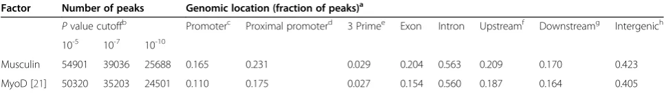
To compare the binding pattern of the bHLH myogenic inhibitor MSC to that of the myogenic activator MyoD, ChIP-seq for endogenous MSC was performed in RD cells under growth conditions. MSC binds at a compar-able number of sites as MyoD and with a similar gen-omic distribution, although there was a slightly greater enrichment of MSC binding in the region surrounding the transcription start site (TSS) compared to MyoD (Table 1), possibly reflecting the GC-rich nature of pro-moters and the preferred MSC E-box (see below). As with MyoD, MSC was found to bind widely at regions outside of those generally thought of as gene related, binding to a high degree (approximately 40% of all sites)
in intergenic regions. A number of sites identified as be-ing specifically and strongly enriched for either MyoD or MSC by ChIP-seq were tested with biologically inde-pendent site-specific ChIP, and factor-specific enrich-ment in agreeenrich-ment with the ChIP-seq data found at all sites (Additional file 1: Figure S1).
MSC heterodimerizes with proteins to bind to E-boxes [10], and we have previously shown by LC-MS/ MS that the E-protein E12 associates with MSC in RD cells, while MyoD does not associate with MSC [18]. To further confirm that the ChIP-seq data represent distinct MyoD or MSC bHLH dimers, a TAP-tagged MSC was created. This was shown to maintain biological activity as measured by its ability to repress myogenic reporters and bind E-boxes in electrophoretic mobility shift assays (EMSAs) (data not shown). The tagged MSC was then introduced stably into RD cells through retroviral trans-duction and MSC-associated complexes pulled down and subjected to LC-MS/MS. As expected, all E-proteins were found to associate with MSC, while there was no indication of a MSC: MyoD interaction (Additional file 2: Table S1).
A motif analysis of the binding site preferred by MSC found strong enrichment for binding at a GC core E-box (Figure 1A, top), one of the two E-box cores we previously identified as being preferred by MyoD (Figure 1A, bottom). In contrast to MyoD, MSC exhibits
a strong nucleotide preference for a ‘G’ at the first
nucleotide after the E-box (CAGCTGG), designated position +1 relative to the E-box. Also notable was a dif-ference in the sequences enriched at the two positions
immediately before the E-box, designated positions −1
and−2 relative to the E-box. We have previously shown
that MyoD:E and NeuroD2:E heterodimers show a
flanking preference for G or A in the −1 and −2
posi-tions [3,24], whereas the MSC motif does not demon-strate a similarly strong preference at these positions (Figure 1A, positions 2 and 3).
As anticipated from the motif analysis, MyoD and MSC showed overlapping but not identical binding
Table 1 Number and genomic location of musculin and MyoD ChIP-seq peaks in RD cells
Factor Number of peaks Genomic location (fraction of peaks)a
Pvalue cutoffb Promoterc Proximal promoterd 3 Primee Exon Intron Upstreamf Downstreamg Intergenich 10-5 10-7 10-10
Musculin 54901 39036 25688 0.165 0.231 0.029 0.204 0.563 0.209 0.170 0.423
MyoD [21] 50320 35203 24501 0.110 0.175 0.027 0.154 0.560 0.187 0.164 0.405
a
The fraction of MyoD and musculin peaks found in each listed type of genomic region are given. Note that categories are not mutually exclusive, and a single peak may be included in multiple categories.
b
ThreePvalue cutoffs were used to evaluate whether ChIP-seq reads are considered a‘peak’and included in the count of the total number of peaks.
c
+/−500 bp from the transcription start site (TSS).
d
+/−2 kb from the TSS.
e
+/−500 nucleotides from the end of the transcript.
f
–2 kb to−10 kb upstream of the TSS.
g
+2 kb to +10 kb from the end of the transcript.
h
locations in the genome. MyoD and MSC peaks were assigned to sequential cumulative bins of 3,000 peaks
based on rank by P value and the percentage overlap
ranged from approximately 40% to 80% (Figure 1B). A motif analysis of sites that were found to bind only MSC (MSC-specific) in comparison to sites bound either solely by MyoD (MyoD-specific) or by both MyoD and MSC (shared) identified a strong enrichment for C at the−1 position and G at the +1 position, giving an 8-bp
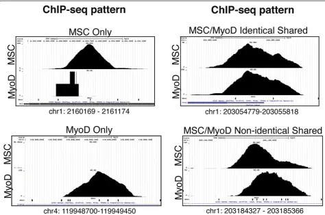
motif of CCAGCTGG (Figure 1C). Examination of the ChIP-seq data at specific loci identified sites bound only by one of the factors, sites bound by both factors in an apparently identical pattern, and sites bound by each factor in closely overlapping but non-identical binding patterns (Figure 2). The closely overlapping but distinct patterns suggests each factor is binding to a distinct E-box in the region; however, this is identified as an‘ over-lap’in the analysis shown in Figure 1B.
Figure 1MSC has similar, but non-identical, DNA binding characteristics to MyoD and binds at many of the same genomic locations.
Musculin binding is enriched at DNase hypersensitive genomic regions and regions with higher levels of histone acetylation
We have previously shown that MyoD binding induces histone acetylation at binding sites throughout the gen-ome [3]. To test the hypothesis that the gengen-ome-wide binding of MSC might inhibit acetylation in either a glo-bal manner or at some subset of MyoD-bound locations, we performed ChIP-seq for acetylated histone H4 (AcH4) from RD cells under conditions similar to the MyoD and MSC ChIP-seq data. AcH4 enrichment was examined at peaks identified as MSC-specific, MyoD-specific and shared. Surprisingly, the highest levels of AcH4 enrichment showed a stronger association with MSC peaks, both MSC specific and shared (Figure 3A). This trend became even more evident when peaks were grouped based on distance from the nearest gene TSS. While MyoD-specific peaks showed essentially identical AcH4 enrichment regardless of their location relative to a TSS, MSC-specific and shared peaks showed a strong shift to higher AcH4 enrichment at peaks located closer
to a TSS (<2 kb from the nearest TSS) (P value of the difference between MSC-specific and MyoD-specific peaks: 1 × 10-45,P value for MSC-specific versus shared peaks: 1 × 10⁻⁷) (Figure 3B). MSC binding did not cor-relate, either positively or negatively, with genes that we have previously identified as being differentially regu-lated in RD cells compared to normal myogenic cells [21] (data not shown).
Given the lack of a global effect on gene expres-sion, we hypothesized that the association with AcH4 might simply reflect binding of MSC at regions of open chromatin. The MyoD-specific, MSC-specific and shared peaks in the RD cells were compared to publicly available DNase hypersensitivity data from human myoblasts. Shared peaks had the highest proportion of peaks that overlapped with DNase
hypersensitive sites (shared: approximately 80%,
MSC-specific: approximately 70%, MyoD-specific: ap-proximately 50%) (Figure 3C), and this relation held across the entire range of hypersensitive values (Additional file 3: Figure S2A).
MyoD-specific peaks seemed to have a surprisingly low level of association with hypersensitive sites, but subcategorizing the MyoD-specific peaks based on whether they were unique to RD cells, or common to RDs and human myotubes [21] revealed that common peaks were generally associated with hypersensitive sites, and peaks unique to RDs were not (Additional file 3: Figure S2B). We have previously shown that differences in MyoD binding between myotubes and RD cells can be correlated with differences in E-box accessibility be-tween the cell types [21]. This suggests that the MyoD peaks specific to RMS, that is, not present in primary skeletal muscle cells, represent binding by MyoD to E-boxes that are normally inaccessible to bHLH binding in primary muscle cells and were therefore not identified as
lying in HSS regions in the primary muscle cell dataset. Taken as a whole, the above data identify MSC binding as largely occurring in the context of areas of open and accessible chromatin.
Musculin dimers have less restrictive binding site preferences at flanking nucleotides than MyoD dimers
Electrophoretic mobility shift assays with in vitro trans-lated proteins were performed to further investigate the sequence preference of MyoD and MSC dimers using the sequence from a MSC-specific peak at the SKI gene that contained the MSC-specific consensus 8-bp motif (CCAGCTGG). Shifts comparing binding of MyoD:E and MSC:E heterodimers demonstrated that MSC het-erodimers could bind to the 8-bp motif or a probe in
which the −1 position was changed from a ‘C’ to a ‘G’
(−1 mut), making it more reflective of the core MSC
motif from Figure 1A (Figure 4A, compare lane 4 to 5). Binding of MSC to a probe in which both−1 (‘C’to‘G’)
and +1 (‘G’ to ‘C’) had been changed (−1/+1 mut) still occurred, but at reduced levels (Figure 4A, compare lanes 4 and 5 to 6). In contrast, the ability of MyoD:E heterodimers to form complexes improved as the probe
was shifted away from the 8-bp MSC motif (Figure 4A, compare lane 10 to 12). Taken together with the motif analysis identifying the differences at positions −1 and −2, this suggests that the sequence preference for dimer binding is more stringent for dimers containing MyoD than those containing MSC, even with a common dimer partner. Similar results were observed with homodimers of both MSC and MyoD, though both types of homodi-mers formed more weakly compared to their heterodi-mer counterparts (Figure 4A, compare lane 1 to 3 and 7 to 9, data not shown).
To further test this hypothesis, competition assays were performed on MSC and MyoD heterodimers. As expected, MSC was competed off the MSC-specific 8-bp motif equally well by cold competitors with the
consen-sus 8-bp motif, –1 mut, or −1/+1 mut (Figure 4B, left
panel, compare lanes 3 and 4 to 5 and 6, and to 7 and 8), suggesting relatively similar affinity. MSC was not competed with a sequence in which the core nucleotides
of the E-box were inverted to a ‘CG’ from ‘GC’
(Figure 4B, left panel, compare lanes 9 and 10 to 2), dem-onstrating sequence specificity of the competition assays.
In contrast, MyoD heterodimers were only effectively competed by sequences at which it had formed visible complexes (Figure 4B, right panel, compare lanes 16 and 17 to 12 and 13, and to 14 and 15), and even a single nucleotide change had a notable impact on competition
(compare−1/+1 mut to−1 mut). As with MSC, MyoD:E
heterodimers failed to form on the CG core E-box (data not shown).
In addition to the relaxed preference at the positions immediately flanking the E-box, relative to MyoD, MSC also exhibited a sharp difference in response to sequence changes at the−2 position. The inclusion of a‘T’at the−2 position is permissive for MSC heterodimer binding (Figure 4C, compare lane 3 to 4), but absolutely abolishes binding of MyoD heterodimers (Figure 4C, compare lane 7 to 8), with similar results seen with the homodimers (Figure 4C, compare lane 1 to 2 and 5 to 6).
Discussion
Our genome-wide comparison of the DNA binding characteristics of MyoD and MSC reveals that, even though MSC is one of multiple myogenic inhibitors and might be expected to bind at only a subset of all MyoD binding locations, it binds at a comparable number of sites as MyoD, with a similar, but non-identical binding site preference. Even though MSC heterodimerizes with the same E-proteins as MyoD and shares the same se-quence preference at the central dinucleotide of E-boxes, it has less sequence preference for the positions that flank E-boxes than other bHLH dimers we have re-ported [3,24]. It should be noted that the electrophoretic mobility shift assays were performed using in vitro
translated proteins, and thus would not reflect the effect of any post-translational modifications that may occur in vivo. However, the in vitro binding preferences reflect those preferences seen in the in vivo ChIP-seq results. Additionally, work with other bHLH factors has demon-strated excellent correlation between binding sites iden-tified by ChIP-seq and binding seen with EMSA [24]. Overall, the broad overlap of MyoD and MSC binding indicates a potential for MSC to buffer the binding and activity of MyoD broadly, as well as other E-box binding factors.
We have previously shown that MSC can inhibit MyoD-mediated activation of myogenic targets by oc-cluding specific E-boxes [19], in addition to competing for a limiting pool of E-proteins [18]. We have proposed
that this activity controls the
growth-versus-differentiation decision point in myogenic cells, serving as a component of interlocking oscillating regulatory cir-cuits that keep myogenic cells balanced between prolif-eration and terminal differentiation [19].
This model suggests that the relationship between bHLH proteins and target sites is highly dynamic, in which dimers form and dissociate, from both their pro-tein partners and DNA binding sites, resulting in a fluc-tuating expression of gene targets. In turn, some subset of such targets feed back on the process to regulate growth and differentiation appropriately. MSC has a very similar core binding motif as MyoD dimers but a greater degree of flexibility for flanking nucleotides, which could reflect a lower need for tight regulation of the specific E-boxes MSC can bind to compared to MyoD and be the mechanism by which MSC acts to broadly sequester E-proteins and occupy potential MyoD binding sites. Other factors have also been suggested as having an in-hibitory function during myogenesis by binding at E-boxes [26,27], further potentially increasing the complex nature of the interplay occurring at bHLH binding sites. The similar core motif requirements for MyoD and MSC ensure that MSC binds at many sites regulated by
MyoD, and the +1 ‘G’ preference of the MSC motif
in-creases the likelihood of it targeting E-boxes located in GC-rich gene promoter regions. While the gene regula-tion analysis did not identify a global role in gene sup-pression, MSC does modulate MyoD activity at the myogenic microRNA miR-206 [19], and it may have a similar role at many other MyoD regulated genes, an ef-fect that would not be discernible with our current analysis.
individual cellular context, potentially interfering with the binding of inhibitory complexes. It is not known what histone modification enzymes MSC might recruit, nor is it clear how MSC activity would differ depending on the extent of competition by other bHLH proteins for binding partners and sites, and these additional pa-rameters might contribute to a context-specific ability to serve as a positive or negative regulator.
Both the finding that MSC is associated with regions enriched for acetylated histone H4 in RD cells and DNase hypersensitive sites in normal myotubes suggests MSC generally binds at areas of open chromatin. It is unclear at this point why the notable enrichment is seen at sites closest to transcription start sites (<2 kb), though it is possible that part of this enrichment is due to the GC-rich nature of promoters and the binding site prefer-ence of MSC for an additional flanking ‘G’compared to MyoD (Figure 1). While an effect by MSC on histone acetylation cannot be formally ruled out, mass spectrom-etry data did not identify any association with histone acetyltransferases (KLM, unpublished observations), sug-gesting that, in skeletal muscle cells, MSC is opportunis-tic in binding to areas of open chromatin, rather than instructing changes in chromatin structure. This would be consistent with the model proposed above, serving to assist MSC acting in a role as a dynamic competitor of MyoD function in the differentiation of skeletal muscle.
Conclusions
The myogenic bHLH inhibitor musculin binds widely throughout the genome in RD rhabdomyosarcoma cells and has a broadly overlapping, but non-identical, set of binding sites and peaks as MyoD. Compared to the pre-ferred MyoD E-box sequence, MSC has slightly less stringency for flanking sequence preference, permitting binding to a slightly broader set of E-boxes and poten-tially overlapping with other bHLH factors. Together with prior studies showing the ability of MSC to modu-late MyoD activity at overlapping sites at specific pro-moters, these results suggest a broad potential for MSC to modulate the activity of MyoD, and perhaps other bHLH proteins, during normal development and in cancers.
Additional files
Additional file 1: Figure S1.MyoD and MSC site-specific ChIP confirms the ChIP-seq results. Biologically independent site-specific ChIP was per-formed at three sites indicated by the ChIP-seq to be MyoD-specific bind-ing sites, three sites indicated as MSC-specific, and one control location with no significant binding of either factor, as indicated by both the chart and the screenshots. The enrichment was calculated for each location as the percentage of input amplified in qPCR with antibody divided by the percentage of input amplified with no antibody, and the value is indi-cated at the top of each bar. Note that they-axis is non-linear.
Screenshots are from the UCSC genome browser, and the identity of the factor used in the ChIP, and the number of reads at the peak of occu-pancy are indicated along the side. ChIP, chromatin immunoprecipitation; ChIP-seq, chromatin immunoprecipitation coupled to high-throughput sequencing; MSC, musculin.
Additional file 2: Table S1.LC-MS/MS identification of MSC-associated transcription factors in RD cells.
Additional file 3: Figure S2.MSC binds sites associated with DNase hypersensitivity, and MyoD peaks found only in RD cells, not in normal myotubes, are associated with areas identified in myotubes as DNase-resistant. (A) Shared MyoD and MSC binding peaks are associated strongly with DNase hypersensitive (HSS) sites in human myoblasts. The overlap between ChIP-seq peaks and HSS data is graphed for the entirety of the range of HSS values. Values for a DNase signal of‘0’are equal to 1 –the fraction graphed in Figure 3C. The data are plotted as a cumulative distribution function, where a value on they-axis represents the fraction of data that has a value equal to or less than the correspondingx-axis DNase HSS value. (B) MyoD-specific sites bound by MyoD only in RD cells, and not in human myotubes, overlap poorly with HSS sites in human myotubes. The MyoD-specific peaks from Figure 3C and (A) were further grouped into those peaks that were found both in RD cells and normal human myotubes (RD/myotube shared), and those found only in RD cells (RD-specific). As in Figure 3C, the data for each category (for example, RD-specific) are plotted as the fraction of peaks that overlap with sites that have some signal in the HSS data (that is, the graphed fraction = 1– fraction of peaks at HSS score of‘0’). ChIP-seq, chromatin immunoprecipi-tation coupled to high-throughput sequencing; HSS, hypersensitive; MSC, musculin.
Abbreviations
AcH4:acetylated histone H4; bHLH: basic helix-loop-helix; bp: base pair; ChIP: chromatin immunoprecipitation; ChIP-seq: chromatin
immunoprecipitation coupled to high-throughput sequencing;
EMSA: electrophoretic mobility shift assay; HSS: hypersensitivity; kb: kilobase; LC-MS/MS: liquid chromatography coupled to tandem mass spectrometry; MRF: myogenic regulatory factor; MSC: musculin; mut: mutation; qPCR: quantitative PCR; RMS: rhabdomyosarcoma; TAP: tandem affinity purification; TEV: tobacco etch virus; TSS: transcription start site.
Competing interests
The authors declare they have no potential competing interests.
Authors’contributions
KLM contributed to all experimental designs, performed all non-computational experiments and drafted the manuscript. ZY performed the ChIP-seq and all other computational analyses and contributed to both ex-perimental design and data interpretation. APF performed the RD cell-type control ChIP-seq. SJT conceived the project, contributed to all experimental designs and edited the manuscript. All authors read and approved the final manuscript.
Acknowledgements
KLM was supported by a Developmental Biology Predoctoral Training Grant (T32HD007183). ZY was supported by the NIH Interdisciplinary Training Grant in Cancer Research (T32CA080416). APF was supported by a grant from the University of Washington Child Health Research Center (NIH
U5K12HD043376-10) and Hyundai Hope on Wheels. SJT was supported by NIH NIAMS (R01AR045113).
Author details
1
Human Biology Division, Fred Hutchinson Cancer Research Center, 1100 Fairview Ave N C3-168, Seattle WA 98109, USA.2Molecular and Cellular Biology Program, University of Washington, Seattle WA 98105, USA.3Clinical Research Division, Fred Hutchinson Cancer Research Center, Seattle WA 98109, USA.4Department of Pediatrics, University of Washington School of Medicine, Seattle WA 98105, USA.5Department of Neurology, University of Washington, Seattle WA 98105, USA.
References
1. MacQuarrie KL, Fong AP, Morse RH, Tapscott SJ:Genome-wide transcription factor binding: beyond direct target regulation.Trends Genet2011,27:141–148.
2. Davis RL, Weintraub H, Lassar AB:Expression of a single transfected cDNA converts fibroblasts to myoblasts.Cell1987,51:987–1000.
3. Cao Y, Yao Z, Sarkar D, Lawrence M, Sanchez GJ, Parker MH, MacQuarrie KL, Davison J, Morgan MT, Ruzzo WL, Gentleman RC, Tapscott SJ: Genome-wide MyoD binding in skeletal muscle cells: a potential for broad cellular reprogramming.Dev Cell2010,18:662–674.
4. Berkes CA, Tapscott SJ:MyoD and the transcriptional control of myogenesis.Semin Cell Dev Biol2005,16:585–595.
5. Benezra R, Davis RL, Lockshon D, Turner DL, Weintraub H:The protein Id: a negative regulator of helix-loop-helix DNA binding proteins.Cell1990,
61:49–59.
6. Spicer DB, Rhee J, Cheung WL, Lassar AB:Inhibition of myogenic bHLH and MEF2 transcription factors by the bHLH protein Twist.Science1996,
272:1476–1480.
7. St-Pierre B, Flock G, Zacksenhaus E, Egan SE:Stra13 homodimers repress transcription through class B E-box elements.J biol chem2002,
277:46544–46551.
8. Lemercier C, To RQ, Carrasco RA, Konieczny SF:The basic helix-loop-helix transcription factor Mist1 functions as a transcriptional repressor of myoD.EMBO J1998,17:1412–1422.
9. Hamamori Y, Wu HY, Sartorelli V, Kedes L:The basic domain of myogenic basic helix-loop-helix (bHLH) proteins is the novel target for direct inhib-ition by another bHLH protein, Twist.Mol Cell Biol1997,17:6563–6573. 10. Lu J, Webb R, Richardson JA, Olson EN:MyoR: a muscle-restricted basic helix-loop-helix transcription factor that antagonizes the actions of MyoD.Proc Natl Acad Sci USA1999,96:552–557.
11. Lu J-R, Bassel-Duby R, Hawkins A, Chang P, Valdez R, Wu H, Gan L, Shelton JM, Richardson JA, Olson EN:Control of facial muscle development by MyoR and capsulin.Science2002,298:2378–2381.
12. Moncaut N, Cross JW, Siligan C, Keith A, Taylor K, Rigby PW, Carvajal JJ:
Musculin and TCF21 coordinate the maintenance of myogenic regulatory factor expression levels during mouse craniofacial development.Development2012,139:958–967.
13. Lee GH, Chang MY, Hsu CH, Chen YH:Essential roles of basic helix-loop-helix transcription factors, capsulin and musculin, during craniofacial myogenesis of zebrafish.Cell Mol Life Sci2011,68:4065–4078. 14. Ismat A, Schaub C, Reim I, Kirchner K, Schultheis D, Frasch M:HLH54F is
required for the specification and migration of longitudinal gut muscle founders from the caudal mesoderm ofDrosophila.Development2010,
137:3107–3117.
15. Hishikawa K, Marumo T, Miura S, Nakanishi A, Matsuzaki Y, Shibata K, Ichiyanagi T, Kohike H, Komori T, Takahashi I, Takase O, Imai N, Yoshikawa M, Inowa T, Hayashi M, Nakaki T, Nakauchi H, Okano H, Fujita T:Musculin/ MyoR is expressed in kidney side population cells and can regulate their function.J Cell Biol2005,169:921–928.
16. Yu L, Sangster N, Perez A, McCormick PJ:The bHLH protein MyoR inhibits the differentiation of early embryonic endoderm.Differentiation2004,
72:341–347.
17. Yu L, Mikloucich J, Sangster N, Perez A, McCormick PJ:MyoR is expressed in nonmyogenic cells and can inhibit their differentiation.Exp Cell Res 2003,289:162–173.
18. Yang Z, MacQuarrie KL, Analau E, Tyler AE, Dilworth FJ, Cao Y, Diede SJ, Tapscott SJ:MyoD and E-protein heterodimers switch rhabdomyosar-coma cells from an arrested myoblast phase to a differentiated state.
Genes Dev2009,23:694–707.
19. MacQuarrie KL, Yao Z, Young JM, Cao Y, Tapscott SJ:miR-206 integrates multiple components of differentiation pathways to control the transition from growth to differentiation in rhabdomyosarcoma cells.
Skeletal Muscle2012,2:7.
20. Saab R, Spunt SL, Skapek SX:Chapter 7–myogenesis and
rhabdomyosarcoma: the Jekyll and Hyde of skeletal muscle.Current Top Dev Biol Cancer Dev2010,94:197–234.
21. MacQuarrie KL, Yao Z, Fong AP, Diede SJ, Rudzinski ER, Hawkins DS, Tapscott SJ:Comparison of genome-wide binding of MyoD in normal hu-man myogenic cells and rhabdomyosarcomas identifies regional and local suppression of pro-myogenic transcription factors.Mol Cell Biol 2013,33:773–784.
22. Tapscott SJ, Davis RL, Thayer MJ, Cheng PF, Weintraub H, Lassar AB:MyoD1: a nuclear phosphoprotein requiring a Myc homology region to convert fibroblasts to myoblasts.Science1988,242:405–411.
23. Davis RL, Cheng PF, Lassar AB, Weintraub H:The MyoD DNA binding domain contains a recognition code for muscle-specific gene activation.
Cell1990,60:733–746.
24. Fong AP, Yao Z, Zhong JW, Cao Y, Ruzzo WL, Gentleman RC, Tapscott SJ:
Genetic and epigenetic determinants of neurogenesis and myogenesis.
Dev Cell2012,22:721–735.
25. Palii CG, Perez-Iratxeta C, Yao Z, Cao Y, Dai F, Davison J, Atkins H, Allan D, Dilworth FJ, Gentleman R, Tapscott SJ, Brand M:Differential genomic tar-geting of the transcription factor TAL1 in alternate haematopoietic line-ages.EMBO J2011,30:494–509.
26. Genetta T, Ruezinsky D, Kadesch T:Displacement of an E-box-binding re-pressor by basic helix-loop-helix proteins: implications for B-cell specifi-city of the immunoglobulin heavy-chain enhancer.Mol Cell Biol1994,
14:6153–6163.
27. Soleimani VD, Yin H, Jahani-Asl A, Ming H, Kockx CE, van Ijcken WF, Grosveld F, Rudnicki MA:Snail regulates MyoD binding-site occupancy to direct enhancer switching and differentiation-specific transcription in myogen-esis.Mol Cell2012,47:457–468.
doi:10.1186/2044-5040-3-26
Cite this article as:MacQuarrieet al.:Genome-wide binding of the basic helix-loop-helix myogenic inhibitor musculin has substantial overlap with MyoD: implications for buffering activity.Skeletal Muscle20133:26.
Submit your next manuscript to BioMed Central and take full advantage of:
• Convenient online submission
• Thorough peer review
• No space constraints or color figure charges
• Immediate publication on acceptance
• Inclusion in PubMed, CAS, Scopus and Google Scholar
• Research which is freely available for redistribution