R E S E A R C H A R T I C L E
Open Access
Intracisternal tuberculoma: a refractory type
of tuberculoma indicating surgical
intervention
Fanfan Chen
1†, Lei Chen
2†, Yongfu Cao
1, Yongjun Yi
1, Jingwen Zhuang
1, Wuhua Le
1, Wei Xie
1, Lanbo Tu
1,
Peng Li
1, Yimin Fang
3, Ling Li
4, Yuqing Kou
5, Kaikai Fu
5, Hua He
6*and Hongbin Ju
7*Abstract
Background:Central nervous system (CNS) tuberculoma is a rare disease with severe neurological deficits. This retrospective research is to review the data of patients diagnosed as CNS tuberculoma. Surgeries were performed in all patients. The clinical features especially the neurological image and the anatomical characters of the tuberculomas were concerned.
Methods:Totally 11 patients diagnosed as CNS tuberculoma were admitted in Guangzhou First People’s Hospital (7cases) and Changzheng Hospital (4 cases) during 2006–2015. The data including preoperative condition, neurological imaging, and surgical findings was collected and analyzed.
Results:The lesions of nine patients (9/11) were totally or subtotally excised and two (2/11) were partially excised. Neurological functions of all patients were improved after surgery without secondary infection. Lesions of nine (9/11) patients preoperatively progressed as a result of paradoxical reaction. Of the 9 patients demonstrated paradoxical progression, all lesions were partially or totally located at the cisterns or the subarachnoid space. Preoperative ATTs lasted 2 to 12 months and tuberculomas were not eliminated. The arachnoid was found thickened and tightly adhered to the lesions during surgeries. Of the 2 cases that paradoxical reaction were excluded, both patients (case 6, intramedullary tuberculoma; case 11, intradural extramedullary tuberculoma) were admitted at onset of the disease. ATTs were preoperatively given for 1 week as neurological deficits aggravated. The tuberculous lesions of CNS or other system showed no obvious change and paradoxical reaction could not be established in both cases.
Conclusions: Exudates of tuberculosis is usually accumulated in the cisterns and frequently results in the paradoxical formation of tuberculoma. Intracisternal tuberculoma is closely related to paradoxical reaction and refractory to anti-tuberculosis therapy. Micro-surgical excision is safe and effective. Early surgical intervention may be considered in the diagnosis of intracisternal tuberculoma especially when paradoxical reaction participates in the development of tuberculoma.
Keywords: Central nervous system, Paradoxical response, Tuberculosis, Spine, Tuberculoma
* Correspondence:hehua1624@smmu.edu.cn;bin7810@126.com †Equal contributors
6Neurosurgery Department, Changzheng Hospital, The Second Military
Medical University;State key Laboratory of Drug Research, Shanghai Institute of Material Medical, Chinese Academy of Sciences, 415# Fengyang Road, Shanghai 200003, China
7Spinal Surgery Department, Guangzhou First People’s Hospital, Guangzhou
Medical University, 1# Panfu Road, Guangzhou, Guangdong 510180, China Full list of author information is available at the end of the article
Background
Tuberculosis (TB) with central nervous system (CNS) in-volvement occurs in approximately 1% of all TB patients and causes the highest morbidity and mortality [1]. CNS tuberculosis has various forms. Tuberculous meningitis (TM) is the most frequent form of CNS TB and CNS tuberculoma is the type next to TM [2, 3]. Spinal intradural tuberculomas including intramedullary and intradural extramedullary tuberculoma are exceptionally rare and ac-count for approximate 2%–5% of all CNS tuberculoma [4]. Furthermore, Intradural extramedullary tuberculoma of the spinal cord is the most unusual type [5].
Although CNS tuberculoma is an uncommon disease, it usually presents with severe neurological deficits including altered mental status, hydrocephalus, cranial nerve palsies, hemiparesis and seizures et al. [6]. Anti-tuberculosis treat-ments (ATT) combined with surgeries in the treatment of CNS tuberculoma have been reported occasionally [7, 8]. However, most of the articles mainly described the rarity of this disease and clinical situation including diagnosis, medications and outcomes of surgeries [9–12]. The loca-tions, possible pathogenesis and the relation between them were not mentioned in those reported cases, except for the optochiasmatic tuberculoma [13].
Of the patients in this study, we were aware of the characteristics that the subarachnoid space including cerebral cisterns and spinal subarachnoid space were susceptible regions for the formation of tuberculomas following TM and/or tuberculous arachnoiditis. This situation was also noticed by other doctors [4, 14]. Para-doxical reaction, defined as an phenomenon of effective medical treatment demonstrating opposite effect in cer-tain lesions [15, 16], is prone to occur at the cisterns and frequently results in the formation of intracisternal tuberculoma [17, 18]. In most cases, ATT is effective in treating the CNS tuberculoma [19]. Unfortunately, when paradoxical reaction participates in the etiology of tuber-culoma, the effectiveness of ATT is usually limited and additional corticosteroids is indicated [20]. Nevertheless, long period of medical therapy for resolving the lesion results in limited improvement or even deterioration of the impaired neurological function, especially for the spinal paradoxical tuberculoma [2]. From the above in-formation, intracisternal tuberculoma, paradoxical reac-tion and their intimate relareac-tion result in the severity and difficulty of medicine treatment. Accordingly, rapid elimination of the tuberculoma by surgical intervention should be considered.
Methods
Patients’data
This retrospective research was approved by the ethics committee of Guangzhou First People’s Hospital and Changzheng Hospital. From 2006 to 2015, 11 patients
diagnosed as CNS tuberculoma requiring surgical inter-vention were admitted in the neurosurgery department of Guangzhou First People’s Hospital, Guangdong, China and Changzheng Hospital, Shanghai. All patients underwent surgical excision of tuberculoma. Preoperative sputum smear was negative of tubercle bacillus of all patients. The diagnosis of tuberculosis of nine cases (pulmonary or CNS) was established in specialized hospital for tubercu-losis. Two patients were diagnosed as spinal tuberculoma at onset of the disease and adequate ATT was adminis-tered for 1 week before sugeries. The age of patients ranged from 7 to 52 years old. Six patients were female and five were male. Of the nine patients with preoperative diagnosis of tuberculosis, effective ATT was applied. Effectiveness of ATT was evidenced by the improvement of the clinical symptom and radiological findings (includ-ing pulmonary or most part of the CNS lesions). However, new lesions or progression of one lesion was found in these nine patients in later period during the ATT. Para-doxical reactions were identified in these nine patients and additional corticosteroid was administered. Paradoxical re-action was not considered in the patient presenting intra-medullary tuberculoma and the patient with intradural extramedullary tuberculoma of the fifth thoracic vertebra as both patients presented tuberculomas initially. For the two patients, surgeries were performed 1 week after ATT initiated as negative sputums for tubercle bacillus were confirmed. As shown in Table 1, the location of symptom related tuberculoma included the cerebral hemispheres (three cases), posterior fossa (three cases), and spinal (five cases, including one case of intramedullary tuberculoma and four cases of intradural extramedullary tuberculoma.).
Preoperative preparations
All patients were given routine examinations of blood, blood electrolyte, liver function and kidney function. Electrocardiography and chest radiography were per-formed to exclude cardiac or pulmonary contraindications for surgeries. Sputum smears were performed repetitively assuring negative result of tubercle bacillus. MRI was acquired for a preoperative evaluation. ATT based on the regimen of specialized hospital for treatment of tubercu-losis and was continued during the entire hospital stay of the patient.
Surgical management
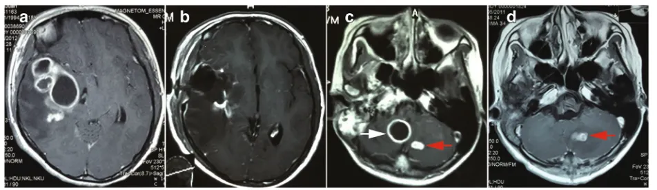
without surgical intervention) underwent two surgeries for the intradural extramedullary tuberculoma. Other patients underwent operations once respectively. The symptom-inducing lesion was the surgical target in pa-tient with multiple lesions. For example, papa-tient 2 (Fig. 2c, d), the lesion locating at right cisterna magna (Fig. 2c, white arrow) was responsible for the hydroceph-alus and hemiparesis. This lesion was the target of sur-gery. The lesion of left cerebellum was small and asymptomatic (Fig. 2c, d, red arrow). This lesion with other supratentorial lesions was resolved by the effective ATT. Precautions were taken to protect the surrounding
normal brain tissue from surgical contamination. For the spinal tuberculomas, pedicle screw fixation surgeries were performed if necessary. All patients were followed until the present time. Follow-ups were performed by telephone or at outpatient department in all patients.
Results
Preoperative MRI findings
For CNS tuberculoma, MRI is the most important examination for preoperative preparation. The lesions showed hypo- to iso-intense on T1WI (Fig. 1a) and mixed signal intensity on T2WI (Fig. 1b). Contrast-Table 1Clinical data of patients
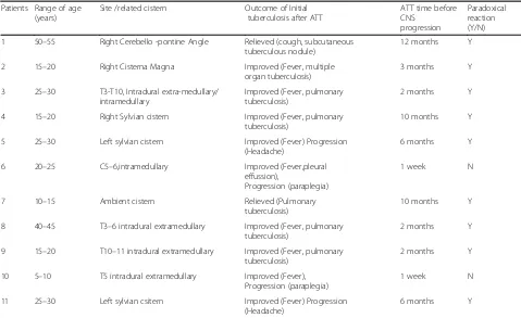
Patients Range of age (years)
Site /related cistern Outcome of Initial tuberculosis after ATT
ATT time before CNS
progression
Paradoxical reaction (Y/N) 1 50–55 Right Cerebello -pontine Angle Relieved (cough, subcutaneous
tuberculous nodule)
12 months Y
2 15–20 Right Cisterna Magna Improved (Fever, multiple organ tuberculosis)
3 months Y
3 25–30 T3-T10, Intradural extra-medullary/ intramedullary
Improved (Fever, pulmonary tuberculosis)
2 months Y
4 15–20 Right Sylvian cistern Improved (Fever, pulmonary tuberculosis)
10 months Y
5 25–30 Left sylvian cistern Improved (Fever) Progression (Headache)
6 months Y
6 20–25 C5–6,intramedullary Improved (Fever,pleural effussion),
Progression (paraplegia)
1 week N
7 10–15 Ambient cistern Relieved (Pulmonary tuberculosis)
10 months Y
8 40–45 T3–6 intradural extramedullary Improved (Fever, pulmonary tuberculosis)
2 months Y
9 15–20 T10–11 intradural extramedullary Improved (Fever, pulmonary tuberculosis)
2 months Y
10 5–10 T5 intradural extramedullary Improved (Fever), Progression (paraplegia)
1 week N
11 25–30 Left sylvian csitern Improved (Fever) Progression (Headache)
6 months Y
Tthoracic,Ccervical,Yyes,Nno
Fig. 1Typical MRI of intracisternal tuberculoma.aThe lesions showed iso- and hypo-intense on T1WI.bMixed signal intensity on T2WI.c
enhanced T1WI sequence displayed isolated or conglomer-ated ring enhancing lesions accompanied with hypo-intense non-enhancing content inside in most situations (Fig. 1c, 2a, c).
This group of patients contained various locations of the CNS including temporal lobe, frontal lobe, cerebello-pontine angle, cerebella and intravertebral canal. It is note-worthy that except one intramedullary tuberculoma case, all other cases were associated with the subarachnoid space, including the sylvian fissure (Fig. 2a), cerebellopontine angle (Fig. 1c), ambient cistern (Fig. 5a), cerebellomedullary cistern (Fig. 2c) and the subarachnoid space of the spinal cord (Fig. 3a).
Surgical findings
Patient 3 presented with coexisting intramedullary (Fig. 3a-d, red arrows) and intradural extramedullary le-sions (Fig. 3,the continuous enhancing intradural extrame-dullary lesion from T3-T9). Preoperative evaluation identified that the lesions responsible for the symptoms were the multiple intradural extramedullary tuberculomas. Given that the intramedullary lesion was located at the ventral spinal cord, a direct excision of the intramedullary lesion might cause further spinal injury to the spinal func-tion that was already seriously damaged, the intramedul-lary lesion was not resected. A laminectomy of T3-T4, T7-T9 or the excision of the corresponding extramedullary lesions was performed (Fig. 3b). However, the lesion of T9 was missed(Fig. 3b, white arrow). After 2 months of ATT, the following situations were displayed: extramedullary lesion of T9 enlarged (Fig. 3c, white arrow); the residual lesion of T5-T6 disappeared; the intramedullary lesion showed no change (Fig. 3a-c). A second time surgery was performed to remove the progressing extramedullary lesion of T9. At the same time, a pedicle screw fixation
was performed to maintain the stability of the spinal col-umn (Fig. 3d).
The intramedullary tuberculoma was revealed to be well-defined lesions (Fig. 4a) which is totally different for the intradural extramedullary tuberculoma. As for the latter, the abscess extended on the surface of spinal cord (Fig. 4b). The irregular lesion adhere to surrounding tissues intimately. Similar situation was oberserved in the cerebral tuberculoma. Even for the same tubercu-loma, the interface between the tuberculoma and paren-chyma was relatively loose and easy to be separated (Fig. 5e, white arrow displayed the interface of the tuber-culoma and the parenchyma of brain) while the adhesion of tuberculoma in the subarachnoid space was tight (Fig. 5f, white arrow displayed the adhesion of tubercu-loma and arachnoid). This kind of close relation between subarachnoid space and the cisternal part of tubercu-loma was due to the thickening arachnoid, the hyper-plastic fibrous tissue closely surrounding the lesion. Coagulation and sharp dissection is necessary for separ-ation. Furthermore, manipulation in the cisternal part of tuberculoma should be more careful as vital vessel may course in the cistern. Decisions of cutting or separation and reservation the vessel should be done based on clear identification (Fig. 5g, red arrow indicates the vessel after separation of arachnoid).
None of the patients experienced secondary infection following the surgical procedures. All of the patients were followed from the date of surgery until the present time. The outcomes of all patients were evaluated with Karnofsky Performance Scale in different neurological recovery periods. The recovery times of patients in this study were different. For the most severe case as case 3, 40 months had passed before the patient finally carried out daily activities (KPS was 90). For the other patient, it took about 2–6 months for recovery (KPS were 90–100).
Fig. 2Pre and post-surgical image of intracisternal tuberculoma.aPreoperative enhancing MRI of a tuberculoma locating at right sylvian fissure.
Pathology
All specimens were pathologically analyzed. The patho-logical characteristics including granulomas, necrosis, caseation, cell types (lymphocyte, Langerhans-type giant cell), and the result of acid-fast bacillus staining were re-corded. Not all the pathological features were demon-strated in one case. Typical tuberculomas showed a granulomatous reaction consisting of epithelioid cells and giant cells mixed with mononuclear inflammatory cells (predominantly lymphocytes) that form a granu-loma (Fig. 6b, c) [6, 21].
Discussion
CNS tuberculoma is a devastating disease with high morbidity and mortality. Although the outcome of the patients in this study was encouraging, the periods of
preoperative ATT (for the paradoxical tuberculoma) were 2 to 12 months (average 5.9 ± 4.0 months). Neuro-logical functions in most cases were aggravated during the ATT treatment as a result of a paradoxical reaction which limited the effect of ATT. Surgical excision of the lesion was a turning point for this group of patients. To our opinion, early surgical excision is an appropriate procedure when a paradoxical lesion formed. This is par-ticularly critical for spinal tuberculoma [1, 22].
Although the clinical situations of the patients were different, for example the sites of the lesions and the response to treatment, we noticed that the tubercu-loma of nine (9/11) patients who were considered paradoxical tuberculoma were correlated with the subarachnoid space. Furthermore, it was intriguing that the relapsed lesions of the two patients who Fig. 3MRI of a patient with intradural extramedullary tuberculoma and intramedullary tuberculoma (two operations).aPreoperative MRI of the first operation of the patient manifesting intradural extramedullary tuberculoma from T3 to T9 (the enhancing lesion from T3 to T9) and intramedullary tuberculoma (red arrow).bPostoperative MRI of the first operation of the patient manifesting intradural extramedullary tuberculoma from T3 to T9 and intramedullary tuberculoma (red arrow). The intramedullary tuberculoma of T7 was left untouched while the intradural extramedullary lesion of T9 was missed (white arrow).cPreoperative MRI of the second operation of the patient. The intradural extramedullary tuberculoma of T9 was the target of operation (white arrow).dPostoperative MRI of the second operation of the patient. The intradural extramedullary tuberculoma of T9 was excised (white arrow)
experienced local recurrence were located at the dor-sal ambient cistern or spinal subarachnoid space, re-spectively. We identified the thickening arachnoid adhered to the lesion during the surgery as described in previous literature [8]. The subarachnoid space in-volvement accompanied with the thickening arachnoid seems to be a potential risk factor for the occurrence and recurrence of tuberculoma. Meanwhile, frequent occurence of paradoxical reaction in the subarachnoid space furtherly proved the refractory nature of intra-cisternal tuberculoma.
Tuberculoma is frequently developed in subarachnoid space as a result of paradoxical reaction
The classical model of TM pathology suggests two steps of the pathological process of tuberculosis meningitis. Firstly, the mycobacteria begins to grow at the paren-chyma or meninges of the brain or spinal cord, and then develops and matures into a tuberculous abscess. Sec-ondly, the abscess breaks up and releases its contents into the subarachnoid space which causes TM [23]. TM may also be the result of the direct hematogenous spread of pulmonary tuberculosis [1]. Based on above Fig. 5The MRI and intraoperative situation of a tuberculoma associated with ambient cistern.aPreoperative MRI of the lesion.bDrainage surgery was performed for the first time operation at local hospital with residue mainly at the ambient cistern.cThe tuberculoma was relapsed with effective ATT and corticosteroids.dA total resection of the lesion was done.eIntraoperative image showed the clear boundary of the part of tuberculoma situating at cerebellum parenchyma (white arrow).fThe intracisternal part of the same tuberculoma demonstrated tightly adhesion to the thickening arachnoid which was relatively difficult for separation (white arrow).gExposure of a vessel coursed in the subarachnoid space (red arrow)
Fig. 6The specimen of an intracisternal tuberculoma and the pathological examination.aThe specimen of an intracisternal tuberculoma (patient 7).bTypical image showed the caseous necrosis of the specimen (arrow), epithelial cell and lymphocyte.cTypical image showed the
knowledge, tuberculoma and TM may be considered as the initial manifestations of the CNS tuberculous path-ology. More importantly, the subarachnoid space is a pathological and anatomical region where mycobacteria and its products frequently exist. The exudates of TM and the arachnoiditis usually accumulate at the sub-arachnoid space including the interpeduncular cisterns, optochiasmatic cistern, the ambient and the suprasellar cisterns, the sylvian fissures as well as the subarachnoid space of spine [17]. Spinal subarachnoid space is ana-tomically similar to the cerebral cisterns. This explained the facts that intradural extramedullary tuberculoma was closely related to the arachnoiditis as well [14]. Patho-logical progression following cisternal exudates includes arachnoiditis and tuberculoma [4, 11, 24]. This kind of cisternal tuberculoma displayed its own features other than the parenchymal tuberculoma. The gelatinous ex-udate is constrained by the hyperplastic thickened arach-noid whereas the exudate still could spread in the cistern. This illustrates multiple lobular tuberculoma is common in the images of published articles and in our cases which is different from the parenchymal tubercu-loma (Fig. 1c, 2a) [8]. The adhesion of arachnoid led to the formation of the inter-septum of the tuberculoma. More importantly, the exudates in the cistern was con-sidered a form of the paradoxical response [17]. Accord-ingly, tuberculoma secondary to the cisternal exudate was a product of paradoxical reaction too. It was re-ported the optochiasmatic region had a high propensity for accumulating a quantity of exudates, which was a frequent type of paradoxical reaction [25].
Intracisternal tuberculoma is refractory to ATT and an indication for surgical intervention
Based on the above information, because the cisternal tuberculoma shows a high rate of paradoxical develop-ment, the effectiveness of ATT is limited [23]. The im-munological reaction based paradoxical reaction results in the progression of the lesion which calls for additional cor-ticosteroids but no needs of change of ATT regimen [20]. Thus, prolonged therapeutic time is inevitable. At the same time, the paradoxical progression of the intracisternal le-sion furtherly damages the neurological function which in-dicates surgery. Natarajan et al. believed that paradoxical intraduaral extramedullary tuberculoma was an indication for surgery [7]. According to our findings, the relatively enclosed space of a tuberculoma-occupying cistern which impedes the flow of CSF may result in a low concentration of medicine in the area. In the two relapsed cases reported in our study, the lesions were both situated in the sub-arachnoid space. As for the case of the tuberculoma locat-ing at the ambient cistern to the cerebellum, even the drainage surgery had a limited effect and did not change the microenvironment of the intracisternal tuberculoma
(case 7). Finally, the safety of microsurgical excision had been proven as no secondary infection or dissemination was observed in literatures or our cases [9, 26]. All these factors ensure us that surgical resection should be early considered for the intracisternal tuberculoma.
Intracisternal tuberculoma is accompanied with serious neurological deficits requiring early resolving of the lesions
As mentioned above, Intracisternal tuberculoma is fre-quently progressed from paradoxical reaction and fast elimination of the lesions was usually difficult. It is worse that the exudates, arachnoiditis and/or tubercu-loma developing in the cistern and adjacent parenchyma furtherly damage the neurological functions [23]. Tuber-culoma in the cistern blocks the circulation of CSF resulting in hydrocephalus. The mass effect and sur-rounding edema may lead to the occurrence of hernia or the compression of vital structures of the CNS in certain areas. The exudates and tuberculoma can affect the blood vessels arranged in the area causing vasculitis and subsequently stroke [27]. In a univariate analysis, sylvian fissure exudates were predictors of stroke [28]. The crit-ical site of the intracisternal tuberculoma also requires more attention especially surgical consideration.
Conclusions
Intracisternal and intradural extramedullary tuberculoma are specific forms of tuberculoma requiring particular at-tention. Exudates accumulation and arachnoiditis are the pathological changes occurring in these areas. Meanwhile, paradoxical reaction frequently participates in the forma-tion of tuberculoma in these regions. This kind of intracis-ternal tuberculoma is refractory to ATT and corticosteroids which may take a long period to get the lesion resolved and potentially induce severe neurological injury. Surgical exci-sion is an inevitable and necessary procedure for intracis-ternal tuberculoma for improving neurological function.
Abbreviations
ATT:Anti-tuberculosis treatment; CNS: Central nervous system; CSF: Cerebral spinal fluid; MRI: Magnetic resonance imaging; TB: Tuberculosis;
TM: Tuberculous meningitis
Acknowledgements
We acknowledge Dr. Bing Luo, Jian Su, Hengchang Li and Zeng Yang for the clinical help; Dr. Xiangdong Xu for the MRI.
Funding
This work including the data collection, analysis and interpretation, manuscript revising was supported by National Natural Science Fund of China (81302187), PLA logistics research project (CWS14C063) and Fund of State Key Laboratory of Drug Research (SIMM1705KF-10).
Availability of data and materials
Authors’contributions
HBJ, HH, FFC and LC participated in the design of this research. HH offered part of the data. FFC, LC wrote and revised the manuscript. WX, YYJ, YFC, WHL, JWZ, LBT, PL participated in the operation, treatment and data collection. YMF administered the ATT regimen. LL,YQ K, KKF carried out the data analysis. All authors have read and approved the final manuscript.
Ethics approval and consent to participate
This retrospective research was approved by the ethics committee of Guangzhou First People’s Hospital and Changzheng Hospital.
Consent for publication
Informed consents to publish were obtained from the patients with individual data involved in this article.
Competing interests
The authors declare that they have no competing interests.
Publisher’s Note
Springer Nature remains neutral with regard to jurisdictional claims in published maps and institutional affiliations.
Author details
1
Neurosurgery Department, Guangzhou First People’s Hospital, Guangzhou Medical University, 1# Panfu Road, Guangzhou, Guangdong 510180, China.
2Neurosurgery Department, Shenzhen Second People’s Hospital, Shenzhen
University, 3002# Sungang Road, Shenzhen 518037, China.3Tuberculosis
Department, Guangzhou Chest Hospital, 62# Hengzhi Gang Road, Guangzhou, Guangdong 510095, China.4Record Department, Guangzhou
First People’s Hospital, Guangzhou Medical University, 1# Panfu Road, Guangzhou, Guangdong 510180, China.5Department of Navy Medicine, The
Second Military Medical University, 800# Xiangyin Road, Shanghai 200433, China.6Neurosurgery Department, Changzheng Hospital, The Second Military
Medical University;State key Laboratory of Drug Research, Shanghai Institute of Material Medical, Chinese Academy of Sciences, 415# Fengyang Road, Shanghai 200003, China.7Spinal Surgery Department, Guangzhou First People’s Hospital, Guangzhou Medical University, 1# Panfu Road, Guangzhou, Guangdong 510180, China.
Received: 4 May 2017 Accepted: 29 November 2017
References
1. DeLance AR, Safaee M, MC O, Clark AJ, Kaur G, Sun MZ, Bollen AW, Phillips JJ, Parsa AT. Tuberculoma of the central nervous system. J Clin Neurosci. 2013;20(10):1333–41.
2. Skendros P, Kamaria F, Kontopoulos V, Tsitouridis I, Sidiropoulos L. Intradural, eextramedullary tuberculoma of the spinal cord as a complication of tuberculous meningitis. Infection. 2003;31(2):115–7.
3. Das KK, Jaiswal S, Shukla M, Srivastava AK, Behari S, Kumar R. Concurrent cerebellar and cervical intramedullary tuberculoma: paradoxical response on antitubercular chemotherapy and need for surgery. J Pediatr Neurosci. 2014; 9(2):162–5.
4. Luo L, Pino J. An intradural extramedullary tuberculoma of the spinal cord in a non-HIV-infected patient: case report and review of the literature. Lung. 2006;184(3):187–93.
5. Dastur HM. Diagnosis and neurosurgical treatment of tuberculous disease of the CNS. Neurosurg Rev. 1983;6(3):111–7.
6. Thwaites GE, van Toorn R, Schoeman J. Tuberculous meningitis: more questions, still too few answers. The Lancet Neurology. 2013;12(10):999–1010. 7. Muthukumar N, Sureshkumar V, Ramesh VG. En plaque intradural
extramedullary spinal tuberculoma and concurrent intracranial
tuberculomas: paradoxical response to antituberculous therapy. Case report. Journal of neurosurgery Spine. 2007;6(2):169–73.
8. Rajshekhar V. Surgery for brain tuberculosis: a review. Acta Neurochir. 2015; 157(10):1665–78.
9. Li H, Liu W, You C. Central nervous system tuberculoma. J Clin Neurosci. 2012;19(5):691–5.
10. Botturi A, Prodi E, Silvani A, Gaviani P, Vanoli G, Carbone A, Salmaggi A. Brain tuberculoma (Mycobacterium Africanum): high index of suspicion helps in avoiding biopsy/surgery. Neurol Sci. 2012;33(2):363–5.
11. Roca B. Intradural extramedullary tuberculoma of the spinal cord: a review of reported cases. The Journal of infection. 2005;50(5):425–31.
12. Shingade RG, Prakashchandra SP. Role of advanced diagnostic imaging in intracranial Tuberculoma: MR spectroscopy. J Clin Diagn Res. 2015;9(8):TJ03–4. 13. Akhaddar A, El Hassani MY, Chakir N, Jiddane M. Optochiasmatic
tuberculoma: complication of tuberculous meningitis. Report of a case and review of the literature. Journal of neuroradiology Journal de
neuroradiologie. 2001;28(2):137–42.
14. Chang KH, Han MH, Choi YW, Kim IO, Han MC, Kim CW. Tuberculous arachnoiditis of the spine: findings on myelography, CT, and MR imaging. AJNR Am J Neuroradiol. 1989;10(6):1255–62.
15. Afghani B, Lieberman JM. Paradoxical enlargement or development of intracranial tuberculomas during therapy: case report and review. Clin Infect Dis. 1994;19(6):1092–9.
16. Nicolls DJ, King M, Holland D, Bala J, del Rio C. Intracranial tuberculomas developing while on therapy for pulmonary tuberculosis. Lancet Infect Dis. 2005;5(12):795–801.
17. Schoeman JF, Donald PR. Tuberculous meningitis. Handb Clin Neurol. 2013; 112:1135–8.
18. Verma R, Patil TB, Lalla R. Tuberculous optochiasmatic arachnoiditis. J Global Infect Dis. 2014;6(2):89.
19. Idris MN, Sokrab TE, Arbab MA, Ahmed AE, El Rasoul H, Ali S, Elzubair MA, Mirgani SM. Tuberculoma of the brain: a series of 16 cases treated with anti-tuberculosis drugs. The international journal of anti-tuberculosis and lung disease. 2007;11(1):91–5.
20. Dheda K, Barry CE 3rd, Maartens G. Tuberculosis. Lancet. 2016;387(10024):1211–26. 21. Garg RK. Tuberculosis of the central nervous system. Postgrad Med J. 1999;
75(881):133–40.
22. Fox GJ, Mitnick CD, Benedetti A, Chan ED, Becerra M, Chiang CY, Keshavjee S, Koh WJ, Shiraishi Y, Viiklepp P, et al. Surgery as an adjunctive treatment for multidrug-resistant tuberculosis: an individual patient data Metaanalysis. Clin Infect Dis. 2016;62(7):887–95.
23. Garg RK, Malhotra HS, Kumar N. Paradoxical reaction in HIV negative tuberculous meningitis. J Neurol Sci. 2014;340(1–2):26–36.
24. Garg RK, Paliwal V, Malhotra HS. Tuberculous optochiasmatic arachnoiditis: a devastating form of tuberculous meningitis. Expert Rev Anti-Infect Ther. 2011;9(9):719–29.
25. Sinha MK, Garg RK, Anuradha HK, Agarwal A, Parihar A, Mandhani PA. Paradoxical vision loss associated with optochiasmatic tuberculoma in tuberculous meningitis: a report of 8 patients. The Journal of infection. 2010;60(6):458–66.
26. Jha D, Khatri P, Choudhary A, Sethi R, Kumar S. Endoscopic third ventriculostomy in prepontine-suprasellar tuberculoma with tuberculous meningitis hydrocephalus: a case report. Pediatr Neurosurg. 2007;43(1):42–6. 27. Schoeman JF, Van Zyl LE, Laubscher JA, Donald PR, Serial CT. Scanning in
childhood tuberculous meningitis: prognostic features in 198 cases. J Child Neurol. 1995;10(4):320–9.
28. Cherian A, Thomas SV. Central nervous system tuberculosis. Afr Health Sci. 2011;11(1):116–27.
• We accept pre-submission inquiries
• Our selector tool helps you to find the most relevant journal
• We provide round the clock customer support
• Convenient online submission
• Thorough peer review
• Inclusion in PubMed and all major indexing services
• Maximum visibility for your research
Submit your manuscript at www.biomedcentral.com/submit