Open Access
Research article
Androgen responsive intronic non-coding RNAs
Rodrigo Louro, Helder I Nakaya, Paulo P Amaral, Fernanda Festa,
Mari C Sogayar, Aline M da Silva, Sergio Verjovski-Almeida and
Eduardo M Reis*
Address: Departamento de Bioquimica, Instituto de Quimica, Universidade de São Paulo, 05508-900 São Paulo, Brazil
Email: Rodrigo Louro - [email protected]; Helder I Nakaya - [email protected]; Paulo P Amaral - [email protected];
Fernanda Festa - [email protected]; Mari C Sogayar - [email protected]; Aline M da Silva - [email protected]; Sergio Verjovski-Almeida - [email protected]; Eduardo M Reis* - [email protected]
* Corresponding author
Abstract
Background: Transcription of large numbers of non-coding RNAs originating from intronic regions of human genes has been recently reported, but mechanisms governing their biosynthesis and biological functions are largely unknown. In this work, we evaluated the existence of a common mechanism of transcription regulation shared by protein-coding mRNAs and intronic RNAs by measuring the effect of androgen on the transcriptional profile of a prostate cancer cell line.
Results: Using a custom-built cDNA microarray enriched in intronic transcribed sequences, we found 39 intronic non-coding RNAs for which levels were significantly regulated by androgen exposure. Orientation-specific reverse transcription-PCR indicated that 10 of the 13 were transcribed in the antisense direction. These transcripts are long (0.5–5 kb), unspliced and apparently do not code for proteins. Interestingly, we found that the relative levels of androgen-regulated intronic transcripts could be correlated with the levels of the corresponding protein-coding gene (asGAS6 and asDNAJC3) or with the alternative usage of exons (asKDELR2 and asITGA6) in the corresponding protein-coding transcripts. Binding of the androgen receptor to a putative regulatory region upstream from asMYO5A, an androgen-regulated antisense intronic transcript, was confirmed by chromatin immunoprecipitation.
Conclusion: Altogether, these results indicate that at least a fraction of naturally transcribed intronic non-coding RNAs may be regulated by common physiological signals such as hormones, and further corroborate the notion that the intronic complement of the transcriptome play functional roles in the human gene-expression program.
Background
Non-coding RNAs (ncRNAs), particularly those tran-scribed from genomic regions spanning introns of spliced genes, have been proposed as a fundamental advance in the genetic operating system of higher organisms by
influ-encing the genomic program of differentiation and devel-opment in individuals and species [1,2]. Many functions, such as transcriptional or translational regulation, RNA splicing, gene silencing, imprinting and dosage compen-sation, have been assigned to mammalian ncRNAs [3].
Published: 30 January 2007
BMC Biology 2007, 5:4 doi:10.1186/1741-7007-5-4
Received: 1 September 2006 Accepted: 30 January 2007
This article is available from: http://www.biomedcentral.com/1741-7007/5/4
© 2007 Louro et al; licensee BioMed Central Ltd.
Most functionally characterized ncRNAs belong to classes of small RNAs such as microRNAs [4] and small nucleolar RNAs (snoRNAs) [5]. While several novel long ncRNAs have been described in mammalian organisms [6,7] few have been characterized in more detail, the ~15 kb Xist RNA involved in inactivation of chromosome X being an exception [8].
Recent studies based on computational analysis and experimental validation have revealed that both introns and intergenic regions constitute major sources of ncRNAs [3,9,10]. Extensive analyses of the human expressed sequence tags (EST) database have focused par-ticularly on spliced mRNAs, and ESTs that overlap at least one exon have identified an abundant number of sense-antisense transcript pairs in humans [7,11,12], mice [13] and plants [14], pointing to a well-conserved mechanism of post-transcriptional regulation of gene expression in eukaryotes.
Recently, microRNAs have been shown to be consistently altered in normal and tumor cells, allowing the identifica-tion of tissue-specific expression signatures [15]. Using a custom-built cDNA microarray enriched in ESTs corre-sponding to fragments of intronic transcripts, we previ-ously detected transcription of a set of totally intronic ncRNAs, which are long (0.6–1.1 Kb), unspliced, and ori-ented in the antisense direction relative to the correspond-ing protein-codcorrespond-ing transcript [16]. Moreover, expression levels of 23 intronic ncRNAs were shown to correlate with the degree of tumor differentiation in prostate cancer [16]. The potential role of long ncRNAs in human cancer is also exemplified by MALAT-1, a 7-kb RNA that was reported to be associated with metastatic non-small cell lung cancer [17]. Other reports have shown that totally or partially intronic long antisense transcripts may have regulatory functions, such as modulating the methylation status of promoters [18] or the alternative splicing pattern of the corresponding protein-coding gene [19]. However, little attention has been given to the mechanism used by the cell to control the expression of these long intronic RNAs.
Androgens have been implicated in central events govern-ing the regulation of distinct and diverse physiological processes in normal and neoplastic prostate cells. These hormones are known to promote cell division and prolif-eration of epithelial cells, to modulate programmed cell death and cell quiescence [20,21] and to regulate cellular metabolism [22]. More recently, several studies have char-acterized the temporal transcriptional program reflecting the cellular response to androgens, which led to the iden-tification of novel androgen-regulated protein-coding genes [23-25].
In the present study, to identify androgen-responsive non-coding intronic RNAs, we used a custom-built spotted cDNA microarray enriched in intronic transcribed sequences and a prostate cancer cell line (LNCaP) cul-tured in the presence of a synthetic androgen. Using ori-entation-specific reverse transcription followed by PCR (RT-PCR) we determined the sense or antisense direction of a subset of intronic transcripts. We found that the levels of these intronic RNAs in androgen-treated and control cells correlate with the levels of message or to the alterna-tive usage of exons in the corresponding protein-coding transcript. Additional approaches such as rapid amplifica-tion of the cDNA ends (RACE) and chromatin immuno-precipitation assays (ChIP) demonstrated that these intronic messages represent long and unspliced RNA tran-scripts that may be directly regulated by the androgen receptor (AR) in vivo. These findings indicate that tran-scription of long intronic ncRNAs may be controlled by mechanisms common to protein-coding transcripts, such as those involving hormonal control of gene-promoter activation. In addition, our results indicate that a fraction of this class of long unspliced intronic RNAs may have a role in post-transcriptional regulation of gene expression by modulating transcript stability and alternative splicing.
Results and discussion
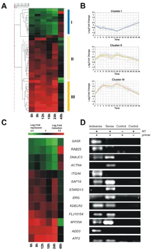
Identification of intronic RNAs regulated by androgen We used a spotted cDNA microarray platform enriched in intronic transcripts [16] to identify intronic RNAs and exonic protein-coding messages that had significant changes in transcription levels when hormone-responsive LNCaP prostate cancer cells were treated with the syn-thetic androgen R1881. Expression levels of KLK3 (PSA, prostate-specific antigen) [24] and TMEPAI [26], two genes previously shown to be upregulated by androgens in prostate epithelium, were determined by real-time PCR, confirming the effectiveness of androgens to induce responsive genes in our experimental conditions (see Additional file 1). Next, we used significance analysis of microarrays (SAM) [27] to identify transcripts that showed statistically significant changes in levels after exposure to androgen for periods of 6–48 hours. Only transcripts showing significant expression changes (≥ 1.5-fold change and false discovery rate < 5%) for at least three consecutive time points were selected for further analysis. We found 168 protein-coding exonic messages (see Addi-tional file 2) and 39 intronic RNAs (Figure 1A) that had statistically different patterns of temporal expression in androgen-treated cells compared with untreated controls (the complete list of androgen-regulated exonic tran-scripts coding for proteins is shown in Additional file 3).
Androgen-responsive intronic RNAs Figure 1
contained RNAs whose levels were 2–3-fold downregu-lated within 24 h after androgen exposure and were restored at 48 h. Clusters II and III included intronic RNAs that were upregulated by androgen within 24 h and later restored. Cluster II grouped RNAs that were 2-t3-fold increased, and cluster III grouped RNAs that were upregu-lated 3–6-fold. All of these intronic RNAs are unspliced messages, and apparently have no coding potential as determined by ESTScan analysis [28] (a complete list of androgen-regulated intronic RNAs is provided in Addi-tional file 4).
Most mammalian snoRNAs [5] and a large fraction of microRNAs [29] are derived from intronic sequences of protein-coding and noncoding genes. To investigate the possibility that some of the androgen-regulated intronic transcripts might be primary transcripts that, after process-ing, could generate known small RNAs of those classes, we compared the sequence of the 39 androgen-regulated intronic RNAs with those of 346 snoRNAs [30] and 383 microRNAs [31]; no similarity was found.
We then investigated the enrichment of specific gene ontology (GO) categories for androgen-regulated exonic and intronic transcripts using BiNGO, a gene ontology comparison tool [32]. We postulated that intronic tran-scripts represent a new class of RNAs that might act as
cis-regulatory factors [33]. Thus, for each androgen-regulated intronic transcript we used the GO annotation assigned to the corresponding protein-coding mRNA mapping to the same genomic locus. We found no significant enrichment of specific GO categories (p < 0.05) among the selected sets of exonic or intronic androgen-regulated transcripts when a correction for multiple testing was applied. How-ever, we noted that 13 of the set of 39 androgen-regulated intronic transcripts belonged to the signal-transduction GO category (GO no. 0007165). Indeed, some of these loci have already been implicated in cellular events related to prostate cell growth and differentiation. For example, the protein encoded by GAS6 mRNA is a ligand of the Axl receptor tyrosine kinase, and the Gas6/Axl complex has been shown to exhibit mitogenic activity in human pros-tatic cancer cell lines by modulating the PI3K/AKT and MEK signal-transduction pathways [34]. Similarly, the transcription factor encoded by ERG, a member of the ETS family of central genes involved in integrating signals that regulate cell growth and differentiation, stress responses and tumorigenesis, has been previously identified as the most frequently overexpressed proto-oncogene in malig-nant prostate epithelial cells [35].
Characterization of androgen-regulated intronic RNAs Orientation-specific RT-PCR was performed (see Methods for details) on a selected set of androgen-regulated intronic RNAs (Figure 1C). Sense or antisense orientation
of the intronic RNA relative to the corresponding protein-coding gene could be unambiguously assigned in most cases (13 of 17). For most of these (10 of 13) an intronic antisense RNA could be detected (Figure 1D). For six of these antisense RNAs (DNAJC3, SAP18, KDELR2,
FLJ10154, MYO5A and ATF2) we detected an overlapping
intronic sense message transcribed in the opposite strand. Four RNAs (GAS6, RAB25, ITGA6 and ADD3) were only detected in the antisense orientation, and three intronic messages (ACTN4, STARD13 and ERG) were only detected in the sense orientation (Figure 1D). The intronic sense transcripts of ACTN4, STARD13 and ERG did not show coding potential as determined by ESTScan. With-out further experimentation, we could not determine if these intronic sense transcripts are novel non-coding exons present in alternative splicing forms, or if they rep-resent ncRNAs originated from independent intronic tran-scriptional units located in the sense strand. However, the latter explanation concurs with sense intronic transcrip-tional units that have been observed elsewhere [9,36].
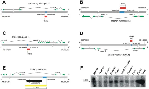
Next, RACE-PCR experiments with cDNA from prostate adenocarcinoma cells were performed for a selected set of intronic RNAs to obtain their full-length sequences. This selected set comprised intronic RNAs transcribed in the antisense (GAS6 and ITGA6), sense (STARD3) or both antisense and sense orientations (MYO5A and DNAJC3) as determined by orientation-specific RT-PCR. RACE experiments revealed that antisense intronic RNAs from
DNAJC3,MYO5A and ITGA6 were long (0.5–0.8 kb),
unspliced transcripts that mapped totally inside the intronic regions of the corresponding protein-coding gene (Figure 2A–C). An unspliced, totally intronic sense tran-script from STARD3 of approximately 1.4 kb was obtained (Figure 2D). Further analysis of the coding-potential by ESTScan indicated that extended intronic STARD3 could generate a 74-residue peptide, but subsequent BLASTp searches [37] did not show any similarity of this small pre-dicted product to known proteins in GenBank or to con-served protein domains at CDD [38]. We found in GenBank a 3.2-kb mRNA cloned from a brain cDNA library [GenBank: AK123478] that confirmed and extended the intronic antisense RNA mapping to the
GAS6 locus. Next, we performed PCR with primers
com-plementary to the cDNA probe mapping to an intron of
GAS6 deposited in the microarray and to the AK123478
mRNA. We obtained a 4.2-kb amplicon, demonstrating that the GAS6 antisense intronic RNA transcribed in pros-tate cells was even longer (at least 4.2 kb) than the previ-ously known RNA (Figure 2E, yellow bar).In silico analysis using ESTScan [28] failed to identify open reading frames for extended intronic sequences transcribed from
A strand-specific northern blot confirmed the expression of a long (~5 kb) GAS6 antisense transcript in various human tissues (Figure 2F). The observation that andro-gen-regulated intronic RNAs could be detected in tissues that are not particularly responsive to this hormone (Fig-ure 2, panel F) suggests that regulation of the steady-state levels of these transcripts may also proceed through an androgen-independent pathway.
Effect of androgen in the levels of intronic RNAs and corresponding protein-coding transcripts
Of the androgen-regulated transcripts identified in our analysis, only KDELR2 and ADD3 showed detectable changes in expression levels of both intronic and exonic transcripts. The intronic ADD3 transcript increased 4.9-fold (log2 ratio = 2.3) in androgen-treated cells, whereas the corresponding protein-coding transcript decreased 3.3-fold (log2 ratio = -1.7) in the same experiment.
Intronic KDELR2 transcript levels increased 2-fold (log2 ratio = 1), and the corresponding exonic transcript increased 2.3-fold (log2 ratio = 1.2) in cells exposed to androgen relative to control cells (see Additional files 3 and 4). These results indicate that for a given intronic-exonic pair transcribed in the same locus, the intronic message may be directly or inversely modulated with respect to the exonic message after androgen treatment.
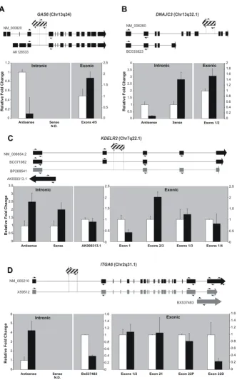
Orientation-specific reverse transcription, followed by quantitative real-time PCR using RNA from treated and control untreated cells, was used to confirm the change in levels of intronic transcripts after androgen exposure. We tested two intronic RNAs that were detected only in the antisense orientation (GAS6 and ITGA6), and two intronic messages in which both antisense and sense tran-scription were measured (DNAJC3 and KDELR2) (Figure 3A–D, "intronic" panels). In each case, we quantified in
Characterization of androgen-responsive non-coding intronic RNAs Figure 2
Antisense intronic transcription may regulate the levels and the post-transcriptional processing of sense protein-coding mRNAs
Figure 3
parallel the expression levels of exons of the correspond-ing protein-codcorrespond-ing gene (Figure 3A–D, "exonic" panels). We confirmed by quantitative PCR a 10-fold decrease of the intronic antisense transcript to GAS6 after androgen stimulation (Fig 3A, intronic panel). Interestingly, pro-tein-coding messages for GAS6 that contain exons 4 and 5 were upregulated (1.8-fold) under identical experimental conditions (Figure 3A, exonic panel). We observed that the levels of sense and antisense intronic RNAs at the
DNAJC3 locus were modulated in opposite directions
after androgen treatment (Figure 3B, intronic panel). The upregulation observed for the sense intronic RNA from
DNAJC3 after androgen exposure was in agreement with
the results from the microarray experiments (Figure 3B, intronic panel), and was also observed for the protein-coding DNAJC3 transcript (Figure 3B, exonic panel). Modification in the relative levels of intronic RNAs from
KDELR2 and ITGA6 after androgen treatment was
con-firmed (Figure 3C and 3D, intronic panels). For KDELR2, both antisense and sense intronic RNA levels were upreg-ulated by androgen (2.5-fold and 2.0-fold increase, respectively; Figure 3C, intronic panel). Likewise, the anti-sense intronic RNA at the ITGA6 locus was 4.2-fold increased in the presence of androgen (Figure 3D, intronic panel). However, in these cases we could not detect nota-ble changes in the relative levels of the respective protein-coding transcripts when primers designed to measure con-stitutive exons were used (Figure 3C and 3D, exonic pan-els).
To accumulate further evidence of functional involvement of antisense intronic non-coding transcripts on the splic-ing mechanism [19,33], we investigated the existence of correlation between their relative levels and the levels of the corresponding protein-coding mRNAs, using primers designed to probe regulated exons, that is, exons that are absent in at least one documented transcript isoform. At
the KDELR2 locus, we found that exons 2 and 3 were
upregulated (2-fold) after androgen treatment (Figure 3C, exonic panel). Notably, exon 2 was absent in one KDELR2
transcript isoform [GenBank: BP269541]. Therefore, it is conceivable that the observed increase in the level of intronic antisense RNA for KDELR2 after androgen treat-ment may induce increased retention of exon 2. We could not detect any notable androgen-driven change in the lev-els of transcripts containing exons 1 and 3 or exons 1 and 4 (Figure 3C, exonic panel). Notwithstanding, we observed that exon 1 for KDELR2 was downregulated 2.4-fold after androgen treatment when a pair of primers designed to probe only this exon was used (Figure 3C, exonic panel). This result can only be accounted for by the accumulation after androgen treatment of an as yet uncharacterized alternative KDELR2 transcript with a shorter 3' end.
At the ITGA6 locus we found that the levels of a protein-coding transcript isoform containing a longer terminal exon (exon 22D) were downregulated 4.2-fold after androgen treatment (Figure 3D, exonic panel), and thus inversely correlated with the 4.2-fold increase observed in the antisense intronic RNA under the same conditions (Figure 3D, intronic panel). However, it was not possible to determine whether this result was actually due to downregulation of the ITGA6 mRNA isoform containing exon 22D, as we also detected downregulation of another intronic transcript naturally transcribed in the ITGA6
locus [GenBank: BX537483] that spans the 3' terminal exon of the gene (Figure 3D).
Non-coding RNAs have been shown to affect splicing of protein-coding genes [19,39]. HBII-52, a snoRNA was found to regulate alternative splicing of the serotonin receptor gene 5-HT2CRA in trans, by binding to a silencing element in exon Vb of 5-HT2CRA [39]. Lack of HBII-52
expression causes defective 5-HT2CRA pre-mRNA processing and probably accounts for the behavioral problems observed in patients with congenital Prader-Willi syndrome [39]. A previous study on the FAS locus has identified a partially intronic antisense RNA that is naturally transcribed from the first intron of the gene, which was named Saf [19]. In vitro overexpression of Saf
induced skipping of several 3' exons of Fas mRNA, result-ing in a Fas protein that was no longer able to anchor to the cell membrane or to induce Fas-mediated apoptosis [19]. Our results identify novel intronic antisense RNAs that might be involved in regulatory mechanisms of alter-native splicing of protein-coding genes, in analogy to those observed for HBII-52 and Fas/Saf.
Increased binding of the androgen receptor to an androgen response element motif upstream of asMYO5A
Androgen-mediated transcriptional regulation involves direct hormone interaction with the AR protein and the translocation of this complex to the nucleus, promoting specific interactions with DNA (i.e. androgen response element; ARE) motifs located near or within the target sequences [40]. To support a mechanism of direct tran-scriptional control of androgen-regulated intronic RNAs, we used a bioinformatics approach [24] to look for puta-tive ARE motifs located upstream of the identified andro-gen-regulated intronic RNAs. At least one putative ARE motif was identified for 24 androgen-regulated intronic transcripts (a complete list of identified putative ARE motifs is presented in Additional file 5).
ChIP assays were performed to verify functional binding of AR to a putative regulatory ARE motif located upstream of the intronic antisense RNA mapping to the MYO5
bound to ARs (Figure 4B and 4C, asMYO5A ARE). The
asMYO5A ARE motif is located at a 5' genomic region 812
bp upstream of the region where the antisense MYO5A
intronic transcript maps. Binding of AR to a previously described functional ARE that regulates transcription of the PSA gene was tested in parallel as a positive control for the ChIP assay (Figure 4B, PSA ARE). A genomic region upstream from TUBA6, a gene that does not contain puta-tive ARE motifs in its promoter region, was used as a neg-ative control for non-specific immunoprecipitation of DNA fragments (Figure 4B, TUBA6).
The presence of a weak band for asMYO5A ARE in control cell lysates immunoprecipitated with anti-AR (Figure 4B,
asMYO5A ARE), indicating that binding of AR to
asMYO5A ARE may occur to some extent in untreated
cells. This may be explained by the presence of trace amounts of androgen remaining in charcoal-stripped fetal calf serum added to the cell-culture medium. An alterna-tive explanation for the background signal of asMYO5A ARE measured in control cells may be the existence of small amounts of AR constitutively bound to the
chroma-tin, as described for another transcription factor, cyclic AMP-responsive element-binding protein (CREB) [41].
The increased binding of AR to the asMYO5A ARE motif is in agreement with the androgen-induced increased expression of the antisense intronic MYO5A RNA
(asMYO5A) detected by microarray (Figure 1), and
con-firmed by orientation-specific reverse transcription fol-lowed by real-time PCR (Figure 4A). MYO5A encodes an actin-based processive motor involved in intracellular trafficking and exocytosis [42]. Tissue-specific Myo5a iso-forms are originated from alternative exon usage of the primary transcript, which in turn is thought to modulate the protein specificity for different cargoes within the cell [43]. It is conceivable that the increase in asMYO5A levels in prostate cells in response to androgen affects the rela-tive levels of the alternarela-tively spliced forms of MYO5A, thus modulating protein activity or specificity. Together, these results suggest that functional binding of androgen-AR activated complexes to regulatory androgen-ARE regions is required to modulate the levels of intronic antisense RNAs after androgen treatment of prostate cells.
Increased levels of antisense intronic MYO5A transcript correlates to binding of androgen receptor to a putative upstream androgen response element
Figure 4
Conclusion
Although there is still no conclusive evidence about the molecular mechanisms involved in response to androgen, we postulate that intronic transcripts exert their regulatory functions by acting in cis through Watson-Crick base-pair-ing to complementary sense pre-mRNA [33]. The results presented here corroborate the notion that at least a frac-tion of these intronic transcripts may directly affect the levels and/or splicing of the protein-coding transcript originating in the opposite strand, and concur with previ-ous reports in the literature [44,45].
It is not yet clear if the intronic transcripts identified in this work exert their potential regulatory functions as long (0.5–5 kb) RNAs, or alternatively, as shorter RNAs result-ing from processresult-ing of the primary transcript. Processresult-ing of these androgen-regulated ncRNAs into shorter RNAs of defined sizes would also raise the possibility that some may modulate transcriptional or post-transcriptional events in messages from different loci, in a manner similar to microRNAs [4]. The identification of possible short RNAs modulated by androgen will require further work, such as 15% denaturing polyacrylamide gel electrophore-sis or northern blots of prostate cells exposed to androgen in time-course experiments to document the accumula-tion of the long intronic RNAs, the time course of their conversion to short RNAs, and the accumulation of possi-ble short RNA intermediates.
Previous reports have shown that a large number of tran-scription-factor binding sites (TFBSs) for well-character-ized transcription factors such as Sp1, c-Myc, p53 and Creb lie upstream to transcriptionally active intronic regions [41,46], some of which generate transcripts that are oriented antisense relative to the known gene [46]. Notably, Cawley et al [46] found that comparable frac-tions of protein-coding genes and ncRNAs having an experimentally determined upstream TFBS region were regulated after exposure to retinoic acid. The results pre-sented here provide further evidence that ncRNAs tran-scribed from intronic segments of the human genome may be regulated by physiological signals that commonly act on protein coding-transcripts such as hormones. Sim-ilar results were observed in mice when the effect of lipopolysaccharide stimulation on the levels of several long ncRNAs was measured [47]. Together, these results strongly suggest that at least in mammalian cells intronic ncRNAs are transcribed in a regulated fashion and there-fore must exert physiological roles, which corroborates the existence of a yet underappreciated complex RNA-based regulatory system involving non-coding messages in higher organisms [2,3,48].
Methods
Cell culture and RNA isolation
The experimental design was based on recent published work [24]. In summary, prostate carcinoma cell lines LNCaP, DU145 and PC3 were obtained from the Ameri-can Type Culture Collection and maintained using the suggested medium supplemented with 10% (v/v) fetal calf serum (FCS), 3 mM L-glutamine, 100 μg/ml strepto-mycin and 100 U/ml penicillin. LNCaP cells were grown in RPMI 1640, and DU145 and PC3 cells were cultured in DMEM. For the androgen-response experiments, LNCaP cells were cultured for 24 hours in androgen-deprived medium (RPMI 1640) with 5% (w/v) charcoal-stripped fetal calf serum (FCS; Invitrogen, Carlsbad, CA, USA). After 24 hours, cells were placed in fresh RPMI 1640 medium, with 10% (w/v) charcoal-treated FCS, and either 1 nM of the synthetic androgen R1881 (Perkin Elmer, Wellesley, MA, USA) or equivalent volume of vehicle (eth-anol) was added. Cells were harvested after 0, 6, 9, 12, 18, 24, and 48 hours for RNA isolation. Two independent cul-tures of androgen-treated cells and of control cells (inde-pendent biological replicates), were used for RNA isolation.
In parallel, cells were harvested after 4 hours of hormone treatment for isolation of DNA that was subsequently used in ChIP assays, as described below. Total RNA was purified from experimental and control cells by caesium chloride cushion [49] after cell lysis (4 M guanidine iso-thiocyanate; 0.1 M β-mercaptoethanol; 25 mM sodium citrate pH 7.0). Total RNA (100 μg sample) was treated with RNase-free DNase (RNeasy kit; Qiagen, Hilden, Ger-many) for 15 min in the medium recommended by the manufacturer to minimize genomic DNA contaminants, and kept at -80°C until use.
Microarray experiments
DNAse-treated total RNA from LNCaP, DU145 and PC3 cells.
For each independent biological replicate hybridization, fluorescently labeled targets from each experimental time point and the reference pool were combined and hybrid-ized on an automated slide processor (GE Healthcare) using protocols recommended by the manufacturer. For each time point, two independent biological replicate hybridizations were performed. Each hybridization used one RNA, isolated from a different biological replicate of androgen-treated or untreated control cells. Processed slides were scanned in a microarray scanner (GenePix 4000 B; Molecular Devices, Sunnyvale, CA, USA). More detailed information about microarray experiments and analysis is provided as supplementary material (see Addi-tional file 6) following MIAME guidelines [51].
Data acquisition and statistical analysis
Background-subtracted artifact-removed median intensi-ties of both Cy3 and Cy5 emissions were extracted for each spot from raw images using image analysis software (ArrayVision version 8.0; GE Healthcare). Only spots with signal intensity above the average plus 3 SD from a set of negative controls (plant and bacterial DNA) were used in subsequent analyses. As each microarray contains two replicates of each spotted cDNA, a total of four replicate measurements was obtained for each spot at each time point. We evaluated the average coefficient of variation between replicates of expression ratios between treated and control cells calculated in two different ways. For each time point, expression ratios between treated and control cells were reconstructed either from (i) ratios relative to the reference pool or (ii) Cy5 intensities obtained from androgen-treated and control cells using a one-color approach [52]. A smaller average coefficient of variation between replicates was obtained with the latter approach, and therefore we opted to use only the Cy5-labeled ple measurements to calculate the treatment:control sam-ple ratios. We used the mean intensity signal (40% trimmed) of each dataset for normalization between experiments. Raw and normalized microarray intensities were deposited in the Gene Expression Omnibus (GEO) database [53] (accession number GSE5345).
Transcripts with statistically significant changes in expres-sion in response to androgen stimulation were identified using the statistical analysis of microarrays (SAM) method [27]. Only transcripts showing significant expression changes in at least three consecutive time points were selected for further analysis, using as parameters the two-class response (paired data), 1000 permutations, K-near-est neighbors imputer, fold change ≥ 1.5 and FDR < 5%. Expression profiles of SAM-selected transcripts were grouped using hierarchical clustering (UPGMA with
Eucli-dean distance) and visualized using software (Spotfire Decision Site; Spotfire, Somerville, MA, USA).
Orientation-specific RT-PCR and orientation-specific real-time quantitative RT-PCR
of the target transcript in the control (ethanol vehicle), which was set at unity.
Rapid amplification of cDNA ends-PCR and northern blot For RACE-PCR we used a commercial cDNA library pre-pared from poly(A)+ RNA isolated from a human pros-tatic adenocarcinoma cell line (Human XG marathon-ready cDNA, catalog no. 7498-1; BD Biosciences, Palo Alto, CA, USA). Two rounds of PCR reactions were per-formed for each intronic transcript using two pairs of gene-specific primers in nested PCR reactions. To obtain a long (~5 kb) transcript corresponding to asGAS6, a gene-specific PCR was performed using a forward primer com-plementary to the partial asGAS6 cDNA sequence depos-ited in the microarray and a reverse primer complementary to the antisense transcript deposited in [GenBank: AK123478]. The PCR product was sequenced to confirm the identity. To confirm the size and orienta-tion of the full-length asGAS6 obtained by PCR, a north-ern blot was performed using a commercial membrane containing 2 μg poly(A)+ RNA from 10 human tissues (First Choice Human Blot 4; Ambion, Austin, TX, USA), using a probe complementary to the antisense strand of
GAS6 intronic transcript spotted onto the microarray.
Sense α-[32P]-CTP-labeled RNA probes were obtained by
in vitro transcription using T7 RNA polymerase
(Ribo-probe; Promega, Madison, WI, USA). Hybridization and wash procedures were carried out as described in the human blot manufacturer's protocol (Ambion). The probed blot was processed in a phosphorimager (Storm; GE Healthcare).
Putative identification of androgen-responsive elements and chromatin immunoprecipitation assay
Using a bioinformatics approach, we searched for regions with similarity to the palindromic ARE consensus AGAA-CAnnnTGTTCT [40], obtained from the TRANSFAC data-base of eukaryotic cis-acting regulatory DNA elements [55]. The search was conducted within a 3-kb region of genomic sequence, upstream of the putative transcrip-tional start sites of the 39 androgen-regulated intronic transcripts identified in the microarray analysis. When available, RACE-extended fragments were used. ChIP assays were performed as described in the literature [56-58], and specific PCR primers were designed to detect binding of the androgen receptor to the putative ARE pro-moter sequence of asMYO5A, identified by in silico analy-sis. A chromatin immunoprecipitation assay kit (catalog no. 17–295, Millipore, Billerica, MA, USA) was used, with the protocols recommended by the manufacturer. Briefly, LNCaP cells treated with androgen or vehicle as described above were fixed with 1% formaldehyde for 10 min at room temperature and lysed with sodium dodecyl sulfate lysis buffer. Cell lysates were sonicated to shear the DNA between 200 and 1000 bp using a sonic dismembrator
(Model 500; Fischer Scientific, Waltham, MA, USA). Aliq-uots of the lysate (100 μL). containing approximately 2 × 10⁶ cells were diluted in 900 μL dilution buffer, and 10 μL from each aliquot (1% of total lysate) were separated and used subsequently as loading controls for input DNA. Par-allel chromatin immunoprecipitation reactions were per-formed with either 3 μg polyclonal antibody anti-AR (H-280; Santa Cruz Technologies, Santa Cruz, CA, USA) or 1
μg normal mouse IgG (negative control). Immunoprecip-itated chromatin fractions and respective loading control fractions were treated for cross-link reversal, treated with RNase A and proteinase K, and DNA fragments were puri-fied using Spin Columns. An aliquote (2 μl) from each eluted DNA was used as template in PCR reactions with the following primers flanking the putative asMYO5A
ARE: forward, TTTGCTGGATAAGGATTCCCA; reverse, AGTTCCTTAAACTTTAGCTTAGAAAAAGGA. Similar reac-tions were performed with control primers designed to the known AREIII motif of the PSA (prostate-specific anti-gen) enhancer (positive control) (forward primer: GCT-CAGCCTTTGTCTCTGATGA, reverse: TGCAAGATGATATCTCTCTCAGATCC) and to the TUBA6
non-related upstream genomic sequence (negative con-trol) (forward primer: GACTACAGGTGTGCGCCATCAT, reverse: TGCCGTGTTCCAGGCAGTAG). For each DNA target, semi-quantitative PCR was performed by running parallel reactions with increasing numbers of PCR cycles (25, 30 or 40 cycles). Cycle runs of 25 and 30 PCR cycles resulted in non-saturated amplification of each DNA tar-get in immunoprecipitated and input DNA fractions from androgen-treated or control cells (data not shown). Equal volumes from each PCR reaction were loaded and sepa-rated in a 3% agarose gel. The intensity of PCR-amplified bands was determined by gel densitometry (Image Mas-ter; GE Healthcare).
Accession numbers
We have deposited in GenBank the following sequence data under the indicated accession numbers (in parenthe-ses): antisense intronic ITGA6 [GenBank: DQ866759], antisense intronic DNAJC3 [GenBank: DQ866760], anti-sense intronic MYO5A [GenBank: DQ866761], intronic
STARD13 [GenBank: DQ866762], antisense intronic
GAS6 [GenBank: DQ866763], antisense intronic RAB25
[GenBank: DQ866751], antisense intronic ADD3 [Gen-Bank: DQ866752], intronic ACTN4 [GenBank: DQ866753], intronic SAP18 [GenBank: DQ866754], intronic ERG [GenBank: DQ866755], intronic KDELR2
[GenBank: DQ866756], intronic FLJ10154 [GenBank: DQ866757], and intronic ATF2 [GenBank: DQ866758].
Authors' contributions
EMR wrote the manuscript. MCS, AMDS, SVA and EMR contributed reagents, materials or analysis tools. All authors read and approved the final manuscript.
Additional material
Acknowledgements
Authors thank Junio Cota Silva and Vinícius Coutinho for technical help with RACE experiments. This work was mainly funded by grant 02/13283-6 from Fundação de Amparo à Pesquisa do Estado de São Paulo (FAPESP) to SVA and AMDS, by grant 03-465 RG/BIO/LA from TWAS to EMR, by grant 01/10707-7 to MCS from FAPESP and by fellowships to RL and HIN from FAPESP. Fellowships from Conselho Nacional de Desenvolvimento Científico e Tecnológico (CNPq) supported PPA and FF. MCS, AMDS and SVA were partially supported by CNPq.
References
1. Mattick JS: RNA regulation: a new genetics? Nat Rev Genet 2004,
5:316-323.
2. Mattick JS, Makunin IV: Non-coding RNA. Hum Mol Genet 2006,
15(Suppl 1):R17-29.
3. Willingham AT, Gingeras TR: TUF love for "junk" DNA. Cell
2006, 125:1215-1220.
4. Bartel DP: MicroRNAs: genomics, biogenesis, mechanism, and function. Cell 2004, 116:281-297.
5. Kiss T: Small nucleolar RNAs: an abundant group of noncod-ing RNAs with diverse cellular functions. Cell 2002,
109:145-148.
6. Okazaki Y, Furuno M, Kasukawa T, Adachi J, Bono H, Kondo S, Nikaido I, Osato N, Saito R, Suzuki H, Yamanaka I, Kiyosawa H, Yagi K, Tomaru Y, Hasegawa Y, Nogami A, Schonbach C, Gojobori T, Baldarelli R, Hill DP, Bult C, Hume DA, Quackenbush J, Schriml LM, Kanapin A, Matsuda H, Batalov S, Beisel KW, Blake JA, Bradt D, Brusic V, Chothia C, Corbani LE, Cousins S, Dalla E, Dragani TA, Fletcher CF, Forrest A, Frazer KS, Gaasterland T, Gariboldi M, Gissi C, Godzik A, Gough J, Grimmond S, Gustincich S, Hirokawa N, Jackson IJ, Jarvis ED, Kanai A, Kawaji H, Kawasawa Y, Kedzierski RM, King BL, Kona-gaya A, Kurochkin IV, Lee Y, Lenhard B, Lyons PA, Maglott DR, Maltais L, Marchionni L, McKenzie L, Miki H, Nagashima T, Numata K, Okido T, Pavan WJ, Pertea G, Pesole G, Petrovsky N, Pillai R, Pontius JU, Qi D, Ramachandran S, Ravasi T, Reed JC, Reed DJ, Reid J, Ring BZ, Ringwald M, Sandelin A, Schneider C, Semple CA, Setou M, Shi-mada K, Sultana R, Takenaka Y, Taylor MS, Teasdale RD, Tomita M, Verardo R, Wagner L, Wahlestedt C, Wang Y, Watanabe Y, Wells C, Wilming LG, Wynshaw-Boris A, Yanagisawa M, Yang I, Yang L, Yuan Z, Zavolan M, Zhu Y, Zimmer A, Carninci P, Hayatsu N, Hirozane-Kishikawa T, Konno H, Nakamura M, Sakazume N, Sato K, Shiraki T, Waki K, Kawai J, Aizawa K, Arakawa T, Fukuda S, Hara A, Hashizume W, Imotani K, Ishii Y, Itoh M, Kagawa I, Miyazaki A, Sakai K, Sasaki D, Shibata K, Shinagawa A, Yasunishi A, Yoshino M, Waterston R, Lander ES, Rogers J, Birney E, Hayashizaki Y: Analysis of the mouse tran-scriptome based on functional annotation of 60,770 full-length cDNAs. Nature 2002, 420:563-573.
7. Yelin R, Dahary D, Sorek R, Levanon EY, Goldstein O, Shoshan A, Diber A, Biton S, Tamir Y, Khosravi R, Nemzer S, Pinner E, Walach S, Bernstein J, Savitsky K, Rotman G: Widespread occurrence of antisense transcription in the human genome. Nat Biotechnol
2003, 21:379-386.
8. Penny GD, Kay GF, Sheardown SA, Rastan S, Brockdorff N: Require-ment for Xist in X chromosome inactivation. Nature 1996,
379:131-137.
9. Bertone P, Stolc V, Royce TE, Rozowsky JS, Urban AE, Zhu X, Rinn JL, Tongprasit W, Samanta M, Weissman S, Gerstein M, Snyder M:
Global identification of human transcribed sequences with genome tiling arrays. Science 2004, 306:2242-2246.
10. Kampa D, Cheng J, Kapranov P, Yamanaka M, Brubaker S, Cawley S, Drenkow J, Piccolboni A, Bekiranov S, Helt G, Tammana H, Gingeras TR: Novel RNAs identified from an in-depth analysis of the transcriptome of human chromosomes 21 and 22. Genome Res 2004, 14:331-342.
11. Shendure J, Church GM: Computational discovery of sense-antisense transcription in the human and mouse genomes. Genome Biol 2002, 3:research0044.0041-research0044.0014. 12. Chen J, Sun M, Kent WJ, Huang X, Xie H, Wang W, Zhou G, Shi RZ,
Rowley JD: Over 20% of human transcripts might form sense-antisense pairs. Nucleic Acids Res 2004, 32:4812-4820.
13. Kiyosawa H, Yamanaka I, Osato N, Kondo S, Hayashizaki Y: Anti-sense transcripts with FANTOM2 clone set and their impli-cations for gene regulation. Genome Res 2003, 13:1324-1334. 14. Ma J, Morrow DJ, Fernandes J, Walbot V: Comparative profiling
of the sense and antisense transcriptome of maize lines. Genome Biol 2006, 7:R22.
15. Lu J, Getz G, Miska EA, Alvarez-Saavedra E, Lamb J, Peck D, Sweet-Cordero A, Ebert BL, Mak RH, Ferrando AA, Downing JR, Jacks T, Horvitz HR, Golub TR: MicroRNA expression profiles classify human cancers. Nature 2005, 435:834-838.
16. Reis EM, Nakaya HI, Louro R, Canavez FC, Flatschart AV, Almeida GT, Egidio CM, Paquola AC, Machado AA, Festa F, Yamamoto D, Alvarenga R, Da Silva CC, Brito GC, Simon SD, Moreira-Filho CA, Leite KR, Camara-Lopes LH, Campos FS, Gimba E, Vignal GM, El-Dorry H, Sogayar MC, Barcinski MA, Da Silva AM, Verjovski-Almeida S: Antisense intronic non-coding RNA levels correlate to the degree of tumor differentiation in prostate cancer. Oncogene
2004, 23:6684-6692.
Additional File 1
Supplementary Figure 1. Induction of androgen-responsive genes in pros-tate cells.
Click here for file
[http://www.biomedcentral.com/content/supplementary/1741-7007-5-4-S1.pdf]
Additional File 2
Supplementary Figure 2. Temporal expression profile of androgen-respon-sive exonic RNAs.
Click here for file
[http://www.biomedcentral.com/content/supplementary/1741-7007-5-4-S2.pdf]
Additional File 3
Supplementary Table 1. Androgen-responsive exonic transcripts.
Click here for file
[http://www.biomedcentral.com/content/supplementary/1741-7007-5-4-S3.pdf]
Additional File 4
Supplementary Table 2. Androgen-responsive intronic transcripts.
Click here for file
[http://www.biomedcentral.com/content/supplementary/1741-7007-5-4-S4.pdf]
Additional File 5
Supplementary Figure 3. In silico identification of ARE motifs in upstream regions of androgen-regulated intronic RNAs.
Click here for file
[http://www.biomedcentral.com/content/supplementary/1741-7007-5-4-S5.pdf]
Additional File 6
MIAME file.
Click here for file
17. Ji P, Diederichs S, Wang W, Boing S, Metzger R, Schneider PM, Tidow N, Brandt B, Buerger H, Bulk E, Thomas M, Berdel WE, Serve H, Muller-Tidow C: MALAT-1, a novel noncoding RNA, and thy-mosin beta4 predict metastasis and survival in early-stage non-small cell lung cancer. Oncogene 2003, 22:8031-8041. 18. Imamura T, Yamamoto S, Ohgane J, Hattori N, Tanaka S, Shiota K:
Non-coding RNA directed DNA demethylation of Sphk1 CpG island. Biochem Biophys Res Commun 2004, 322:593-600. 19. Yan MD, Hong CC, Lai GM, Cheng AL, Lin YW, Chuang SE:
Identi-fication and characterization of a novel gene Saf transcribed from the opposite strand of Fas. Hum Mol Genet 2005,
14:1465-1474.
20. Isaacs JT, Lundmo PI, Berges R, Martikainen P, Kyprianou N, English HF: Androgen regulation of programmed death of normal and malignant prostatic cells. J Androl 1992, 13:457-464. 21. Geck P, Szelei J, Jimenez J, Lin TM, Sonnenschein C, Soto AM:
Expression of novel genes linked to the androgen-induced, proliferative shutoff in prostate cancer cells. J Steroid Biochem Mol Biol 1997, 63:211-218.
22. Swinnen JV, Verhoeven G: Androgens and the control of lipid metabolism in human prostate cancer cells. J Steroid Biochem Mol Biol 1998, 65:191-198.
23. Xu LL, Su YP, Labiche R, Segawa T, Shanmugam N, McLeod DG, Moul JW, Srivastava S: Quantitative expression profile of androgen-regulated genes in prostate cancer cells and identification of prostate-specific genes. Int J Cancer 2001, 92:322-328.
24. Nelson PS, Clegg N, Arnold H, Ferguson C, Bonham M, White J, Hood L, Lin B: The program of androgen-responsive genes in neoplastic prostate epithelium. Proc Natl Acad Sci USA 2002,
99:11890-11895.
25. DePrimo SE, Diehn M, Nelson JB, Reiter RE, Matese J, Fero M, Tib-shirani R, Brown PO, Brooks JD: Transcriptional programs acti-vated by exposure of human prostate cancer cells to androgen. Genome Biol 2002, 3:RESEARCH0032.
26. Xu LL, Shanmugam N, Segawa T, Sesterhenn IA, McLeod DG, Moul JW, Srivastava S: A novel androgen-regulated gene, PMEPA1, located on chromosome 20q13 exhibits high level expression in prostate. Genomics 2000, 66:257-263.
27. Tusher VG, Tibshirani R, Chu G: Significance analysis of micro-arrays applied to the ionizing radiation response. Proc Natl Acad Sci USA 2001, 98:5116-5121.
28. Iseli C, Jongeneel CV, Bucher P: ESTScan: a program for detect-ing, evaluatdetect-ing, and reconstructing potential coding regions in EST sequences. Proc Int Conf Intell Syst Mol Biol 1999:138-148. 29. Rodriguez A, Griffiths-Jones S, Ashurst JL, Bradley A: Identification
of mammalian microRNA host genes and transcription units. Genome Res 2004, 14:1902-1910.
30. Lestrade L, Weber MJ: snoRNA-LBME-db, a comprehensive database of human H/ACA and C/D box snoRNAs. Nucleic Acids Res 2006, 34:D158-162.
31. Griffiths-Jones S: The microRNA Registry. Nucleic Acids Res 2004,
32:D109-111.
32. Maere S, Heymans K, Kuiper M: BiNGO: a Cytoscape plugin to assess overrepresentation of gene ontology categories in biological networks. Bioinformatics 2005, 21:3448-3449. 33. Reis EM, Louro R, Nakaya HI, Verjovski-Almeida S: As antisense
RNA gets intronic. Omics 2005, 9:2-12.
34. Sainaghi PP, Castello L, Bergamasco L, Galletti M, Bellosta P, Avanzi GC: Gas6 induces proliferation in prostate carcinoma cell lines expressing the Axl receptor. J Cell Physiol 2005, 204:36-44. 35. Petrovics G, Liu A, Shaheduzzaman S, Furasato B, Sun C, Chen Y, Nau M, Ravindranath L, Dobi A, Srikantan V, Sesterhenn IA, McLeod DG, Vahey M, Moul JW, Srivastava S: Frequent overexpression of ETS-related gene-1 (ERG1) in prostate cancer transcrip-tome. Oncogene 2005, 24:3847-3852.
36. Kapranov P, Drenkow J, Cheng J, Long J, Helt G, Dike S, Gingeras TR:
Examples of the complex architecture of the human tran-scriptome revealed by RACE and high-density tiling arrays. Genome Res 2005, 15:987-997.
37. Altschul SF, Madden TL, Schaffer AA, Zhang J, Zhang Z, Miller W, Lip-man DJ: Gapped BLAST and PSI-BLAST: a new generation of protein database search programs. Nucleic Acids Res 1997,
25:3389-3402.
38. Marchler-Bauer A, Anderson JB, Cherukuri PF, DeWeese-Scott C, Geer LY, Gwadz M, He S, Hurwitz DI, Jackson JD, Ke Z, Lanczycki CJ, Liebert CA, Liu C, Lu F, Marchler GH, Mullokandov M, Shoemaker
BA, Simonyan V, Song JS, Thiessen PA, Yamashita RA, Yin JJ, Zhang D, Bryant SH: CDD: a Conserved Domain Database for protein classification. Nucleic Acids Res 2005, 33:D192-196.
39. Kishore S, Stamm S: The snoRNA HBII-52 regulates alternative splicing of the serotonin receptor 2C. Science 2006,
311:230-232.
40. Shaffer PL, Jivan A, Dollins DE, Claessens F, Gewirth DT: Structural basis of androgen receptor binding to selective androgen response elements. Proc Natl Acad Sci USA 2004, 101:4758-4763. 41. Euskirchen G, Royce TE, Bertone P, Martone R, Rinn JL, Nelson FK,
Sayward F, Luscombe NM, Miller P, Gerstein M, Weissman S, Snyder M: CREB binds to multiple loci on human chromosome 22. Mol Cell Biol 2004, 24:3804-3814.
42. Watanabe M, Nomura K, Ohyama A, Ishikawa R, Komiya Y, Hosaka K, Yamauchi E, Taniguchi H, Sasakawa N, Kumakura K, Ushiki T, Sato O, Ikebe M, Igarashi M: Myosin-Va regulates exocytosis through the submicromolar Ca2+-dependent binding of syntaxin-1A. Mol Biol Cell 2005, 16:4519-4530.
43. Au JS, Huang JD: A tissue-specific exon of myosin Va is respon-sible for selective cargo binding in melanocytes. Cell Motil Cytoskeleton 2002, 53:89-102.
44. Lee JT, Davidow LS, Warshawsky D: Tsix, a gene antisense to Xist at the X-inactivation centre. Nat Genet 1999, 21:400-404. 45. Katayama S, Tomaru Y, Kasukawa T, Waki K, Nakanishi M, Nakamura
M, Nishida H, Yap CC, Suzuki M, Kawai J, Suzuki H, Carninci P, Hay-ashizaki Y, Wells C, Frith M, Ravasi T, Pang KC, Hallinan J, Mattick J, Hume DA, Lipovich L, Batalov S, Engstrom PG, Mizuno Y, Faghihi MA, Sandelin A, Chalk AM, Mottagui-Tabar S, Liang Z, Lenhard B, Wahl-estedt C: Antisense transcription in the mammalian tran-scriptome. Science 2005, 309:1564-1566.
46. Cawley S, Bekiranov S, Ng HH, Kapranov P, Sekinger EA, Kampa D, Piccolboni A, Sementchenko V, Cheng J, Williams AJ, Wheeler R, Wong B, Drenkow J, Yamanaka M, Patel S, Brubaker S, Tammana H, Helt G, Struhl K, Gingeras TR: Unbiased mapping of transcrip-tion factor binding sites along human chromosomes 21 and 22 points to widespread regulation of noncoding RNAs. Cell
2004, 116:499-509.
47. Ravasi T, Suzuki H, Pang KC, Katayama S, Furuno M, Okunishi R, Fukuda S, Ru K, Frith MC, Gongora MM, Grimmond SM, Hume DA, Hayashizaki Y, Mattick JS: Experimental validation of the regu-lated expression of large numbers of non-coding RNAs from the mouse genome. Genome Res 2006, 16:11-19.
48. Johnson JM, Edwards S, Shoemaker D, Schadt EE: Dark matter in the genome: evidence of widespread transcription detected by microarray tiling experiments. Trends Genet 2005,
21:93-102.
49. Chirgwin JM, Przybyla AE, MacDonald RJ, Rutter WJ: Isolation of biologically active ribonucleic acid from sources enriched in ribonuclease. Biochemistry 1979, 18:5294-5299.
50. Dias-Neto E, Correa RG, Verjovski-Almeida S, Briones MR, Nagai MA, da Silva W Jr, Zago MA, Bordin S, Costa FF, Goldman GH, Car-valho AF, Matsukuma A, Baia GS, Simpson DH, Brunstein A, de Oliveira PS, Bucher P, Jongeneel CV, O'Hare MJ, Soares F, Brentani RR, Reis LF, de Souza SJ, Simpson AJ: Shotgun sequencing of the human transcriptome with ORF expressed sequence tags. Proc Natl Acad Sci USA 2000, 97:3491-3496.
51. Brazma A, Hingamp P, Quackenbush J, Sherlock G, Spellman P, Stoeckert C, Aach J, Ansorge W, Ball CA, Causton HC, Gaasterland T, Glenisson P, Holstege FC, Kim IF, Markowitz V, Matese JC, Parkin-son H, RobinParkin-son A, Sarkans U, Schulze-Kremer S, Stewart J, Taylor R, Vilo J, Vingron M: Minimum information about a microarray experiment (MIAME)-toward standards for microarray data. Nat Genet 2001, 29:365-371.
52. Peixoto BR, Vencio RZ, Egidio CM, Mota-Vieira L, Verjovski-Almeida S, Reis EM: Evaluation of reference-based two-color methods for measurement of gene expression ratios using spotted cDNA microarrays. BMC Genomics 2006, 7:35.
53. Gene Expression Omnibus (GEO) [http:// www.ncbi.nlm.nih.gov/geo/]
54. Pfaffl MW: A new mathematical model for relative quantifica-tion in real-time RT-PCR. Nucleic Acids Res 2001, 29:e45. 55. Matys V, Fricke E, Geffers R, Gossling E, Haubrock M, Hehl R,
Publish with BioMed Central and every scientist can read your work free of charge
"BioMed Central will be the most significant development for disseminating the results of biomedical researc h in our lifetime."
Sir Paul Nurse, Cancer Research UK
Your research papers will be:
available free of charge to the entire biomedical community
peer reviewed and published immediately upon acceptance
cited in PubMed and archived on PubMed Central
yours — you keep the copyright
Submit your manuscript here:
http://www.biomedcentral.com/info/publishing_adv.asp
BioMedcentral regulation, from patterns to profiles. Nucleic Acids Res 2003,
31:374-378.
56. Chen H, Toyooka S, Gazdar AF, Hsieh JT: Epigenetic regulation of a novel tumor suppressor gene (hDAB2IP) in prostate can-cer cell lines. J Biol Chem 2003, 278:3121-3130.
57. Shim M, Eling TE: Protein kinase C-dependent regulation of NAG-1/placental bone morphogenic protein/MIC-1 expres-sion in LNCaP prostate carcinoma cells. J Biol Chem 2005,
280:18636-18642.