REVIEW
Unsolved matters in leprosy: a
descriptive review and call for further research
Carlos Franco‑Paredes
1,2*and Alfonso J. Rodriguez‑Morales
3Abstract
Leprosy, a chronic mycobacterial infection caused by Mycobacterium leprae, is an infectious disease that has ravaged human societies throughout millennia. This ancestral pathogen causes disfiguring cutaneous lesions, peripheral nerve injury, ostearticular deformity, limb loss and dysfunction, blindness and stigma. Despite ongoing efforts in interrupt‑ ing leprosy transmission, large numbers of new cases are persistently identified in many endemic areas. Moreover, at the time of diagnosis, most newly identified cases have considerable neurologic disability. Many challenges remain in our understanding of the epidemiology of leprosy including: (a) the precise mode and route of transmission; (b) the socioeconomic, environmental, and behavioral factors that promote its transmission; and (c) strategies to achieve early diagnosis and prevent neurologic impairment to reduce the large burden of disability among newly identified cases; and among those who endure long‑term disability in spite of completing multidrug therapy.
Keywords: Leprosy, Mycobacterium leprae, Hansen’s disease, Sequelae, Peripheral nerve, Schwann cell, Histiocytes, Leprosy reactions, Disability, Elimination
© 2016 The Author(s). This article is distributed under the terms of the Creative Commons Attribution 4.0 International License
(http://creativecommons.org/licenses/by/4.0/), which permits unrestricted use, distribution, and reproduction in any medium,
provided you give appropriate credit to the original author(s) and the source, provide a link to the Creative Commons license,
and indicate if changes were made. The Creative Commons Public Domain Dedication waiver (http://creativecommons.org/
publicdomain/zero/1.0/) applies to the data made available in this article, unless otherwise stated.
Background
Leprosy is a chronic mycobacterial infection caused by
Mycobacterium leprae leading to a plethora of clinical manifestations ranging from cutaneous manifestations to disfigurement, deformity, stigma, and disability (neu-rologic and blindness). The burden of disease associated with M. leprae infection in humans stems from the abil-ity of this bacterial pathogen to induce severe injury of peripheral nerves (Schwann cells) and skin (keratinocytes and histiocytes) [1–7]. The clinical spectrum of disease of leprosy is further defined by the immune response to the leprosy bacillus ranging from tuberculoid, to borderline, and to lepromatous forms (Ridley-Jopling) [1, 2]. Once the infection is established, the occurrence of leprosy reactions, because of their inflammatory impact on the peripheral nerve, constitutes an important contributor to sensory loss and dysfunction [2, 3, 8, 9].
Leprosy trends
Leprosy does not constitute the ancestral plague that once used to be. However, the elimination of leprosy as a public health problem as defined by the World Health Organization, has not been achieved in any meaningful and sustainable manner [6, 7]. Besides its measurable medical consequences, leprosy hampers the freedoms and capabilities of individuals and affected communities [10]; and often excludes individuals from social life due to the often associated stigma [11–13]. The early tales of fear and pity that leprosy in its severe forms elicited among many human groups, continues to transpire to a similar degree into modern societies [6, 7, 13].
Leprosy continues to be an important infectious dis-ease in many endemic settings as demonstrated by: (a) a growing number of new cases [7, 14, 15]; (b) many patients completing multi-drug therapy but subsequently developing leprosy reactions [16, 17]; or (c) microbio-logically treated individuals but with long-term neuro-logic dysfunction and disability originated by irreversible peripheral nerve injury [2, 16].
Since 1981, multi-drug therapy (MDT) has been uni-versally instituted through active case finding in highly affected communities [6, 14]. These programs have
Open Access
*Correspondence: [email protected]
1 Infectious Diseases Clinic, Phoebe Putney Memorial Hospital, 507 3rd
Avenue, Albany, GA 31721, USA
achieved some degree of success by decreasing the preva-lence of the disease [14], however, there are many contin-uing challenges including: (a) yearly, new cases continue to be detected in highly endemic areas [7]; (b) since 2005, the number of reported new cases has remained consist-ently stable despite continuous use of MDT concomi-tantly with a substantial decrease in the prevalence of the disease [7]; (c) a rising number of new cases are expected to reach the 4 million mark by the year 2020 [7]; and (d) From 2007 to 2013, new cases continue to be identified with grade 2 disability with no evidence of this indicator decreasing [7].
There are two major potential reasons for the persis-tent detection of new cases of leprosy in endemic areas. The first one is that the “elimination phase” has transi-tioned into an era of complacency [6, 7, 14]. The reported rate of new case detections suggests that the rate of new cases decreased by 60 % from 2000 to 2005 [7]. How-ever, there is evidence to suggest that the detection of cases did not truly decreased to such degree during this period; and that current reports may actually represent an underestimation of newly detected cases [7]. Sec-ondly, persistent transmission of M. leprae calls for reas-sessing our long-held notions about its mechanism and routes of transmission [18–20]. Current epidemiologic trends reinforce old disagreements regarding the portal of entry and the pathways of M. leprae into the human body [19, 20]. Neither person-to-person transmission nor host-susceptibility explains the patchy distribution of leprosy, and new cases are detected in persons who have had no know contact with human leprosy (30–60 % of cases) [5, 6]. Transmission of leprosy to close contacts has been documented and it is considered a major risk factor for developing leprosy among susceptible individ-uals [21–23]. Nonetheless, the precise mode and route of transmission has not been satisfactorily defined [22, 23]. It has been assumed that person-to-person transmission occurs by nasal secretions or cutaneous lesions under circumstances such as overcrowding, inadequate housing and lack of hygiene [21–23].
There is sufficient ecological data to suggest that the transmission of leprosy is potentially influenced by envi-ronmental factors such as soil and water exposures, insect vectors playing a role [24–35], and the free-living amoebas (e.g., Acanthamoeba spp.) may participate in the environmental viability of leprosy in some biotopes [30, 31]. Zoonotic transmission from natural infection of armadillos in the Southeast United States has been confirmed as responsible for the majority of autochtho-nous transmission of cases in this area [32]. It is likely that armadillos may also play an important role in the transmission of leprosy in some areas of Latin America such as in Colombia, Venezuela, Mexico, and Brazil [33].
Understanding how environmental factors influence host-pathogen interactions in complex natural systems [34, 35], where multiple feedbacks between biotic and abiotic factors take place, is especially important in the context of environmentally persistent pathogens such as
M. leprae.
Human migration and the spread of Mycobacterium leprae
The mycobacterial ancestor of M. leprae diverged from the tuberculosis bacilli approximately 66 million years ago, long before the origin of the Homo genus [36–42]. Estimates of the intracellular adaptation of M. leprae
events indicate the crucial role of humans as reservoirs of disease and potentially transmitting to their close con-tacts, it is also feasible that nasal discharges or cutaneous lesions of populations migrating into previously leprosy-free biotopes may have caused a spillover of M. leprae
into environmental niches with optimal biotic and abiotic factors that subsequently amplified the cycle of transmis-sion of leprosy.
In modern times, it is likely that the clustering of cases of leprosy occurs among individuals living in resource-poor areas with favorable ecological niches for M. leprae
to thrive [21–23]. In turn, the human host acquires M. leprae by an increased exposure to mycobacteria by their low socioeconomic standing combined with their bio-logic susceptibility to acquire the infection and develop the disease. In these settings, poverty operates by pro-moting low schooling, poor housing in often-unstruc-tured settlements with overcrowding, lack safe water, absence of water management systems and sewage, and, as a result most experience poor hygienic practices [21–23]. Additionally, most individuals who have been diagnosed with leprosy have also experienced food short-ages and malnutrition. Suffering from leprosy and other neglected tropical diseases becomes part of their biologi-cal destiny and their way of life. Therefore, it is important to consider the larger social drivers that underlie the une-qual distribution of life choices of individuals living in the highest endemic areas that place them at risk of suffering from leprosy and other neglected diseases.
Mycobacterial ecology
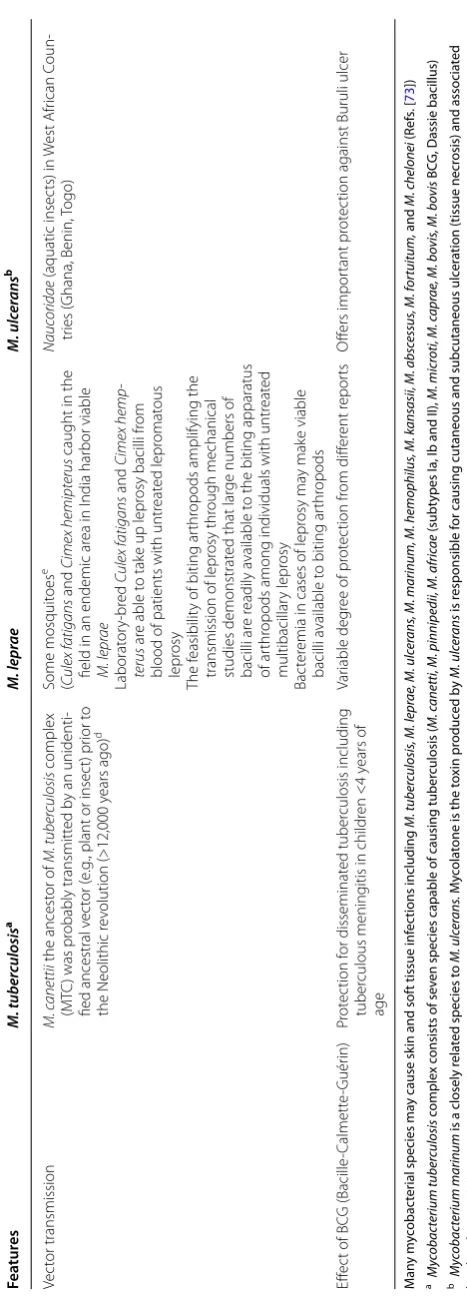
Humanity is irremediably imbedded in a matrix of nat-ural and man-made ecologies of living organisms [44]. Mycobacteria are ubiquitous microorganisms that live in natural waters, soils, and engineered water systems that have role in nutrient cycling. A major determinant of the ecology and epidemiology of mycobacterial species is the presence of a lipid-rich outer membrane leading to bio-film formation, antibiotic/disinfectant resistance, aero-solization, and surface adherence [20, 44]. A few species have evolved from this environmental pools to become major human pathogens such as M. tuberculosis, M. lep-rae and M. ulcerans [13, 45–49]. Searching for common ecological patterns and transmission dynamics among these three closely phylogenetically related species may assist in identifying environmental sources of persistent infection [45–49]. For the two major human pathogens,
M. tuberculosis and M. leprae, it is crucial to adapt to the intracellular lifestyle and to modulate the lipid metabo-lism of sanctuary cells [44] (Table 1). M. tuberculosis and
M. leprae have evolved pathogenic mechanisms through complex evolutionary negotiations between these path-ogens and their hosts, while the acquisition of a large
plasmid encoding the toxin mycolactone relates to the underlying mechanism of pathogenicity of M. ulcerans
[48–50]. This mycobacterial pathogen is causative agent of Buruli ulcer, which is a chronic destructive necrotizing infection of subcutaneous tissue that has been reported to occur in more than 30 countries [48–50]. In contrast to M. tuberculosis and M. leprae, M. ulcerans adaptation mechanisms have involved the selection of certain genes that facilitate its livelihood occupying aquatic aerobic, dark, and osmotically stable environments and its ability to reside in the extracellular matrix of the subcutaneous tissues where it unleashes the production of its toxin [50]. Genetic analyses of M. ulcerans have shown that it had a common ancestor with M. marinum and that it diverged around a million years ago [45, 49, 50]. M. marinum pro-duces a relatively milder nodular cutaneous lesions com-pared with Buruli ulcer [50].
Proverbial human-to-human transmission via respira-tory droplets of M. leprae infection has been traditionally considered the driving engine of transmission of leprosy [18, 19, 51, 52]. While leprosy bacilli are present in the nasopharynx of individuals with multibacillary leprosy [51] and from cutaneous lesions [52], and that these bacilli are able to infect other susceptible human hosts [18, 19], the precise mechanism and route of transmis-sion remain to be completely elucidated. Indeed, the current epidemiology of the persistent transmission of leprosy along with collected evidence made since the 19th Century suggest that environmental factors such as soil and water, vegetation, arthropods [20], free-living amoebas [30, 31], and animal reservoir host such as the nine-banded armadillo (Dasypus novemcintus) play an influential role in the ongoing transmission of M. leprae
[32, 33].
In 1895, Hansen and Looft made the initial observa-tion regarding the possibility of environmental factors involved in the transmission of leprosy [24]. They sug-gested that the initial site of cutaneous lesions often involved sites with direct contact with environmental surfaces (e.g., wading in streams and rivers in patients with lesions in calves). Subsequently, 27 years after Hansen’s description of M. leprae, Sand proposed that the transmission of leprosy between humans takes place indirectly. His findings were the result of analyzing 1221 patients in the Norwegian leprosarium of Reitgjaerdet in whom the transmission within household was relatively low and most cases occurred in men who had more con-tact with environmental sources. He further proposed that perhaps a living organism or ground containing decomposing material were factors involved in the trans-mission cycle [25].
Table
1
C
omparison of the thr
ee major pa
tho genic sp ecies within the My cobac terium genus Fea tur es M. tub er culosis a M. lepr ae M. ulc er ans b G enome G
enome of 4.4
Mb with 4000 genes and only six
pseudogenes (91
% coding capacit
y)
No e
vidence of major r
educ tiv e e volution Reduc tiv e e
volution and lar
ge pseudogene (1116)
for
mation r
esulting in a 3.31
‑Mb genome
Only 50
% coding capacit
y b
y 1605 genes
M. ulc er ans do wnsiz ed fr om 6.6 MB ( M. marinum ) t o 5.8 Mb genome M. marinum and M. ulc er ans shar e 97
% of genes
since the
y shar
e a common ancest
or and e
volv ed from M. marinum b y lat
eral gene transf
er and
reduc
tiv
e e
volution (~771 pseudogenes)
Selec
ted genes facilitat
e occup
ying an aer
obic
niche en
vir
onment, osmotically stable
, dar k, and ex tracellular en vir onments Tar
get cell of inf
ec tion Intr ac ellular M acr ophages (alv
eolar) and in the r
eticuloen
‑
dothelial syst
em
Non
‑classical immune cells (
epithelial cells , endothelial cells , fibr oblasts , adipoc yt es
, glia and
neur ons) [ 70 ] Intr ac ellular Sch wann cells H istioc yt es Keratinoc yt es Ex tr ac ellular Ex tracellular matr
ix in subcutaneous tissues wher
e M. ulc er ans dir ec
ts the pr
oduc
tion of the polyk
e‑
tide t
oxin m
ycolac
tone
c leading t
o tissue necr
osis
and local tissue and syst
emic immunosuppr
ession
Pathogenic mechanisms
Intracellular persist
ence in macr
ophages causes
necr
otizing g
ranulomas that cause tissue
destruc
tion in the lung or other or
gans
Inf
ec
tion of S
ch
wann cells leads t
o per ipheral ner ve dysfunc tion secondar y t o dem yelination Repr og
ramming of S
ch
wann cells link
ed t o dis ‑ seminat ed disease D estruc tiv e ulcerativ
e ulcers (Buruli ulcer) with
subcutaneous fat necr
osis En vir onmental det er minants M
embers of the
M
yc
obacterium tuber
culosis
complex can be shed b
y inf
ec
ted hosts t
o the
en
vir
onment in sputum, f
eces
, and ur
ine b
y
humans; in milk fr
om dair
y animals (
cattle) and
inf
ec
ted body tissues fr
om other domestic and
wild animals
Fr
ee
‑living amoebas ma
y ac
t as macr
ophage
‑lik
e
niches in the en
vir
onment
Clust
er
ing of cases b
y geospatial assessments of
ther mal ‑h ydr olog ical det er
minants in Ethiopia
and I
ndia
Ar
madillo is a natural host in the
W est er n Hemi ‑ spher
e and causes a z
oonosis in the S
outher
n
USA
In some settings some pr
imat
es species ma
y become r eser voirs of M. lepr ae Ther
e is e
vidence of viable
M. lepr
ae
found in soil
,
wat
er
, and fr
ee
‑living amoebas (
Ac
anthamboeba
spp
.)
Sphag
num and other moss v
egetation ma
y ha
ve
facilitat
ed transmission in the second half o the
19th and the beg
inning of the 20th centur
y liv
‑
ing in isolat
ed far
ms in coastal Nor
wa
y
Ecolog
ical studies ha
ve link ed contaminat ed wat er with M. lepr ae in pollut ed sur face wat
er in the
tr
opics
M. lepr
ae
contaminat
es clothes b
y washing in
str
eams and incomplet
e dr
ying in the humid
tr
opical climat
e; or b
y bathing and washing
clothes or dishes in a r
epor
t fr
om I
ndonesia
In Brazil
, bathing t
wice w eek ly , infr equent chang ‑
ing of bed linens or hammock among impo
ver
‑
ished populations in Nor
ther n Brazil M yc obacterium ulc er ans transmission c ycle in volv es aquatic insec t v ec tors
, aquatic plants
, and aquatic
animals
A
quatic plants (
e.g ., Rhiz oclonium sp ., H. r eticulatum ) fa
vor the g
ro
wth and biofilm pr
oduc tion of M. ulc er ans Ther e ar e t w o diff er ent wa ys that M. ulc er ans persists
in the en
vir
onment and inf
ec
t aquatic animals
(in the sa
vanna landscape r
esiding in stag
nant or
slo
wly mo
ving wat
ers; and in tr
opical rainf
or
ests
dw
elling in alk
aline wat
ers and its g
ro
wth is
dependent on climat
M an y m yc obac ter
ial species ma
y cause sk
in and sof
t tissue inf
ec
tions including
M. tub
er
culosis
, M. lepr
ae
, M. ulc
er
ans
, M. marin
um, M. hemophilus
, M. k
ansasii, M. absc
essus
, M. for
tuitum, and M. chelonei (R ef s. [ 73 ]) aM yc obac terium tub er culosis c omple x c
onsists of sev
en species capable of causing tuber
culosis (
M. c
anetti, M. pinnip
edii, M. a
fric
ae
(subt
ypes Ia, I
b and II),
M. micr
oti, M. c
apr
ae
, M. b
ovis
, M. b
ovis
BC
G, Dassie bacillus)
bM
yc
obac
terium marin
um
is a closely r
ela
ted species t
o M. ulc er ans . M yc ola
tone is the t
oxin pr oduc ed b y M. ulc er ans is r esponsible f
or causing cutaneous and subcutaneous ulc
er
ation (tissue necr
osis) and associa
ted
local and sy
st
emic immunosuppr
ession
c M
yc
olac
tone pr
oduc
ed inhibition of pr
ot
ein tr
ansloca
tion in
to the endoplasmic r
eticulum r
esulting in a deficit r
elease of inna
te immune sy
st em c yt ok ines , membr ane r ec ept ors
, adhesion molecules
, and specific
immune sy st em c yt ok ines
d O
f the M yc obac terium tub er culosis c omple x, M. b ovis and M. c apr ae ar e f
ound in hosts domestica
ted 10,000–12,000 y
ears ago
, ear
lier anc
estr
al species inf
ec
ted humans man
y y
ears bef
or
e tha
t er
a and subsequen
tly
spr
ead t
o other hosts dir
ec
tly fr
om humans or thr
ough an unk
no
wn v
ec
tor
e T
her
e ar
e sev
er
al biting ar
thr
opods r
esiding in lepr
osy endemic ar
eas of which an
y c
ould pot
en
tially ac
t as a v
ec
tor f
or the tr
ansmission of lepr
osy Table 1 c on tinued Fea tur es M. tub er culosis a M. lepr ae M. ulc er ans b Vec tor transmission M. c anettii the ancest or of M. tuber culosis complex (M TC
) was pr
obably transmitt
ed b
y an unidenti
‑
fied ancestral v
ec
tor (
e.g
., plant or insec
t) pr
ior t
o
the Neolithic r
ev olution (>12,000 y ears ago) d Some mosquit oes e ( Culex f atigans and Cimex hemipterus
caught in the
field in an endemic ar
ea in I
ndia har bor viable M. lepr ae Laborat or y‑ br ed Culex f atigans and Cimex hemp -terus ar
e able t
o tak
e up lepr
osy bacilli fr
om
blood of patients with untr
eat ed lepr omat ous lepr osy The f easibilit
y of biting ar
thr
opods amplifying the
transmission of lepr
osy thr
ough mechanical
studies demonstrat
ed that lar
ge numbers of
bacilli ar
e r
eadily a
vailable t
o the biting apparatus
of ar
thr
opods among individuals with untr
eat ed multibacillar y lepr osy Bac ter
emia in cases of lepr
osy ma
y mak
e viable
bacilli a
vailable t
o biting ar
thr opods Nauc oridae (aquatic insec ts) in W est A frican C oun ‑ tr ies ( Ghana, Benin, Togo) Eff ec
t of BC
G (Bacille ‑C almett e‑ Guér in) Pr ot ec tion f or disseminat ed tuber culosis including tuber culous mening
itis in childr
en <4 y ears of age Var iable deg
ree of pr
ot ec tion fr om diff er ent r epor ts O ffers impor tant pr ot ec
with spillover of M. leprae from human cases (e.g., nasal discharges contaminating soil or water) may facilitate the amplification of the transmission cycle in biotopes with existing suitable ecological abiotic and biotic determi-nants (i.e., tropical and subtropical settings) [34, 35]. In this hypothetical model, we can postulate that chemo-prophylaxis (or preemptive treatment) of contacts of multibacillary cases and effective treatment of leprosy cases decreases spillage of M. leprae into environmental reservoirs (soil, water, plants, or free-living amoebas) [24,
25, 27]. Preliminary evidence from a leprosy-endemic area in India has shown that genetic material of M. lep-rae was detected near washing and bathing areas where cases of leprosy were detected and genetic fingerprinting correlated between human cases and DNA detected in soil samples [24, 29]. The spillover of M. leprae into soil and water may explain the acquisition of this pathogen by armadillos acting as scavengers, and ultimately link-ing a reverse cycle of transmission from armadillos back to humans [32]. Nevertheless, it is possible that there are other unidentified environmental reservoirs or vectors influencing the occurrence of new human infections in highly endemic areas. Zoonotic transmission of M. leprae
from armadillos in the Golf Coast of the United States contributes to endemic human infections detected in this geographic area every year, supporting the fact that lep-rosy is not exclusively transmitted person-to-person [32].
Free‑living amoebas as environmental sanctuaries of M. leprae
There is some evidence that the obligate intracellular M. leprae may spill over onto environmental niches and sur-vive endosymbiotically inside free-living amoebae similar to the mechanism described of Legionella pneumophila
residing inside Acanthamoeba [30, 31]. Large numbers of viable leprosy bacilli are expelled into the environment in the nasal secretions or to a lower degree from skin lesions of individuals diagnosed with multibacillary leprosy [52]. There is also evidence that M. leprae may invade and infect the nasal mucosa or into abraded/punctured skin [52–54]. In this regard, it is feasible that free-living path-ogenic amoebae potentially act as “external” reservoirs capable of ingesting and supporting the environmental viability of M. leprae expelled by infectious patients into the environment and thus acting as a macrophage-like niches [30, 31]. Further evidence has demonstrated that M. leprae remains viable for prolonged periods inside
Acanthamoeba castellani and Acanthamoeba polyphaga; and it is able to survive encystment and retain infectivity in the nu/nu mouse model [33]. It remains to be tested if
M. leprae infected amoebae is able to transport the bacilli through nasal mucosa or through intact or abraded skin to produce clinical disease [31].
Arthropods as vectors of M. leprae transmission The possibility of arthropods as vectors of M. leprae has not been conclusively ruled out [5]. As early as 1915, Adolpho Lutz suggested that “the erratic manner of the propagation of leprosy” might be explained by the bites of biting arthropods, particularly of Culex mosquitoes (i.e., Culex fatigans) [20]. In fact, there are several biting arthropods residing in highly endemic areas of leprosy that theoretically might act as a vector of M. leprae [55–
66]. In some studies, the distribution of single lesions of tuberculoid leprosy correlated with exposed skin areas [60, 61]. Mechanical studies have demonstrated the feasi-bility of biting arthropods to uptake M. leprae since large numbers of bacilli are readily available within cutaneous lesions to the biting apparatus of many species of arthro-pods among individuals with untreated multibacillary leprosy [59, 61–66]. Additionally, it has been shown that patients with lepromatous leprosy by developing bactere-mia may make viable bacilli available to biting arthropods [63–65]. Sandflies have been ruled out as vectors of lep-rosy transmission [66].
There is evidence that mycobacterial species constitut-ing the Mycobacterium tuberculosis complex (i.e., M. can-etti) infected humans before the Neolithic period (< than 12,000 years ago) and that a non-mammalian vector may have played a role (e.g., plants or insects) [44–46]. Tuber-culosis infection later spread to dairy animals as a result of human transmission during their domestication and involving a mechanism of transmission either through direct contact or through an unrecognized vector [44].
Mycobacterium ulcerans transmission cycle involves aquatic insect vectors, aquatic plants, and aquatic ani-mals [45, 47–50]. Similarly, survival of M leprae in envi-ronmental niches may also involve natural reservoirs (e.g., free-living amoebas) or it may be transmitted by arthropods (e.g., mosquitoes). It is also possible that spe-cies of the Mycobacterium tuberculosis complex may use environmental sanctuaries such as free-living amoe-bas to resist the external environment by acting as a macrophage-like niche [20]. Further studies using novel molecular assays need to be conducted to assess the potential contribution of arthropods to the transmission of leprosy in endemic areas.
Early diagnosis and neurologic disability
Peripheral nerve involvement occurs in all patients with
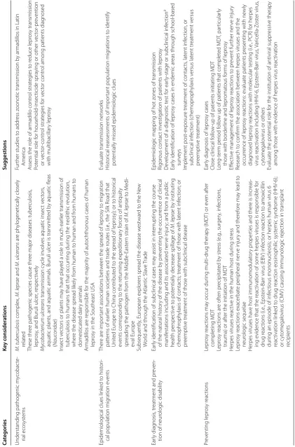
Table 2 A p ot en tial r esear ch and p olic y r oadmap t o r educ e lepr osy tr ansmission aM yc obac terium lepr ae
has the abilit
y t
o r
epr
og
ram the S
ch
w
ann c
ell in
to a st
em ‑c ell ‑like c ell tha t car
ries the bacilli in
to other tissues t
o ensur
e its dissemina
tion. Giv
en this sy
st
emic dissemina
tion it seems f
easible t
o
sear
ch f
or the dev
elopmen
t of assa
ys such as an in
ter
fer
on—assa
y emplo
ying a similar appr
oach t
o the one used f
or det
ec
tion of c
yt ok ine ‑pr oduc tion pa tt er ns b y M. tub er culosis Ca tegories Ke y c onsider ations Suggestions
Understanding pathogenic m
ycobac te ‑ rial ecosyst ems M. tuber culosis complex, M. lepr ae and M. ulc er ans ar e ph ylogenetically closely relat ed These thr
ee pathogenic species cause thr
ee major diseases: tuber
culosis
,
lepr
osy
, and Buruli ulcer
, r espec tiv ely M yc obacterium ulc er ans transmission c ycle in volv
es aquatic insec
t v
ec
tors
,
aquatic plants
, and aquatic animals
. Buruli ulcer is transmitt
ed b
y aquatic fleas
( Nauc oridae ) Insec t v ec
tors or plants ma
y ha
ve pla
yed a r
ole in much ear
lier transmission of
tuber
culosis t
o humans that that occur
ring dur
ing the neolithic r
ev
olution,
wher
e the disease spr
ead lik
ely fr
om human t
o human and fr
om humans t
o domesticat ed dair y animals Ar madillos ar e r esponsible f
or the major
ity of aut
ochthonous cases of human
lepr
osy in the S
outheast USA
Fur
ther studies t
o addr
ess z
oonotic transmission b
y ar
madillos in Latin
Amer
ica
A
ssess the r
ole of ar
madillo contr ol strat eg ies t o r educe lepr osy transmission Pot ential r ole f or household ‑insec ticide spra
ying or other v
ec tor ‑pr ev ention or v ec tor ‑contr ol strat eg ies f or v ec tor contr
ol among patients diag
nosed
with multibacillar
y lepr
osy
Epidemiolog
ical clues link
ed t
o hist
or
i‑
cal population mig
ration e
vents
Ther
e ar
e impor
tant associations bet
w
een the spr
ead of lepr
osy t
o mig
ration
patt
er
ns of ear
lier human societies and trade r
out
es (i.e
., the Silk R
oad that
Unit
ed E
ur
ope t
o China contr
ibut
ed t
o the spr
ead of lepr
osy); or t
o hist or ical ev ents cor responding t
o the r
etur
ning expeditionar
y f
or
ces of antiquit
y
spr
eading the pathogen fr
om the M
iddle
‑East
er
n strain of
M. lepr ae to M edi ‑ eval E ur ope Subsequently , E ur opean explor ers spr
ead the disease w
est
war
d t
o the Ne
w
W
or
ld and thr
ough the A
tlantic Sla
ve
Trade
Evaluat
e transmission net
w or ks H ist or ical r
eassessments of impor
tant population mig
rations t
o identify
pot
entially missed epidemiolog
ic clues
Ear
ly diag
nosis
, tr
eatment and pr
ev
en
‑
tion of neur
olog
ic disabilit
y
Ear
ly identification of subclinical cases ma
y assist in int
er
rupting the course
of the natural hist
or
y of the disease b
y pr
ev
enting the occur
rence of clinical
manif
estations including and its associat
ed ner
ve injur
y; and fr
om a public
health perspec
tiv
e t
o pot
entially decr
ease spillage of
M. lepr ae b y instituting chemophr oph
ylaxis of contac
ts; tr
eatment of those with lat
ent inf ec tion; or pr eemptiv e tr
eatment of those with subclinical disease
Epidemiolog
ic mapping of hot z
ones of transmission
Rigor
ous contac
t in
vestigation of patients with lepr
osy
D
ev
elopment of a diag
nostic t
est f
or ear
ly
‑stage or subclinical inf
ec
tion
a
Ear
ly identification of lepr
osy cases in endemic ar
eas thr ough school ‑based sur ve ys Implement post ‑exposur e tr
eatment of contac
ts , lat ent inf ec tion, or subclinical inf ec tion ( chemopr oph ylaxis v ersus lat ent tr eatment v ersus pr eemptiv e tr eatment) Pr ev enting lepr osy r eac tions Lepr osy r eac tions ma
y occur dur
ing multi
‑drug therap
y (MD
T) or e
ven af ter completing MD T Lepr osy r eac tions ar e of ten pr ecipitat ed b y str ess ( e.g ., sur ger y, inf ec tions ,
trauma) or af
ter the initiation of MD
T
Her
pes viruses r
eac
tiv
e in the human host dur
ing str ess Lepr osy r eac tions exacer bat e per ipheral ner ve injur
y and ther
ef
or
e ma
y lead t
o
neur
olog
ic sequelae
Her
pes viruses ha
ve host immunomodulat
or
y pr
oper
ties and ther
e is incr
eas
‑
ing e
vidence that the r
eac
tivation of some her
pes viruses is r
esponsible f or drug r eac tions (i.e ., Epst ein ‑Bar
r virus (EB
V ) inf ec tion r eac tion t o amo xicillin dur
ing an episode of inf
ec
tious mononucleosis; or her
pes human virus 6
reac
tivation link
ed t
o drug r
eac
tion eosinophilic syst
emic syndr ome (HHV ‑6); or c yt omegalo virus ( CMV
) causing immunolog
ic r
ejec
tion in transplant
recipients
Ear
ly diag
nosis of lepr
osy cases
Close clinical f
ollo
w
‑up of patients initiating MD
T Long ‑t er m per iod f ollo
w up of patients that complet
ed MD
T, par
ticular
ly
those with bor
der
line and lepr
omat
ous f
or
ms of lepr
osy
Eff
ec
tiv
e management of lepr
osy r eac tions t o pr ev ent fur ther ner ve injur y Resear ch t o confir
m the association bet
w
een her
pes viruses and the
occur
rence of lepr
osy r
eac
tions: e
valuat
e patients pr
esenting with ne
wly
diag
nosed lepr
osy r
eac
tions with molecular t
esting (i.e
., PCR) f
or her
pes
virus r
eac
tivation including HHV
‑6, Epst ein ‑Bar r virus , V ar icella ‑Z ost er virus , cyt omegalo
virus or others
Evaluat
e the pot
ential r
ole f
or the institution of antiviral suppr
essiv
e therap
y
among those with e
vidence of her
pes virus r
eac
manifests clinically with decreased sensorimotor func-tion and its associated complicafunc-tions [69, 70]. Addi-tionally, M. leprae leads to a dedifferentiation process of Schwann cells transforming them into Trojan horses for the systemic dissemination of the bacilli [3, 69, 70]. Peripheral nerve sensorimotor dysfunction in patients with leprosy is frequently exacerbated by episodes of leprosy reactions [8, 9]. Indeed, even after effective anti-bacterial therapy, a large number of dead anti-bacterial cells remain within nerves and continue to elicit immuno-logic responses manifested as acute or chronic neuritis [8, 71]. Early detection and treatment of neuropathy in leprosy has important preventive potential. Preventing leprosy reactions or effectively treating them is therefore an important consideration in any strategy attempting to reduce peripheral nerve injury. We need to expand our understanding of factors that predispose individuals to develop leprosy reactions and the mechanisms that trig-ger their occurrence. One important consideration is the potential role of the microbiome in modulating the inflammatory response, particularly of herpes viruses [2]. While there is little research in this area, there is ample evidence in other clinical scenarios to illustrate that herpes viruses modulate inflammatory responses during pathologic conditions [72] (Table 2). Early iden-tification of leprosy cases remains a central priority in controlling this disease. In this regard, school-screening programs employing clinical assessments combined with serological and molecular surveys in endemic areas have been shown to increase the early detection of cases [73]. These programs have the greatest potential for reduc-ing transmission by early institutreduc-ing of treatment early in the course of the disease; and by identifying house-hold contacts and househouse-hold cases. Similarly, geospatial analyses of risk assessments of leprosy based on thermal and hydrological environments have demonstrated useful in predicting clustering of cases in studies conducted in Ethiopia and India [34, 35] (Table 2). Efforts to scale up school-based screenings and geospatial risk assessments based on ecological determinants in hyperendemic set-tings may offer so far, the best opportunity to reduce the occurrence of new cases.
Conclusions
Our understanding of the transmission dynamics of M. leprae is incomplete. While person-to-person transmis-sion may play a role, there is a possibility of other modes of transmission involved. Therefore, there is a need for a fresh reexamination of the historical, phyleogeo-graphic, sociocultural, and environmental factors linked to the spread of M. leprae among human populations. We need to consider mycobacterial ecologies of other pathogenic mycobacteria such as M. ulcerans; and to
expand our exploration for environmental determinants including thermal-hydrological factors (i.e., soil, vegeta-tion, water); intermediate reservoirs or vectors including free-living amoebas, arthropods, and zoonotic transmis-sion. Identifying epidemiologic clues from these analyses may facilitate designing effective control or elimination interventions.
Authors’ contributions
CFP: Participated in the design, review of the literature, and writing of the manuscript. AJRM: Participated in the design, review of the literature, and writing of the manuscript. Both authors read and approved the final manuscript.
Author details
1 Infectious Diseases Clinic, Phoebe Putney Memorial Hospital, 507 3rd
Avenue, Albany, GA 31721, USA. 2 Hospital Infantil de México, Federico Gómez,
Mexico D.F., Mexico. 3 Public Health and Infection Research Group, Faculty
of Health Sciences, Universidad Tecnológica de Pereira, Pereira, Risaralda, Colombia.
Competing interests
The authors declare that they have no competing interests.
Received: 3 February 2016 Accepted: 11 May 2016
References
1. Britton WJ, Lockwood DNJ. Leprosy. Lancet. 2004;363:1209–19. 2. White C, Franco‑Paredes C. Leprosy in the 21st century. Clin Microbiol
Rev. 2015;28(1):80–94.
3. Polycarpou A, Walker SL, Lockwood DN. New findings in the pathogene‑ sis of leprosy and implications for the management of leprosy. Curr Opin Infect Dis. 2013;26:413–9.
4. Rodrigues LC, Lockwood DNJ. Leprosy now: epidemiology, progress, challenges, and research gaps. Lancet Infect Dis. 2011;11(6):464–70. 5. Gay FP. The unsolved problems in leprosy transmission. Science.
1935;81(2099):2083–5.
6. Lockwood DNJ, Shetty V, Oliveira Penna G. Hazards of setting targets to eliminate disease lessons from the leprosy elimination campaign. Brit Med J. 2014;348:g1136.
7. Smith WC, van Brakel W, Gillis T, Saunderson P, Richardus JH. The miss‑ ing millions: a threat to the elimination of leprosy. PLoS Negl Trop Dis. 2015;9(4):e0003658.
8. Scollard DM, Truman RW, Ebenezer GJ. Mechanisms of nerve injury in leprosy. Clin Dermatol. 2015;33:46–54.
9. Scollard DM. The biology of nerve injury in leprosy. Lepr Rev. 2008;79:242–53.
10. Franco‑Paredes C, Santos‑Preciado JI. Freedoms, justice, and the neglected tropical diseases. PLoS Negl Trop Dis. 2011;5(8):e1235. 11. White C. Leprosy and stigma in the context of international migration.
Lepr Rev. 2011;82(2):147–54.
12. Jacob J, Franco‑Paredes C. The stigmatization of leprosy in India and its impact on future approaches to elimination and control. PLoS Negl Trop Dis. 2008;30:e113.
13. Stone AC, Wilbur AK, Buikstra JE, Roberts CA. Tuberculosis and leprosy in perspective. Am J Phys Anthropol. 2009;52:66–94.
14. Lockwood DNJ, Suneetha S. Leprosy: too complex a disease for a simple elimination paradigm. Bull World Health Org. 2005;83(3):230–5. 15. Smith CS, Kahder Noordeen S, Richardus JH, Sansarricq H, Cole ST, Baruaf
S, Soares RS, Savioli L, Aertsh A. A strategy to halt leprosy transmission. Lancet Infect Dis. 2014;14:96–7.
17. Pocaterra L, Jain S, Reddy R, Muzaffarullah S, Torres O, Suneetha S, Lockwood DNJ. Clinical course of erythema leprosum: an 11‑year cohort study in Hyderabad, India. Am J Trop Med. 2006;74(5):868–79.
18. Bratschi MW, Steinmann P, Wickenden A, Gillis TP. Current knowledge on
Mycobacterium leprae transmission: a systematic literature review. Lepr
Rev. 2015;86:142–55.
19. Mensah‑Awere D, Bratschi MW, Steinmann P, Fairley JK, Gillis TP. Developing straegies to block the transmission of leprosy. Lepr Rev. 2015;86:156–64. 20. Blake LA, West BC, Lary CH, Todd JR IV. Environmental nonhuman sources
of leprosy. Rev Infect Dis. 1987;9(3):561–77.
21. Kerr‑Pontes LRS, Barreto ML, Evangelist CMN, Rodrigues LC, Heukelbach J, Feldmeier H. Socioeconomic, environmental, and behavioural risk factors for leprosy in Northeast Brazil: results of a case‑control study. Int J Epidemiol. 2006;35:994–1000.
22. Kerr‑Pontes LRS, Montenegro ACD, Barreto ML, Werneck GL, Feldemeier H. Inequality and leprosy in Northeast Brazil: an ecological study. Int J Epidemiol. 2004;33:262–9.
23. Lockwood DNJ. Commentary: leprosy and poverty. Int J Epidemiol. 2004;33:269–70.
24. Lavania M, Katoch K, Katoch VM, Gupta AK, Chauhan DS, Sharma R, Gan‑ dhi R, Chauhan V, Bansal G, Sachan P, Sachan S, Yadav VS, Jadhav R. Detec‑ tion of viable Mycobacterium leprae in soil samples: insights into possible sources of transmission of leprosy. Infect Genet Evol. 2008;8:627–31. 25. Kazda J, Pavlik I. Obligate pathogenic mycobacteria. In: Kazda J, Pavlik I,
Falkinham JO, Hruska K, editors. The ecology of mycobacteria: impact on animal’s and human’s health. 2nd ed. Berlin: Springer; 2009. p. 1–19. 26. Sreevatsa. Leprosy and arthropods. Indian J Lepr. 1993;65(2):189–200. 27. Desikan KV, Sreevatsa. Extended studies on the viability of M. Leprae
outside the human body. Lepr Rev. 1995;66:287–95.
28. Turankar RP, Lavania M, Singh M, Siva Sai KSR, Jadhav RS. Dynamics of
Myco-bacterium leprae transmission in environmental context: deciphering the
role of environment as potential reservoir. Infect Gen Evol. 2012;12:121–6. 29. Matsuoka M, Izumi S, Budiawan T, Nakata N, Saeki K. Mycobacterium leprae
DNA in daily using water as a possible source of leprosy infection. Indian J Lepr. 1999;71:61–7.
30. Lahiri R, Krahenbuhl JL. The role of free‑living pathogenic amoeba in the transmission of leprosy: a proof of principle. Lepr Rev. 2008;79:401–9. 31. Wheat WH, Casali AL, Thomas V, Spencer JS, Lahiri R, Williams DL, McDon‑
nell GE, Gonzalez‑Juarrero M, Brennan PJ, Jackson M. Long‑term survival and virulence of Mycobacterium leprae in amoebal cysts. PLoS Neglec Trop Dis. 2014;8(12):e3405.
32. Balamayooran G, Pena M, Sharma R, Truman RW. The armadillo as an animal model and reservoir host for Mycobacterium leprae. Clin Dermatol. 2015;33:108–15.
33. Cardona‑Castro N, Beltran JC, Ortiz‑Bernal A, Vissa V. Detection of
Myco-bacterium leprae DNA in nine‑banded armadillos (Dasypus novemcinctus)
from the Andean region of Colombia. Lepr Rev. 2009;80:424–31. 34. Tadesse Argaw A, Sannon EJ, Assefa A, Mikru FS, Mariam BK, Malone JB.
A geospatial risk assessment model for leprosy in Ethiopia based on environmental thermal‑hydrological regime analysis. Geospat Health. 2006;1(1):105–13.
35. Joshua V, Mehendale S, Gupte MD. Bayesian model, ecologial factors and transmission of leprosy in an endemic area of South India. Indian J Med Res. 2016;143:104–6.
36. Gomez‑Valero L, Rocha EPC, Latorre A, Silva F. Reconstructing the ances‑ tor of Mycobacterium leprae: the dynamics of gene loss and genome reduction. Genome Res. 2007;17:1178–85.
37. Trueba G, Dunthorn M. Many neglected tropical diseases may have originated in the Paleolithic or before: new insights from genetics. PLoS Neglect Trop Dis. 2012;6(3):e1393.
38. Monot M, Honore N, Garnier T, Zidane N, Sherafi D, Paniz‑Mondolfi A, Matsuoka M, Taylor GM, Donoghue HD, Bouwman A, Mays S, Wason C, Lockwood D, Khamesipour A, Dowlati Y, Jianping S, Rea TH, Vera‑Cabrera L, Stefani MM, Banu S, Macdonald M, Spakota BR, Spencer JS, Thomas J, Harshman K, Singh P, Busso P, Gattiker A, Rougemont J, Brennan PJ, Cole ST. Comparative genomic and phyleogeographic analysis of
Mycobacte-rium leprae. Nat Genet. 2009;41(12):1282–9.
39. Monot M, Honore N, Garnier T, Araoz R, Coppee JY, Lacroix C, Sow S, Spencer JS, Truman RW, Williams DL, Gelber R, Virmond M, Flageul B, Cho SN, Ji B, Paniz‑Mondolfi A, Convit J, Young S, Fine PE, Rasolofo V, Brennan PJ, Cole ST. On the origin of leprosy. Science. 2005;308(5724):1040–2.
40. Singh P, Cole ST. Mycobacterium leprae: genes, pseudogenes, and genetic diversity. Future Microbiol. 2011;6(1):57–71.
41. Schuenemann VJ, Singh P, Mendum TA, Krause‑Kyora B, Jager G, Bos KI, Herbig A, Economou C, Benjak A, Busso P, Nebel A, Boldsen JL, Kjellstrom A, Wu H, Stewart GR, Taylor M, Bauer P, Lee OYC, Wu HHT, Minnikin DE, Besra GS, Tucker K, Roffey S, Sow SO, Cole ST, Nieselt K, Krause J. Genome‑ wide comparison of medieval and modern Mycobacterium leprae. Sci‑ ence. 2013;341:179–83.
42. Prasanna AN, Mehra S. Comparative phylogenomics of pathogenic and non‑pathogenic Mycobacterium. PLoS One. 2013;8(8):e71248. 43. Cardona‑Castro N, Cortes N, Beltran C, Romero M, Badel‑Mogollon JE,
Bedoya G. Human genetic ancestral composition correlates with the origin of Mycobacterium leprae strains in a leprosy endemic popula‑ tion. PLoS Neglect Trop Dis. 2015;9(9):e0004045. doi:10.1371/journal.
pntd.0004045.
44. Falkinham JO. Surrounded by mycobacteria: nontuberculous mycobacte‑ ria in the human environment. J Appl Microbiol. 2009;107(2):356–67. 45. Garchitorena A, Guegan JF, Lege L, Eyangoh S, Marsollier L, Roche B.
Mycobacterium ulcerans dynamics in aquatic ecosystems are driven by a
complex interplay of abiotic and biotic factors. eLife. 2015;4:e07616. 46. Boritsch EC, Supply P, Honore N, Seeman T, Stinear TP, Brosch R. A glimpse
into the past and predictions for the future: the molecular evolution of the tuberculosis agent. Mol Microbiol. 2014;93(5):835–52.
47. Mostowy S, Behr MA. The origin and evolution of Mycobacterium
tubercu-losis. Clin Chest Med. 2005;26:207–16.
48. Marsollier L, Stinear T, Aubry J, Saint Andre JP, Robert R, Legras P, Manceau AL, Audrain C, Bourdon S, Kouakou H, Carbonnelle B. Aquatic plants stimulate the growth of and biofilm formation by Mycobacterium ulcerans
in axenic culture and harbor these bacteria in the environment. Appl Environ Microbiol. 2004;70(2):1097–103.
49. Röltgen K, Stinear TP, Pluschke G. The genome, evolution and diversity of
Mycobacterium ulcerans. Infect Genet Evol. 2012;12:522–9.
50. Doig KD, Holt KE, Fyfe JAM, Lavender CJ, Eddyani M, Portaels F, Yeboah‑ Manu D, Pluschke G, Seeman T, Stinear TP. On the origin of Mycobacterium
ulcerans, the causative agent of Buruli ulcer. BMC Genomics. 2012;13:258.
51. Davey TF, Res RJW. The nasal discharge in leprosy: clinical and bacterio‑ logical aspects. Lepr Rev. 1974;45:121–34.
52. Job CK, Jayakumar J, Kearney M, Gillis TP. Transmission of leprosy: a study of skin and nasal secretions of household contacts of leprosy patients using PCR. Am J Trop Med Hyg. 2008;78(3):518–21.
53. Chehl S, Job CK, Hastings RC. The nose: a site for the transmission of leprosy in nude mice. Am J Trop Med Hyg. 1985;34:1161–6. 54. Job CK, Harris EB, Allen JL, Hastings RC. Thorns in armadillo ears and
noses and their role in the transmission of leprosy. Arch Pathol Lab Med. 1986;110:1025–8.
55. Dungal N. Is leprosy transmitted by arthropods? Lepr Rev. 1961;32:28–35. 56. Manja KS, Narayanan E, Kasturi G, Kirchheimer WF, Balasubramanyan M.
Non‑cultivable mycobacteria in some field collected arthropods. Lepr India. 1973;45:231–4.
57. Narayanan E, Sreevatsa, Kirchheimer WF, Bedi BMS. Transfer of leprosy bacilli from patients to mouse foot pads by Aedes aegypti. Lepr India. 1977;49:181–6.
58. Narayanan E, Sreevatsa, Raj AD, Kirchheimer WF, Bedi BMS. Persistence and distribution of M. leprae in Aedes aegyptii and Culex fatigans experi‑ mentally fed on leprosy patients. Lepr India. 1978;50:26–36.
59. Saha K, Jain M, Mukherjee MK, Chawla WM, Chaudhary DS, Prakash N. Viability of Mycobacterium leprae within the gut of Aedes aegypti after they feed on multibacillary lepromatous patients: a study by fluorescent and electron microscopes. Lepr Rev. 1985;56:279–90.
60. McDougall AC, Cologlu AS. Lepromatous leprosy in man; depth of the cellular infiltrate and bacillary mass in relation to the possibility of transmission of leprosy by biting arthropods. Ann Trop Med Parasitol. 1983;77(2):187–93.
61. Narayanan E, Manja KS, Bedi BM, Kirchheimer WF, Balasubrahmanyan M. Arthropod feeding experiment in lepromatous leprosy. Lepr Rev. 1972;43(4):188–93.
62. Kirchheimer WF. The role of arthropods in the transmission of leprosy. Int J Lepr Other Mycobact Dis. 1976;44(1–2):104–7.
• We accept pre-submission inquiries
• Our selector tool helps you to find the most relevant journal
• We provide round the clock customer support
• Convenient online submission
• Thorough peer review
• Inclusion in PubMed and all major indexing services
• Maximum visibility for your research
Submit your manuscript at www.biomedcentral.com/submit
Submit your next manuscript to BioMed Central
and we will help you at every step:
64. Sreevatsa, Sengupta U, Ramu G, Desikan KV. Evaluation of bacteraemia in leprosy patients. Lepr India. 1978;50(3):381–7.
65. Reis EM, Araujo S, Lobato J, Neves AF, Costa AV, Gonçalvez MA, Goulart LR, Goulart IMB. Mycobacterium leprae DNA in peripheral blood may indicate a bacilli migration route and high‑risk for leprosy onset. Clin Microbiol Infect. 2014;20:447–52.
66. Sreevatsa, Girdhar BK. Can sandflies be the vector for leprosy? Int J Lepr Other Mycobact Dis. 1992;60(1):94–6.
67. Rambukkana A. Molecular basis for the peripheral nerve predilection of
Mycobacterium leprae. Curr Opin Microbiol. 2001;4:21–7.
68. Lockwood DN, Saunderson PR. Nerve damage in leprosy: a continu‑ ing challenge to scientists, clinicians, and service providers. Int Health. 2012;4(2):77–85.
69. Masaki T, Qu J, Cholewa‑Waclaw J, Burr K, Raaum R, Rambukkana A. Reprogramming adult Schwann cells to stem cell‑like cells by leprosy bacilli promotes dissemination of infection. Cell. 2013;152(1–2):51–67.
70. Randall PJ, Hsu NJ, Quesniaux V, Ryffel B, Muazzam J. Mycobacterium
tuberculosis infection of the ‘non‑classical immune cell’. Immunol Cell Biol.
2015;93:789–95.
71. Van Brakel WH, Nicholls PG, Wilder‑Smith EP, Das L, Barkataki P, Lock‑ wood DNJ. On behalf of the INFIR Study Group. PLoS Negl Trop Dis. 2008;2(4):e212.
72. Sorbara MT, Giardin SE. Emerging themes in bacterial autophagy. Curr Opin Microbiol. 2015;23:163–70.