Cellular mechanisms underlying
steroid-resistant asthma
Ridhima Wadhwa
1,2,6, Kamal Dua
1,2,3,6, Ian M. Adcock
4, Jay C. Horvat
3,
Richard Y. Kim
1,3,5and Philip M. Hansbro
1,3,5Affiliations: 1Centre for Inflammation, Centenary Institute, Sydney, Australia. 2Discipline of Pharmacy, Graduate School of Health, University of Technology Sydney, Sydney, Australia.3Priority Research Centre for Healthy Lungs, University of Newcastle and Hunter Medical Research Institute, Newcastle, Australia. 4The Airways Disease Section, National Heart and Lung Institute, Imperial College London, London, UK.5Faculty of Science, University of Technology Sydney, Sydney, Australia.6Both authors contributed equally.
Correspondence: Philip M. Hansbro, Faculty of Science, University of Technology Sydney, Ultimo, NSW 2007, Australia. E-mail: [email protected]
@ERSpublications
Severe steroid-resistant asthma is a significant clinical problem and recent advances in understanding the mechanisms of pathogenesis enable the identification of novel therapeutic approaches
http://bit.ly/2mQpF3n
Cite this article as:Wadhwa R, Dua K, Adcock IM,et al. Cellular mechanisms underlying steroid-resistant asthma.Eur Respir Rev2019; 28: 190096 [https://doi.org/10.1183/16000617.0096-2019].
ABSTRACT Severe steroid-resistant asthma is clinically important, as patients with this form of the disease do not respond to mainstay corticosteroid therapies. The heterogeneity of this form of asthma and poor understanding of the pathological mechanisms involved hinder the identification of therapeutic targets and the development of more effective therapies. A major limiting factor in the understanding of severe steroid-resistant asthma is the existence of multiple endotypes represented by different immunological and inflammatory phenotypes, particularly in adults. Several clinical and experimental studies have revealed associations between specific respiratory infections and steroid-resistant asthma in adults. Here, we discuss recent findings from other authors as well as our own studies that have developed novel experimental models for interrogating the association between respiratory infections and severe steroid-resistant asthma. These models have enabled the identification of new therapies using macrolides, as well as several novel disease mechanisms, including the microRNA-21/phosphoinositide 3-kinase/histone deacetylase 2 axis and NLRP3 inflammasomes, and highlight the potential of these mechanisms as therapeutic targets.
Introduction
The Global Initiative for Asthma (GINA) currently defines asthma as a heterogeneous disease that is characterised by inflammation of the airways and a clinical history of wheezing, cough, tightness of chest and shortness of breath varying with time and intensity, as well as expiratory airflow limitation [1]. Asthma is classically considered as a disease driven by increased type 2 immune responses and eosinophilic inflammation [2–5]. However, recent evidence shows that asthma can be associated with elevated type 1/17 immune responses that are associated with non-eosinophilic (i.e.neutrophilic) inflammation and tend to be present in adult patients with moderate to severe asthma [2, 6]. Importantly, gene–environmental interactions play a vital role in the progression of the disease, with specific insults to the respiratory epithelium [7] inducing the release of pro-inflammatory mediators that recruit inflammatory cells to the airways. Exogenous factors such as respiratory viral and bacterial infections, a high-fat diet and/or obesity, air pollution and cigarette smoke exposure have all been linked with severe forms of asthma in adults as well as with exacerbations of the disease [2, 8–20]. Here, we review the mechanisms of pathogenesis of severe
Copyright ©ERS 2019. This article is open access and distributed under the terms of the Creative Commons Attribution Non-Commercial Licence 4.0.
Provenance: Commissioned article, peer reviewed.
steroid-resistant (SSR) asthma in adults. Children can also have severe asthma, which may have different pathomechanisms; these have been reviewed recently elsewhere [21–23].
Anti-inflammatory corticosteroids are the mainstay therapy for asthma and are often administered in conjunction with short- or long-actingβ2-adrenoceptor agonists (bronchodilators) [2, 24]. These combination
therapies allow most asthmatics to gain control of their disease symptoms by modulating type 2 cytokine responses and accompanying inflammation, although they have broad-spectrum, nonspecific anti-inflammatory activity. Importantly, however, these therapies are not cures, and long-term, high-dose corticosteroid therapy is associated with many side-effects [4, 24, 25]. Critically, 5–10% of asthmatics do not respond to steroid-based therapies, are more likely to have severe disease and, due to the paucity of effective treatment options, disproportionately account for 50–80% of all asthma-associated healthcare costs [26, 27]. SSR asthma is strongly associated with non-eosinophilic endotypes of the disease, including neutrophilic asthma, and often involves activation of innate immune responses, particularly those mediated by Toll-like receptor (TLR)2 and TLR4 responses, and NLRP3 (nucleotide-binding oligomerisation domain-like receptor family, pyrin domain-containing 3) inflammasome/interleukin (IL)-1βresponses [28–32]. SCHWARTZet al. [33] first described steroid-resistant asthma in a small asthmatic subgroup with poor disease control that did not respond to high-dose oral steroid therapy. With progressive refinement of diagnostic criteria, SSR asthma in adults is now defined as ineffectiveness in achieving >15% improvement in forced expiratory volume in 1 s after 14 days of oral steroid treatment [27, 34]. SSR asthma is the major clinical issue in asthma management and there is an urgent need for therapies that more effectively suppress the symptoms of disease (figure 1).
Mechanisms of steroid resistance
Glucocorticoids (a class of anti-inflammatory corticosteroid) are the mainstay therapy for asthma and are administered through inhaled or oral formulations. Inhaled corticosteroids (ICSs) are synthetic, lipophilic glucocorticoids that, upon inhalation, readily diffuse into the airway tissue and bind and activate cytosolic glucocorticoid receptors (GRs; also called nuclear receptor subfamily 3, group C, member 1 (NR3C1)) [35, 36]. The anti-inflammatory effects are mediated by the GRαisoform vialigand-dependent transcription factors, while GRβ acts as a dominant inhibitor of GRα [37]. GRα homodimers are actively transported into the nucleus by importin-αand importin-13 and bind to a glucocorticoid response element (GRE) to regulate gene transcription [25, 36, 38]. The interaction with transcriptional co-activators, including CBP (cAMP response element binding (CREB)-binding protein), leads to the acetylation of core histones associated with
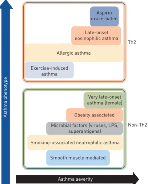
FIGURE 1 Classification of asthma phenotypes and disease severity based on T-helper cell type 2 (Th2) and non-Th2 characteristics. LPS: lipopolysaccharide.
Non-Th2 Th2 Aspirin
exacerbated
Late-onset eosinophilic asthma
Obesity associated
Microbial factors (viruses, LPS, superantigens)
Very late-onset asthma (female) Allergic asthma
Smoking-associated neutrophilic asthma
Smooth muscle mediated Exercise-induced
asthma
Asthma severity
As
anti-inflammatory glucocorticoid response genes and induction of RNA polymerase II-mediated gene transcription. This phenomenon is known as transactivation. Alternatively, activated GR interacts with CBP proteins complexed with promoter regions of pro-inflammatory genes, such as NF-κB and activator protein (AP)-1, causing suppression of pro-inflammatory gene transcriptionviainhibition of histone acetylation and preventing DNA access to RNA polymerase II [39–41]. This process is called transrepression.
A large body of clinical and experimental evidence shows that defective GRαexpression and activity impairs the anti-inflammatory effects of glucocorticoids and plays important roles in the induction of steroid resistance. Interestingly, other studies show that the higher levels of IL-2, IL-4 and IL-13 gene expression result in local cytokine secretion and steroid resistance, which further alters GRαtranslocation [34, 42]. A study by GOLEVA et al.[43] reported that specific silencing of GRβin bronchoalveolar lavage macrophages of individuals with SSR asthma resulted in improved GRαactivity. In another study, IRUSENet al.[44] reported that activation of p38 mitogen-activated protein kinase results in the phosphorylation and impaired function of GRs. Moreover, defective nuclear translocation of GRs results in reduced interaction with GREs and GR:GRE binding is affected by increased NF-κB, AP-1, c-Fos and c-Jun N-terminal kinase responses [34, 42, 45, 46]. Defective nuclear translocation of GRs reduces expression and activity of histone deacetylase (HDAC)2 in patients [47]. Importantly, many patients with SSR asthma have normal nuclear translocation of GR but reduced GR:GRE binding affinity [48, 49], suggesting that SSR asthma can also be induced by dysfunction of a different mechanism. Interestingly, reduced HDAC2 expression and activity is associated with steroid insensitivity and more severe disease in both asthma and COPD [50–52], suggesting that deficiencies in transcriptional co-repressor expression and activity may be important in the pathogenesis of SSR asthma.
Role of respiratory infections in SSR asthma
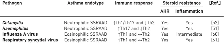
SSR asthma is a serious clinical and socioeconomic problem that is associated with lung function impairment, airway hyperresponsiveness (AHR) and more intense disease exacerbations leading to hospitalisation. Limited treatments are available, including corticosteroid therapy, but none are cures or broadly effective. A crucial drawback is that there are several subsets of SSR asthma with different immunological and inflammatory phenotypes that are likely to drive steroid resistance through different mechanisms [2, 5, 6, 53]. Increasing clinical and experimental evidence strongly implicates specific respiratory infections in the pathogenesis of SSR asthma. We have developed novel and unique animal models of respiratory infection-induced SSR asthma and used them to demonstrate that Chlamydia, Haemophilus influenzae, influenza A virus (IAV) and respiratory syncytial virus (RSV) respiratory infections induce steroid resistance of the key disease features [16–18, 30, 54–56].
We first established Chlamydia, Haemophilus, IAV and RSV respiratory infections and allergic airway disease (AAD) in BALB/c mice separately. Clinical studies show that infections drive more severe disease in established disease [30, 55, 56]. Thus, AAD was first established and then infection was induced. Mice were systemically sensitised with ovalbumin (Ova) and aluminium hydroxide intraperitoneally, followed 10–12 days later by inhaled Ova challenge to induce AAD. Other mouse models of allergic asthma widely use other allergens, such as house dust mite [57–59],Alternaria[60] or cockroach [61] antigen. Our mice were left for 20 days and were re-challenged on days 33–34 of the model, in order to model an allergen-induced exacerbation of disease. To induce severe steroid-resistant AAD (SSRAAD), we induced infections in allergic mice after AAD was initially established, to model the human scenario. Infections were resolved before allergen-induced exacerbation of AAD. To model ICS treatment and examine the response to steroid treatment, mice were administered the steroid dexamethasone (DEX) for the final 3 days of the protocol. Key disease features were assessed 1 day after the final Ova challenge, to assess the impact of resolved infection on the disease phenotype. Importantly, we found that infection drives a severe form of AAD that does not respond to steroid treatment. These protocols have resulted in novel experimental models of Chlamydia-, Haemophilus-, IAV- and RSV-induced SSR asthma that each represents unique endotypes of SSR asthma in humans (table 1).
Chlamydia respiratory tract infection in asthma corresponds to increased airway neutrophils, which predicts the presence of this pathogen and of steroid resistance [55]. It has also been linked to severe asthma in children [62–64].Chlamydiarespiratory infection in mice with AAD results in increased type 1-and 17-associated immune responses, neutrophilic inflammation, suppression of type 2 1-and eosinophilic inflammatory responses [18, 30, 55, 56], and steroid-resistant inflammation and AHR. In early life, Chlamydiarespiratory infections promote more severe experimental asthma in mice by switching type 1 phenotype and inducing IL-13 and TRAIL (tumour necrosis factor-related apoptosis-inducing ligand) responses [17, 65–67]. These models of early-life infection-induced severe asthma are also steroid resistant (P.M. Hansbro, unpublished data).
Experimentally, H. influenzae respiratory infection drives type 17-associated immune responses, airway neutrophilia, steroid resistance and impaired macrophage phagocytosis [16, 27, 54–56].
Viral respiratory tract infections are strongly associated with the induction of asthma in early life and in exacerbations of asthma that are resistant to mainstay therapies. IAV and RSV infections in murine models of asthma result in exaggerated airway eosinophil influx and type 1 and 2 immune responses, mixed eosinophilic and neutrophilic airway inflammation, and steroid resistance of airway inflammation and AHR [56, 68, 69].
Thus, these mouse models are highly representative of the different immune and inflammatory phenotypes of SSR asthma in humans but in each case the inflammation and AHR are steroid resistant. We have interrogated these models to identify critical roles for microRNA (miR)-21/phosphatase and tensin homologue (PTEN)/phosphoinositide 3-kinase (PI3K)/HDAC2 and NLRP3 inflammasome/IL-1βresponses in the pathogenesis of experimental and clinical SSR asthma [27, 55, 56]. Importantly, the infections can activate innate immune responses and induce neutrophilic inflammation and type 1 and 17 immune responses that are also associated with SSR asthma [2]. We have identified potential new treatments using macrolides, miR-21-specific antagomirs, and PI3K and NLRP3 inflammasome inhibitors [30, 55, 56]. Our models can be interrogated to identify potentially important relationships between microbiome load in pulmonary and extrapulmonary compartments and key disease features. Such analyses should be done rigorously to identify novel disease-causing factors.
Novel treatment strategies for SSR asthma
MacrolidesMacrolide antibiotics are a first-line treatment for bacterial infections and exert their effects by blocking bacterial protein synthesis, adherence, motility and biofilm formation [70, 71]. Importantly, recent clinical and experimental evidence shows that they also have anti-inflammatory properties. Macrolides have been shown to be immunomodulatory in severe asthma and can enhance the steroid response by suppressing inflammatory cell infiltration and AHR [72].
We recently used our novel mouse models ofChlamydiaandHaemophilusrespiratory infection-induced SSR asthma to examine the effects of treatment on key disease features with a macrolide (clarithromycin) or standard beta-lactam antibiotic (amoxicillin, which has no known anti-inflammatory properties) in the presence or absence of DEX [55]. Treatment with clarithromycin and amoxicillin had similar antimicrobial effects; however, only clarithromycin suppressed steroid-resistant airway inflammation and AHR in both eosinophilic and neutrophilic AAD. Importantly, clarithromycin exerted its beneficial effects by reducing type 2, tumour necrosis factor (TNF)-α and IL-17 responses. Treatment with amoxicillin had limited effects on key disease features but restored steroid sensitivity, probably by reducing bacterial load. By comparison, the effects of clarithromycin on suppressing features of AAD were independent of its antimicrobial effects. These data show that macrolide-based therapies may be effective in SSR asthma irrespective of the presence of respiratory infection.
A different study showed that treatment with the macrolide roxithromycin significantly improved lung function within 6 weeks in asthmatics with evidence ofChlamydiainfection [73]. Similarly, in asthmatic children with evidence ofChlamydiainfection, clarithromycin treatment reduced the risk and duration of wheezing [74]. Clarithromycin treatment of asthmatics with evidence ofChlamydiainfection also reduced IL-5 levels in the airways [75].
In our study, we showed that treatment with clarithromycin reduced IL-17 responses and suppressed inflammation and AHR in both SSR and steroid-sensitive AAD [54, 55]. Treatment also reduced airway neutrophil count and the levels of sputum IL-8, neutrophil elastase and matrix metalloproteinase-9 [76].
TABLE 1Role of infections in the pathogenesis of severe steroid-resistant asthma and their
response to steroid therapy
These data demonstrate that clarithromycin treatment has antimicrobial as well as anti-inflammatory effects that suppress pro-inflammatory responses in different asthma endotypes with evidence of steroid-resistant disease features, and highlight its potential application in treating infection-induced SSR asthma.
The immunomodulatory effects of macrolides were elegantly demonstrated in a clinical study (AMAZES study) performed by GIBSON et al. [77], who performed a randomised, double-blind, placebo-controlled parallel group trial to assess whether the macrolide azithromycin (500 mg, orally administered three times per week for up to 48 weeks) reduced asthma exacerbations in adults with symptomatic asthma. In this study, azithromycin was used as an add-on therapy to 1) ICS and long-acting β2-adrenoceptor agonist
(n=345; 82%), 2) long-actingβ2-adrenoceptor agonist with long-acting muscarinic antagonist (n=68; 16%),
3) long-acting muscarinic antagonist (n=5; 1.2%), or 4) theophylline (n=2; <1%). A total of 420 randomised asthma patients were assigned, 213 for an azithromycin group consisting of 144 (43%) patients with an eosinophilic endotype and 187 (57%) patients with a neutrophilic endotype, while 207 were treated with placebo control. Azithromycin reduced exacerbations down to 1.07 per patient-year compared to placebo with 1.86 per patient-year. Towards the end of the treatment regimen it was found through sputum culture that 37 patients (17 in the treatment and 20 in the placebo group) had detectable levels of potentially pathogenic microbes. Azithromycin significantly improved quality of life in asthma patients; however, its use was associated with diarrhoea. Nevertheless, these data show that the immunomodulatory effects of azithromycin may be an effective add-on therapy in severe asthma.
miR-21/PI3K/HDAC2 axis
The anti-inflammatory effects of corticosteroids are mediated by activation of GR/NR3C1 and deacetylation of histones by HDAC2, leading to suppression of pro-inflammatory gene transcription and/ or induced expression of anti-inflammatory genes [36, 78]. Importantly, increased PI3K activity is known to decrease HDAC2 responses and diminish steroid responses [52]. Furthermore, increased PI3K activity induces type 17-associated immune responses and reduces interferon (IFN) responses, which allows entry and replication of pathogens in host cells, which can lead to further increases in PI3K activity [79, 80]. Several miRs have been implicated in the pathogenesis of asthma [81, 82] and, of these, miR-21 is known to be important in AAD pathogenesis. miR-21-deficient (miR-21−/−) mice with AAD have reduced eosinophilic inflammation and IL-4 levels, and elevated IFN-γresponses [81, 83]. Biological network-based transcriptome analysis of Ova-challenged miR-21−/− mice determined that dysregulation of IL-12/IFN-γ played an important role in the observed phenotype and implicated miR-21 as a key regulator of IFN-γ signalling and T-cell polarisation. Thus, the loss of miR-21 augments T-helper cell type 1 (Th1)-related delayed hypersensitivity [83]. Interestingly, miR-21 has also been shown to directly downregulate the expression of PTEN, which is an endogenous suppressor of PI3K activity [84]. A study by KWAKet al. [84] demonstrated that mice with Ova-induced AAD had reduced PTEN levels in the epithelial layer of bronchioles and that adenovirus-mediated overexpression of PTEN in AAD resulted in increased IL-4 and IL-5 levels in the airways and reduced AHR. Furthermore, intratracheal administration of pharmacological pan-PI3K inhibitors suppressed bronchial inflammation and AHR in mice with AAD, which highlights the potential for using PI3K inhibitors, such as wortmannin, in the treatment of severe asthma [85].
Recently, we examined the role of miR-21 in the pathogenesis of SSR asthma and assessed its potential for therapeutic targeting using our novel mouse models of Chlamydia, H. influenzae, influenza and RSV respiratory infection-induced SSR asthma [56]. We first profiled the expression of all known mouse miRs using microarray analysis and found that miR-21 was the most highly expressed in our mouse models. We confirmed this result by quantitative PCR and then showed that miR-21 expression in AAD is not reduced by steroid treatment. We measured the expression of several putative targets of miR-21 (identified by multiple target prediction algorithms) and found that PTEN was consistently downregulated in a miR-21-enriched environment and that this was associated with increased PI3K responses (elevated nuclear levels of phosphorylated AKT). Importantly, we showed that these effects were associated with reductions in both nuclear levels of HDAC2 and lung expression of NR3C1. This led us to hypothesise that a novel miR-21/PTEN/PI3K/HDAC2 pathway plays an important disease-causing role in SSRAAD (figure 2).
steroid-resistant AHR in SSRAAD. We also observed similar effects using the pan-PI3K inhibitor LY294002. Notably, all disease features were completely suppressed by antagomiR-21 or LY294002 back to baseline levels achieved with steroid treatment of steroid-sensitive AAD in the absence of infection. Furthermore, we observed similar effects inHaemophilus-, IAV- and RSV-induced SSRAAD.
These data are especially important because Chlamydia/Haemophilus respiratory infections are strongly implicated in type 1/17 immune response-associated, steroid-resistant, neutrophilic asthma, and IAV/RSV infections are strongly implicated in type 1-associated, steroid-resistant, eosinophilic asthma. Collectively, our data show that targeting miR-21 and/or PI3K is likely to be effective across several endotypes of SSR asthma (figure 2) [56].
NLRP3 inflammasome
Inflammasomes are multiprotein signalling complexes that control the maturation and release of potent pro-inflammatory cytokines that are produced in response to endogenous, exogenous and pathogenic risk signals [28, 86]. The NLRP3 inflammasome is composed of an apoptosis-associated speck-like protein containing a pyrin domain, a caspase recruitment domain and a pro-caspase-1 domain [86]. The expression and assembly of inflammasomes occurs as a result of stimulation by microbe-derived pathogen-associated molecular patterns such as lipopolysaccharide and double-stranded DNA. A second signal in the form of damage-associated molecular patterns induces the activation of assembled inflammasomes that cleave and activate pro-caspase-1, which in turn cleaves pro-IL-1β and pro-IL-18 into active IL-1β and IL-18, respectively [28, 31, 86]. Thus, inflammasome activation is essential for caspase-1-dependent IL-1β processing, maturation and secretion (figure 3).
Neutrophilic asthma is associated with increased levels of NLRP3 and caspase-1 in the airways, exaggerated IL-1βresponses, and Th17 differentiation and IL-17 production that are all implicated in AHR and more severe forms of asthma [30, 31]. SIMPSON et al.[87] first demonstrated that neutrophilic asthma is associated with increased sputum expression of TLR2, TLR4, IL-1βand IL-8 and increased levels of lipopolysaccharide. These observations are supported by another clinical study by BAINESet al. [88], which used sputum RNA expression profiling to show that genes in the IL-1 and TNF-α/NF-κB signalling pathways were overexpressed and correlated with key clinical parameters and neutrophilic airway inflammation in asthma. Notably, SIMPSONet al. [89] showed that sputum macrophages are a major source of the elevated NLRP3, caspase-1, caspase-4, caspase-5 and IL-1βlevels they observed. Furthermore, DARVILLEet al. [90] observed that ATP-mediated P2X7R signalling is essential for immunity againstChlamydia, andChlamydia,Haemophilus, IAV and RSV infections all induce NLRP3 inflammasome-induced caspase-1-mediated activation and release of IL-1β[91–94]. We recently showed that NLRP3, caspase-1 and IL-1βresponses are increased in the lung inChlamydia infection-induced experimental SSR asthma [30] and that sputum NLRP3 and IL-1βexpression correlate with key clinical parameters of human SSR asthma, including increased percentage and absolute numbers of neutrophils in the airways, loss of asthma control and reduced lung function [30]. We then interrogated the functional roles and potential for therapeutic targeting of NLRP3, caspase-1 and IL-1βin experimental SSR asthma by intranasally administering MCC950 (a potent and highly selective NLRP3 inflammasome
AAD
miR-21
PTEN Respiratory
infection
Ant-21
PI3K
AKT
HDAC2 Steroid-resistant asthma Pan-PI3K
inhibitor/ LY294002
P
inhibitor [95]), ac-YVAD-cho (a selective caspase-1 inhibitor) or neutralising anti-IL-1β monoclonal antibody duringChlamydiaand Haemophilusinfection-induced SSRAAD. Increased lung IL-1βresponse was suppressed by all three treatments and this effect was associated with the suppression of steroid-resistant neutrophilic airway inflammation and AHR. We then demonstrated that the disease-causing effects of infection are driven by IL-1βand neutrophilic inflammation by recapitulating the key features of neutrophilic SSRAAD in mice with eosinophilic, steroid-sensitive AAD using recombinant mouse IL-1βand anti-Ly6G (blocks recruitment of neutrophils). We propose that specifically targeting the exaggerated NLRP3 inflammasome response component in SSR asthma is a novel and viable therapeutic approach and suggest that this strategy is clinically preferable because it allows for IL-1β processing through other mechanisms for protection against infections [8, 30].
Biologics
There is ample recent evidence that next generation biologics may be steroid sparing or potentially be treatments for SSR disease. IL-5 is well known to be a potent signal for the development, maturation and chemotaxis of eosinophils [4, 5]. Numerous studies suggest that mepolizumab (anti-IL-5 monoclonal antibody) treatment reduces the requirement for oral steroids in severe eosinophilic asthma [96, 97]. Furthermore, there are known links between innate type 2 inducers and mediators IL-33, IL-25 and TSLP (thymic stromal lymphopoietin), and type 2 innate lymphoid and T-helper cells and steroid resistance [98–100]. Antibodies against these factors may prove to be somewhat effective in severe disease [4, 99, 101].
Conclusions
SSR asthma is a clinical problem of significant concern and more effective, and potentially broadly applicable, therapies are urgently required. As a consequence of the underlying mechanisms of pathogenesis, patients do not respond to mainstay steroid-based therapies. Steroid resistance in asthma has been linked to bacterial and viral respiratory infections, infection-induced exacerbations, and a high-fat diet/obesity. Understanding the mechanisms of pathogenesis will enable the identification of novel therapeutic approaches and the development of new and effective treatments. Collectively, clinical and
Allergen/infection
NLRP3
Severe steroid-resistant asthma
ASC specks
Neutrophilic/ eosinophilic inflammation and
airways hyperresponsiveness PAMPs/
DAMPs
Airways
Activation of inflammasome
PYD
CARD
Pro-CASP1
CASP1 Pro-IL-1β/ IL-18 Active caspase-1
Cytokine maturation Pyroptotic cell death and
intracellular content release ASC
MCC 950
YVAD
α-IL-1β
experimental studies have identified macrolides and inhibitors of miR-21, PI3K and inflammasomes as potential new treatments for SSR asthma that may be broadly effective across multiple SSR asthma endotypes. Importantly, our data suggest that aberrant miR-21 and IL-1βresponses drive steroid-resistant airway inflammation and AHR in SSR asthma through mechanisms that are independent of the canonical steroid response pathway and are therefore not amenable to steroid-mediated suppression. The logical extension of these studies would be to identify and characterise the roles, and assess the potential for therapeutic targeting, of other miR-21 targets and downstream mediators of IL-1β responses in SSR asthma. Biologics that target type 2 responses may also have utility in SSR asthma.
Conflict of interest: R. Wadhwa has nothing to disclose. K. Dua has nothing to disclose. I.M. Adcock has nothing to disclose. J.C. Horvat has nothing to disclose. R.Y. Kim has nothing to disclose. P.M. Hansbro reports grants to develop inflammasome inhibitors for therapeutic use.
Support statement: R.Y. Kim is supported by a Fellowship from Lung Foundation Australia and Boehringer Ingelheim. P.M. Hansbro is supported by a Fellowship from the National Health and Medical Research Council of Australia (NHMRC #1079187).
References
1 Global Initiative for Asthma (GINA). Global Strategy for Asthma Management and Prevention. 2018. Available from http://ginasthma.org/
2 Hansbro PM, Kim RY, Starkey MR,et al.Mechanisms and treatments for severe, steroid-resistant allergic airway disease and asthma.Immunol Rev2017; 278: 41–62.
3 Foster PS, Maltby S, Rosenberg HF,et al.Modeling TH2 responses and airway inflammation to understand
fundamental mechanisms regulating the pathogenesis of asthma.Immunol Rev2017; 278: 20–40.
4 Hansbro PM, Scott GV, Essilfie AT,et al.Th2 cytokine antagonists: potential treatments for severe asthma.
Expert Opin Investig Drugs2013; 22: 49–69.
5 Hansbro PM, Kaiko GE, Foster PS. Cytokine/anti-cytokine therapy – novel treatments for asthma? Br J Pharmacol2011; 163: 81–95.
6 Wenzel SE. Asthma phenotypes: the evolution from clinical to molecular approaches. Nat Med 2012; 18: 716–725.
7 Hallstrand TS, Hackett TL, Altemeier WA,et al.Airway epithelial regulation of pulmonary immune homeostasis and inflammation.Clin Immunol2014; 151: 1–15.
8 Kim RY, Rae B, Neal R,et al.Elucidating novel disease mechanisms in severe asthma.Clin Transl Immunology
2016; 5: e91.
9 Moheimani F, Hsu AC, Reid AT,et al.The genetic and epigenetic landscapes of the epithelium in asthma.Respir Res2016; 17: 119.
10 Hansbro PM, Beagley KW, Horvat JC,et al.Role of atypical bacterial infection of the lung in predisposition/ protection of asthma.Pharmacol Ther2004; 101: 193–210.
11 Kaiko GE, Phipps S, Hickey DK,et al. Chlamydia muridarum infection subverts dendritic cell function to promote Th2 immunity and airways hyperreactivity.J Immunol2008; 180: 2225–2232.
12 Starkey MR, Jarnicki AG, Essilfie AT,et al.Murine models of infectious exacerbations of airway inflammation.
Curr Opin Pharmacol2013; 13: 337–344.
13 Wood LG, Li Q, Scott HA,et al.Saturated fatty acids, obesity, and the nucleotide oligomerization domain-like receptor protein 3 (NLRP3) inflammasome in asthmatic patients.J Allergy Clin Immunol2019; 143: 305–315.
14 Rutting S, Xenaki D, Malouf M,et al.Short-chain fatty acids increase TNFα-induced inflammation in primary human lung mesenchymal cells through the activation of p38 MAPK.Am J Physiol Lung Cell Mol Physiol2019; 316: L157–L174.
15 Hirota JA, Gold MJ, Hiebert PR, et al. The nucleotide-binding domain, leucine-rich repeat protein 3 inflammasome/IL-1 receptor I axis mediates innate, but not adaptive, immune responses after exposure to particulate matter under 10μm.Am J Respir Cell Mol Biol2015; 52: 96–105.
16 Essilfie AT, Simpson JL, Dunkley ML,et al.CombinedHaemophilus influenzaerespiratory infection and allergic airways disease drives chronic infection and features of neutrophilic asthma.Thorax2012; 67: 588–599.
17 Horvat JC, Starkey MR, Kim RY,et al. Early-life chlamydial lung infection enhances allergic airways disease through age-dependent differences in immunopathology.J Allergy Clin Immunol2010; 125: 617–625.
18 Horvat JC, Starkey MR, Kim RY,et al. Chlamydial respiratory infection during allergen sensitization drives neutrophilic allergic airways disease.J Immunol2010; 184: 4159–4169.
19 Wood LG, Simpson JL, Hansbro PM,et al.Potentially pathogenic bacteria cultured from the sputum of stable asthmatics are associated with increased 8-isoprostane and airway neutrophilia.Free Radic Res2010; 44: 146–154.
20 Pavord ID, Beasley R, Agusti A,et al.After asthma: redefining airways diseases.Lancet2018; 391: 350–400.
21 Ahmed H, Turner S. Severe asthma in children–a review of definitions, epidemiology, and treatment options in 2019.Pediatr Pulmonol2019; 54: 778–787.
22 Marko A, Ross KR. Severe asthma in childhood.Immunol Allergy Clin North Am2019; 39: 243–257.
23 Saglani S, Fleming L, Sonnappa S,et al.Advances in the aetiology, management, and prevention of acute asthma attacks in children.Lancet Child Adolesc Health2019; 3: 354–364.
24 Rhen T, Cidlowski JA. Antiinflammatory action of glucocorticoids–new mechanisms for old drugs.N Engl J Med2005; 353: 1711–1723.
25 Barnes PJ. Biochemical basis of asthma therapy.J Biol Chem2011; 286: 32899–32905.
26 Wang W, Li JJ, Foster PS,et al.Potential therapeutic targets for steroid-resistant asthma.Curr Drug Targets2010; 11: 957–970.
28 Kim RY, Pinkerton JW, Gibson PG,et al.Inflammasomes in COPD and neutrophilic asthma.Thorax2015; 70: 1199–1201.
29 Green R, Brightling CE, Woltmann G,et al.Analysis of induced sputum in adults with asthma: identification of subgroup with isolated sputum neutrophilia and poor response to inhaled corticosteroids.Thorax2002; 57: 875–879.
30 Kim RY, Pinkerton JW, Essilfie AT,et al.Role for NLRP3 inflammasome-mediated, IL-1β-dependent responses in severe, steroid-resistant asthma.Am J Respir Crit Care Med2017; 196: 283–297.
31 Pinkerton JW, Kim RY, Robertson AAB,et al.Inflammasomes in the lung.Mol Immunol2017; 86: 44–55.
32 Gold M, Hiebert PR, Park HY,et al.Mucosal production of uric acid by airway epithelial cells contributes to particulate matter-induced allergic sensitization.Mucosal Immunol2016; 9: 809–820.
33 Schwartz HJ, Lowell FC, Melby JC. Steroid resistance in bronchial asthma.Ann Intern Med1968; 68: 1141.
34 Adcock IM, Ford PA, Bhavsar P, et al. Steroid resistance in asthma: mechanisms and treatment options.
Curr Allergy Asthma Rep2008; 8: 171–178.
35 Barnes PJ. Inhaled glucocorticoids for asthma.N Engl J Med1995; 332: 868–875.
36 Ito K, Getting SJ, Charron CE. Mode of glucocorticoid actions in airway disease.ScientificWorldJournal2006; 6: 1750–1769.
37 Oakley RH, Jewell CM, Yudt MR,et al.The dominant negative activity of the human glucocorticoid receptorβ isoform. Specificity and mechanisms of action.J Biol Chem1999; 274: 27857–27866.
38 Tao T, Lan J, Lukacs GL,et al.Importin 13 regulates nuclear import of the glucocorticoid receptor in airway epithelial cells.Am J Respir Cell Mol Biol2006; 35: 668–680.
39 Horwitz K, Jackson TA, Bain DL,et al.Nuclear receptor coactivators and corepressors.Mol Endocrinol1996; 10: 1167–1177.
40 Barnes PJ. Glucocorticosteroids: current and future directions.Br J Pharmacol2011; 163: 29–43.
41 Hansbro PM, Starkey MR, Mattes J,et al.Pulmonary immunity during respiratory infections in early life and the development of severe asthma.Ann Am Thorac Soc2014; 11: Suppl. 5, S297–S302.
42 Ito K, Chung KF, Adcock IM. Update on glucocorticoid action and resistance.J Allergy Clin Immunol2006; 117: 522–543.
43 Goleva E, Li LB, Eves PT,et al.Increased glucocorticoid receptor βalters steroid response in glucocorticoid-insensitive asthma.Am J Respir Crit Care Med2006; 173: 607–616.
44 Irusen E, Matthews JG, Takahashi A,et al.p38 Mitogen-activated protein kinase-induced glucocorticoid receptor phosphorylation reduces its activity: role in steroid-insensitive asthma.J Allergy Clin Immunol2002; 109: 649–657.
45 Adcock I, Lane S. Mechanisms of steroid action and resistance in inflammation. Corticosteroid-insensitive asthma: molecular mechanisms.J Endocrinol2003; 178: 347–355.
46 Loke TK, Mallett KH, Ratoff J,et al.Systemic glucocorticoid reduces bronchial mucosal activation of activator protein 1 components in glucocorticoid-sensitive but not glucocorticoid-resistant asthmatic patients.J Allergy Clin Immunol2006; 118: 368–375.
47 Gunawardhana L, Gibson PG, Simpson JL,et al.Activity and expression of histone acetylases and deacetylases in inflammatory phenotypes of asthma.Clin Exp Allergy2014; 44: 47–57.
48 Cosio BG, Tsaprouni L, Ito K,et al.Theophylline restores histone deacetylase activity and steroid responses in COPD macrophages.J Exp Med2004; 200: 689–695.
49 Adcock IM, Barnes PJ. Molecular mechanisms of corticosteroid resistance.Chest2008; 134: 394–401.
50 Barnes PJ. Histone deacetylase-2 and airway disease.Ther Adv Respir Dis2009; 3: 235–243.
51 Adcock IM, Ito K, Barnes PJ. Histone deacetylation: an important mechanism in inflammatory lung diseases.
COPD2005; 2: 445–455.
52 Marwick JA, Caramori G, Stevenson CS, et al. Inhibition of PI3Kδ restores glucocorticoid function in smoking-induced airway inflammation in mice.Am J Respir Crit Care Med2009; 179: 542–548.
53 Gibson PG, Saltos N, Borgas T. Airway mast cells and eosinophils correlate with clinical severity and airway hyperresponsiveness in corticosteroid-treated asthma.J Allergy Clin Immunol2000; 105: 752–759.
54 Essilfie AT, Simpson JL, Horvat JC,et al. Haemophilus influenzae infection drives IL-17-mediated neutrophilic allergic airways disease.PLoS Pathog2011; 7: e1002244.
55 Essilfie AT, Horvat JC, Kim RY,et al.Macrolide therapy suppresses key features of experimental steroid-sensitive and steroid-insensitive asthma.Thorax2015; 70: 458–467.
56 Kim RY, Horvat JC, Pinkerton JW,et al.MicroRNA-21 drives severe, steroid-insensitive experimental asthma by amplifying phosphoinositide 3-kinase-mediated suppression of histone deacetylase 2. J Allergy Clin Immunol
2017; 139: 519–532.
57 Liu G, Cooley MA, Jarnicki AG,et al.Fibulin-1 regulates the pathogenesis of tissue remodeling in respiratory diseases.JCI Insight2016; 1: e86380.
58 Thorburn AN, Foster PS, Gibson PG,et al.Components ofStreptococcus pneumoniaesuppress allergic airways disease and NKT cells by inducing regulatory T cells.J Immunol2012; 188: 4611–4620.
59 Liu G, Cooley MA, Nair PM,et al.Airway remodeling and inflammation in asthma are dependent on the extracellular matrix protein fibulin-1c.J Pathol2017; 243: 510–523.
60 Valladao AC, Frevert CW, Koch LK,et al.STAT6 regulates the development of eosinophilicversusneutrophilic asthma in response toAlternaria alternata.J Immunol2016; 197: 4541–4551.
61 Gong D, Fei F, Lim M,et al.Abr, a negative regulator of Rac, attenuates cockroach allergen-induced asthma in a mouse model.J Immunol2013; 191: 4514–4520.
62 Hahn DL, Schure A, Patel K,et al. Chlamydia pneumoniae-specific IgE is prevalent in asthma and is associated with disease severity.PLoS One2012; 7: e35945.
63 Patel KK, Vicencio AG, Du Z,et al.InfectiousChlamydia pneumoniaeis associated with elevated interleukin-8 and airway neutrophilia in children with refractory asthma.Pediatr Infect Dis J2010; 29: 1093–1098.
64 Patel KK, Webley WC. Evidence of infectious asthma phenotype: Chlamydia-induced allergy and pathogen-specific IgE in a neonatal mouse model.PLoS One2013; 8: e83453.
65 Horvat JC, Beagley KW, Wade MA, et al. Neonatal chlamydial infection induces mixed T-cell responses that drive allergic airway disease.Am J Respir Crit Care Med2007; 176: 556–564.
66 Starkey MR, Essilfie AT, Horvat JC, et al. Constitutive production of IL-13 promotes early-life Chlamydia
67 Starkey MR, Nguyen DH, Essilfie AT,et al.Tumor necrosis factor-related apoptosis-inducing ligand translates neonatal respiratory infection into chronic lung disease.Mucosal Immunol2014; 7: 478–488.
68 Beale J, Jayaraman A, Jackson DJ,et al.Rhinovirus-induced IL-25 in asthma exacerbation drives type 2 immunity and allergic pulmonary inflammation.Sci Transl Med2014; 6: 256ra134.
69 Hansbro NG, Horvat JC, Wark PA, et al. Understanding the mechanisms of viral induced asthma: new therapeutic directions.Pharmacol Ther2008; 117: 313–353.
70 Crosbie P, Woodhead M. Long-term macrolide therapy in chronic inflammatory airway diseases.Eur Respir J
2009; 33: 171–181.
71 Xepapadaki P, Koutsoumpari I, Papaevagelou V, et al. Atypical bacteria and macrolides in asthma. Allergy Asthma Clin Immunol2008; 4: 111.
72 Spahn JD, Fost DA, Covar R,et al.Clarithromycin potentiates glucocorticoid responsiveness in patients with asthma: results of a pilot study.Ann Allergy Asthma Immunol2001; 87: 501–505.
73 Black PN, Blasi F, Jenkins CR,et al.Trial of roxithromycin in subjects with asthma and serological evidence of infection withChlamydia pneumoniae.Am J Respir Crit Care Med2001; 164: 536–541.
74 Esposito S, Blasi F, Bosis F. Efficacy of clarithromycin for the treatment of acute episodes of bronchospasm in children with a history of recurrent wheezing. Tampere, 22nd Annual Meeting of European Society for Paediatric Infectious Diseases (ESPID), 2004.
75 Kraft M, Cassell GH, Pak J,et al. Mycoplasma pneumoniae andChlamydia pneumoniaein asthma: effect of clarithromycin.Chest2002; 121: 1782–1788.
76 Simpson JL, Powell H, Boyle MJ,et al. Clarithromycin targets neutrophilic airway inflammation in refractory asthma.Am J Respir Crit Care Med2008; 177: 148–155.
77 Gibson PG, Yang IA, Upham JW,et al.Effect of azithromycin on asthma exacerbations and quality of life in adults with persistent uncontrolled asthma (AMAZES): a randomised, double-blind, placebo-controlled trial.
Lancet2017; 390: 659–668.
78 Ito K, Ito M, Elliott WM,et al.Decreased histone deacetylase activity in chronic obstructive pulmonary disease.
N Engl J Med2005; 352: 1967–1976.
79 Hsu AC, Starkey MR, Hanish I,et al. Targeting PI3K-p110αsuppresses influenza virus infection in chronic obstructive pulmonary disease.Am J Respir Crit Care Med2015; 191: 1012–1023.
80 Haylock-Jacobs S, Comerford I, Bunting M,et al.PI3Kδdrives the pathogenesis of experimental autoimmune encephalomyelitis by inhibiting effector T cell apoptosis and promoting Th17 differentiation.J Autoimmun2011; 36: 278–287.
81 Foster PS, Plank M, Collison A,et al.The emerging role of microRNAs in regulating immune and inflammatory responses in the lung.Immunol Rev2013; 253: 198–215.
82 Plank MW, Maltby S, Tay HL,et al.MicroRNA expression is altered in an ovalbumin-induced asthma model and targeting miR-155 with antagomirs reveals cellular specificity.PLoS One2015; 10: e0144810.
83 Lu TX, Munitz A, Rothenberg ME. MicroRNA-21 is up-regulated in allergic airway inflammation and regulates IL-12p35 expression.J Immunol2009; 182: 4994–5002.
84 Kwak YG, Song CH, Yi HK,et al.Involvement of PTEN in airway hyperresponsiveness and inflammation in bronchial asthma.J Clin Invest2003; 111: 1083–1092.
85 Ezeamuzie CI, Sukumaran J, Philips E. Effect of wortmannin on human eosinophil responsesin vitroand on bronchial inflammation and airway hyperresponsiveness in guinea pigsin vivo.Am J Respir Crit Care Med2001; 164: 1633–1639.
86 Schroder K, Tschopp J. The inflammasomes.Cell2010; 140: 821–832.
87 Simpson JL, Grissell TV, Douwes J,et al.Innate immune activation in neutrophilic asthma and bronchiectasis.
Thorax2007; 62: 211–218.
88 Baines KJ, Simpson JL, Wood LG, et al. Transcriptional phenotypes of asthma defined by gene expression profiling of induced sputum samples.J Allergy Clin Immunol2011; 127: 153–160.
89 Simpson JL, Phipps S, Baines KJ,et al.Elevated expression of the NLRP3 inflammasome in neutrophilic asthma.
Eur Respir J2014; 43: 1067–1076.
90 Darville T, Welter-Stahl L, Cruz C,et al.Effect of the purinergic receptor P2X7 on Chlamydia infection in
cervical epithelial cells and vaginally infected mice.J Immunol2007; 179: 3707–3714.
91 He X, Mekasha S, Mavrogiorgos N,et al.Inflammation and fibrosis duringChlamydia pneumoniaeinfection is regulated by IL-1 and the NLRP3/ASC inflammasome.J Immunol2010; 184: 5743–5754.
92 Rotta Detto Loria J, Rohmann K, Droemann D,et al.NontypeableHaemophilus influenzaeinfection upregulates the NLRP3 inflammasome and leads to caspase-1-dependent secretion of interleukin-1β–a possible pathway of exacerbations in COPD.PLoS One2013; 8: e66818.
93 Ichinohe T, Pang IK, Iwasaki A. Influenza virus activates inflammasomesviaits intracellular M2 ion channel.
Nat Immunol2010; 11: 404–410.
94 Segovia J, Sabbah A, Mgbemena V,et al.TLR2/MyD88/NF-κB pathway, reactive oxygen species, potassium efflux activates NLRP3/ASC inflammasome during respiratory syncytial virus infection.PLoS One2012; 7: e29695.
95 Coll RC, Robertson AA, Chae JJ, et al. A small-molecule inhibitor of the NLRP3 inflammasome for the treatment of inflammatory diseases.Nat Med2015; 21: 248–255.
96 Bel EH, Wenzel SE, Thompson PJ,et al. Oral glucocorticoid-sparing effect of mepolizumab in eosinophilic asthma.N Engl J Med2014; 371: 1189–1197.
97 Ortega HG, Liu MC, Pavord ID,et al. Mepolizumab treatment in patients with severe eosinophilic asthma.
N Engl J Med2014; 371: 1198–1207.
98 Starkey MR, McKenzie AN, Belz GT,et al.Group 2 innate lymphoid cells: surprises and challenges.Mucosal Immunol2019; 12: 299–311.
99 Robinson D, Humbert M, Buhl R,et al.Revisiting Type 2-high and Type 2-low airway inflammation in asthma: current knowledge and therapeutic implications.Clin Exp Allergy2017; 47: 161–175.
100 Christianson CA, Goplen NP, Zafar I,et al. Persistence of asthma requires multiple feedback circuits involving type 2 innate lymphoid cells and IL-33.J Allergy Clin Immunol2015; 136: 59–68.e14.