*Corresponding author:Khalid L. P ISSN: 0976-3031
Research Article
A COMPLETE PHYTOCHEMICAL ANALYSIS OF ROOT, STEM AND LEAF OF
RUNGIA REPENS
Khalid L. P, Pulate P.V and Kallyani. A. Kavitkar
Department of Botany Vidyabharati Mahavidyalaya camp, amravati (ms) india
DOI: http://dx.doi.org/10.24327/ijrsr.2019.1009.4023
ARTICLE INFO ABSTRACT
The present study deals with the phytochemical analysis by qualitative and quantitative method, anatomical study, extractive value, chemical behavior, antimicrobial activity and chromatography analysis. Powder microscopy showing present of parenchymatous cell, trichome, fiber, stomata etc. Extractive value found that the active constituents in given amount of crude plant material was extracted with respective solvent. Chemical analysis used for the standardization and detection of adulterant in crude drug. Qualitative analysis revealed that present of many phytoconstituents in leaf, stem and root like alkaloid, saponin, carbohydrate, flavonoids glycoside etc.and its amount is calculate by quantitative method. Highest alkaloid is found in stem and root that are 0.97mg and 0.965mg while excess flavonoid and saponin is noted in leaf of Rungia repens is 3.364mg and 1.160mg and it also noted that the anti-microbial efficiency could be due to the presence of variable phytochemical compounds in different extracts.
INTRODUCTION
India is rich in medicinal plant diversity which is distributed in different geographical, environmental conditions and associated tribal & folk knowledge systems. India is one of the eight major centers of origin and diversity of taxonomy which is the part of traditional medicinal system and used pattern of different plants having rich biodiversity and is one of the world’s twelve mega diversity countries. (Principe, P. et.al 2005). In India many undesirable plants so called weeds. Weeds are very usual, superior and wide spread in the crop fields. In India particular in Marathwada region of the Maharashtra state, diversity of undesirable plants in crop fields is seen in day to day life, dominant and easily obtainable.
The genus Rungia repens belongs to family Acanthaceae, It distributed in India and Srilanka. Family is comprising, according to 4300 species in 346 genera. Acanthaceae is a large cosmopolitan family distributed mostly in the tropical and subtropical areas of the world. Rungia repens is an annual erect herb with profuse branching and distributed throughout the warmer parts of India.(J. S Gamble,1921). The herb possesses cylindrical fragile stems. Rungia repens (L.) Nees is a spreading decumbent herb found throughout India mostly as a weed in moist places sides of water channels, bunds of paddy fields, and also under the shadow area of coconut trees. The
plant Rungia repens showed higher water-soluble components than alcohol soluble components. Flavonoids and phenolics are among the major compounds present in the Rungia repens plant (Karuna Modi et.al 2017) and are powerful antioxidants. In Gujarat and Maharastra it is used as parjpataka (Yoganarasimhan, 2000). Whole plant dried and pulverized is given in fevers and cough by the local tribes. Leaf paste also used to cure fungal skin diseases (Vedavathy, 1992).. The herb is used in the treatment of cough and fever and is also credited with vermin-fungal and diuretic properties(Trease GE 2002).Plants contain various important phytoconstituent like terpenoids exhibit various pharmacological activities i.e., anti-inflammatory, anti- cancer, anti-malarial, inhibition of cholesterol synthesis, anti-viral and anti-bacterial activities. Alkaloids are found in medicinal plants used as anesthetic agents. (Mahato SB, Sen, 1997).Hence, there is an urgent need to identify the market sample of Rungia repens macro-and microscopicall.
MATERIAL METHOD
Rungia repens was collected during the winter season from Chikhaldara Ghatang region. Following parameters were done with Rungia repens plant part.
International Journal of
Recent Scientific
Research
International Journal of Recent Scientific Research
Vol. 10, Issue, 09(H), pp. 35050-35055, September, 2019
Copyright © Khalid L. P, Pulate P.V and Kallyani. A. Kavitkar, 2019, this is an open-access article distributed under the terms of the Creative Commons Attribution License, which permits unrestricted use, distribution and reproduction in any medium, provided the original work is properly cited.
DOI: 10.24327/IJRSR CODEN: IJRSFP (USA)
Article History: Received 15th June, 2019
Received in revised form 7th July, 2019 Accepted 13th August, 2019
Published online 28th September, 2019
Key Words:
Qualitative analysis, quantitative analysis, Anatomy, antifungal analysis,
International Journal of Recent Scientific Research Vol. 10, Issue, 09(H), pp. 35050-35055, September, 2019
35051 |
P a g e
Fig 1 Habit: Rungia repens Fig 2 Herbarium of Rungia repens
Anatomical study: The collected fresh material of selected plants was washed 2-3 times with sterile distilled water and preserved 3% formalin solution then used for the anatomical investigation and prepared double stain permanent slide for anatomical study and it observed under Carl Zeiss standard universal Microscope.
Powder microscopy
The plant drug contain some basic cell type.A little quantity of powder was taken onto a microscopic cavity slide.1-2 drops of 0.1% phologlucinol solution and a drop of conc.HCL were added, mount it in glycerol, covered with a cover slip and observed under the microscope with 10x10 magnification and characteristic features of plant powder were recorded. Powder microscopy was carried out by using the method mentioned in ayurvedic Pharmacopeia of India 1966.
Extractive Value
The procedures recommended in ayurvedic Pharmacopeia of India (1966) were followed for calculating extractive values.
Chemical behavioral analysis
This analysis was carried out by using the standard method mentioned in Ayurvedic Pharmacopeia of India (1966). Behavior of powdered plant materials with different chemical reagents i.e. conc. H2SO4, HNO3, ,HCL, 10% NaOH, Iodine solution, Ferric chloride, Potassium iodide,1N H2SO4,HNO3,HCL were observed under day light and UV light.
Preparation of extract
Collected plant was washed 2-3 times with distilled water and separate the plant part i.e. leaves, stem, root and dried under shade condition. These dried plant parts materials were grind and formed a coarse powder and extraction were done by using soxhlet extraction method.
Phytochemical test
The methods used for detection of various phytochemical were followed by qualitative and quantitative chemical test to give general idea regarding the nature of constituents present in crude material of Rungia repens (Kokate, 2005; Harborne, 1998; Sadashivam and Manickam, 2005; Wallis, 1990).
Tests for carbohydrates
Fehling’s Test: 1 ml Fehling’s A solution and 1 ml of Fehling’s B solution were mixed and boiled for one minute. Now the equal volume of test solution was added to the above
mixture. The solution was heated in boiling water bath for 5-10 minutes. First a yellow, the brick red precipitate was observed.
Benedict’s reagent: Equal volumes of Benedict’s reagent and test solution were mixed in a test tube. The mixture was heated in boiling water bath for 5 minutes. Solution appeared green showing the presence of reducing sugar.
Molisch’s test: Equal volumes of Molisch’s reagent and test solution were mixed in a test tube. The mixture was heated in boiling water bath for 5 minutes. Appearance of violet or purple colour ring showing the presence of reducing sugar.
Test for proteins
Biurret Test: To the small quantity of extract 1-2 drops of biurret reagent was added. Formation of violet colour precipitate showed presence of proteins.
Million’s Test: To the small quantity of extract 1-2 drops of Million’s reagent was added. Formation of white colour precipitate showed presence of proteins.
Tests for Anthroquinone glycosides
Borntrager’s Test: To the 3 ml of extracts, dil. H2SO4 was added. The solution was then boiled and filtered. The filtrate was cooled and to it equal volume of benzene was added. The solution was shaken well and the organic layer was separated. Equal volume of dilute ammonia solution was added to the organic layer. The ammonia layer turned pink showing the presence of glycosides. (Prashant Tiwari, 2011)
Tests for Cardiac glycosides
Keller-Killiani Test: To the 5ml of extract, 1ml of
conc.H2SO4,2 ml of Glacial acetic acid and 1 drop of FeCl3 solution was added. Appearance of brown ring shows the presence of cardiac glycosides.
Test for steroids
Salkowski Test:To 2 ml of extract, 2 ml of chloroform and 2
ml of conc.H2SO4 was added. The solution was shaken well. As a result chloroform layer turned red and acid layer showed greenish yellow fluorescence.
Test for alkaloids
Hager’s Test:To the 2-3 ml of filtrate, few drops of dil. HCl and Hager’s reagent was added and shake well. Yellow precipitate was formed showing the presence of alkaloids.
Mayer’s Test: To the 2-3 ml of filtrate, dil.HCl and Mayer’s reagent was added and shake well. Yellow precipitate was formed showing the presence of alkaloids.
Dragendroff’s Test:To the 2-3 ml of filtrate, few dropsof dil.
HCl and Dragendroff’s reagent were added and shake well. Formation of orange-brown precipitate showed the presence of alkaloids.
Test for Tannins and Phenolics compound
FeCl3 solution Test: On addition of 5% FeCl3 solution to the extract, deep blue black colour appeared.(Prashant Tiwari, 2011).
Lead Acetate Test:On addition of lead acetate solution to the extract white precipitate appeared.
Test for Saponin
Foam Test: To 1 ml extract 20 ml distilled water was added and shakes well in measuring cylinder for 15 min. Then 1 cm layer of foam was formed (Prashant Tiwari, 2011).
Test for Xanthoprotein
In 2ml test solution a few drops of conc. nitric acid and 3ml of ammonia were added. Appearance of red precipitate indicates the presence of xanthoprotein.(Prashant Tiwari, 2011).
Test for Glycosides
The extract was mixed with a little anthrons on a watch glass. One drop of conc. sulphuric acid was added and made into a paste and warmed gentle over the water bath. Dark green coloration indicates the presence of glycosides.
Test for Quinone
2ml test solution was treated with a few drops of conc.H2SO4 or aq. NaOH solution. Colour formation indicates the presence of Quinone compound.
Test for fixed oil
A small quantity of powder was pressed between the filter paper. Formation of grease spot indicates the presence of fixed oil and fats.
Quantification of the major phytoconstituents
The crude quantification of major phytochemicals was done using precipitation method. Each sample was analyzed in triplicates. Only alkaloids, flavonoids and saponin from the different parts of the plant under study were quantified according to Kokate, 2005; Harborne, 1998; Sadashivam and Manickam, 2005; Wallis, 1990).
Antimicrobial activity
The antimicrobial activity of the selected methanolic and ethanolic extracts was determined by disc diffusion method (Bauer AW et.al. 1966).Test microorganisms i.e. Aspergillus niger and Aspergillus flavor were collected from mycology lab and culture them on PDA media and incubated at 37°c for 24 hrs. A suspension of tested micro-organisms was spread on Dextrose agar medium by streak plate method under aseptic condition, after incubation the both inhibition were quantified by measuring the diameter of the zone of inhibition in mm.
Chromatographic analysis
The chromatographically study was carried out by using the standard procedure described by Harborne,(1998); Sadashivan and Manickam.,(2005). The qualitative evaluation of the plate was done by determining the migrating behavior of the separated substances given in the form of Rf value.
Distance traveled by the solute from the origin Resolution factor (Rf) = --- Distance traveled by the solvent from the origin
RESULT AND DISCUSSION
In India, the use of different parts of several medicinal plants to cure specific ailments has been in vogue from ancient times (Chopra,1956).Therefore, the present investigation -pharmacognostic studies on Rungia repens is taken to provide the base for the quality control of traditional herbal medicines.
Anatomical study
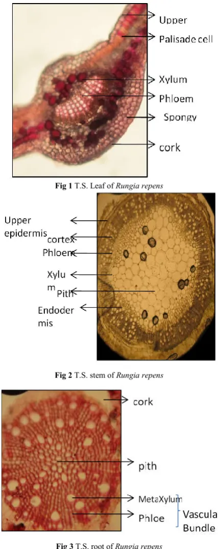
Fig 1 T.S. Leaf of Rungia repens
Fig 2 T.S. stem of Rungia repens
International Journal of Recent Scientific Research Vol. 10, Issue, 09(H), pp. 35050-35055, September, 2019
35053 |
P a g e
Anatomy of leaf of Rungia repens showing upper and lower epidermis, mesophyll tissue which differentiated into palisade and spongy parenchyma which distributed into either side of lamina and centrally located xylum and phloem similar result also observed by Karuna modi and Mamta Shah-2017. Fig.2 T.S. of stem showing outer most epidermis just below it hypodermis and cotex followed by single endodermis and centrally available phloem and xylum it also observed byKaruna modi and Mamta Shah-2017. Fig.3 T.S. of root showed that 2-4 layers of elongated cells, parenchymatous cortex, vascular bundle having xylum, phloem and vessels it also observed byKaruna modi and Mamta Shah-2017
Powder Microscopy
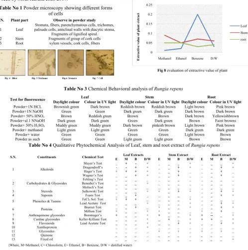
In powder microscopy the aerial parts showed the characteristics cell mention in Table No.1 likewise characters also seen by Modi Karuna et.al. 2017.
Extractive value
The maximum extractive value observed in more polar solvent. The extraction is carried out by using solvents like methanol, ethanol, benzene and distilled water. The extractive values determine the amount of active constituents in given amount of crude plant material. In the present study highest extractive value noted in methanolextract of stem0.9mg according to (Bharat Gami & Farzin Parabia-2011)extractive value in methanol was found higher as compared to acetone. Followed by benzene and Aqouse extract of stem that is 0.7 & 0.6 mg, L.P. Khalid, P.V. Pulate et.al-2017observed benzene leaf extract of M. Tomentosa having 0.13 mg extractive value. Methanolic & ethanolic extract of root showing same amount of extractive value that is 0.5mg and lowest value observed in ethanolic and benzene extract of leaf is 0.2mg mention in Fig. 8.
Fig 8 evaluation of extractive value of plant
0 0.05 0.1 0.15 0.2 0.25
Methanol Ethanol Benzene D/W
Ex
tractive
valu
e
o
f pla
n
t
ex
tract
Leaf
Stem
root
Table No 1 Powder microscopy showing different forms of cells
S.N. Plant part Observe in powder study
1 Leaf
Stomata, fibers, parenchymatous cells, trichomes, palisade cells, anticlinal walls with diacytic stoma,
Fragments of lignified spiral. 2 Stem Fragments of group of cork cells 3 Root xylem vessels, cork cells, fibers
Table No 3 Chemical Behavioral analysis of Rungia repens
Test for fluorescence Leaf Stem Root
Daylight colour Colour in UV light Daylight colour Colour in UV light Daylight colour Colour in UV light
Powder+1N HCL Brownish green Dark brown Reddish brown Reddish brown Light brown Pink brown Powder+1N NaOH Green Green Dark green Dark green Dark brown Dark brown Powder+ 50% HNO3 Brown Reddish green Brown Brown Dark brown Yellowishbrown
Powder+al.1 NNaOH Dark green Dark green Green Dark green Brown Faint browns Powder+ 50% H2SO4 Muddy green Muddy green Dark brown pinkish brown Light brown Pink brown
Powder+ methanol Light green Light green Green Green Dark green Dark green Powder+ water Green Green Green Green Light brown Brown Powder as such Green Green Light green Light green Brown Brown
Table No 4 Qualitative Phytochemical Analysis of Leaf, stem and root extract of Rungia repens
S.N. Constituents Chemical Test Leaf Extracts Stem Extract Root Extract
E M B D/W E M B D/W E M B D/W
1 Alkaloids
Mayer’s Test _ + - + _ _ _ + _ _ _ +
Dragendroff’s - - + + _ _ + + _ _ + +
Hager’s Test + + + + _ _ + _ _ _ + _
Wagner’s Test _ _ + + _ _ + _ _ _ + _
2 Carbohydrates & Glycosides
Fehling’s Test _ _ _ _ _ _ _ _ _ _ _ _
Benedict’s Test _ _ _ + _ _ _ _ _ _ _ _
Molisch’s Test _ _ _ _ _ _ _ _ _ _ _ _
3 Steroids Salkowski Test _ _ _ + + _ + + _ _ + +
4 Saponin Foam Test _ _ + _ _ _ _ _ _ _ + +
5 Phenolics & Tannin FeCl3 Sol. Test + + + _ _ _ _ + _ _ _ _
Lead Acetate Test _ _ _ + _ _ _ + _ _ + +
6 Proteins Biureet Test _ _ + _ + _ _ + _ _ + +
Million Test + _ _ + + _ _ _ + _ + _
7 Anthraquinone glycosides Borntrager’s + _ _ + + _ _ + + -- _ +
8 Cardiac glycosides Keller-Killiani Test _ _ _ + _ + _ + + + _ +
9 Flavonoids Lead Acetate Test _ _ _ + _ _ + + _ _ + +
10 Xanthoprotein _ _ _ + _ _ _ _ _ _ _ _
11 Glycosides _ _ _ _ + _ + _ + _ + _
12 Quinone _ _ _ _ _ _ _ _ _ _ _ _
13 Fixed oil _ _ _ _ _ _ _ _ _ _ _ +
Chemical behavioral analysis
The crude drug of Rungia repens showed various colors when extracts were treated with various chemicals.
consists of various phytoconstituents in different forms gives characteristic reactions with various reagents. phenomenon is used for qualitative examination of crude drugs This technique can be used for the standard
detection of adulterant in crude drug (Wallis,1990).The crude powder of plant under study reacts with various chemicals and behavioral characteristic mention in Table No.3
Qualitative phytochemical screening of Rungia repens stem, leaf and root
The result of preliminary phytochemical screening of leaf in four extracts viz Methanol, Ethanol, Benzene and Distilled water reveals the presence of alkaloids, flavonoids, saponins and phenolic compounds some workers were observed phytochemicals including Flavonoids, Steroids, Saponins and Alkaloids in leaf extract of M.tomentosa (L.P. Khalid, P.V. Pulate et.al-2017). The majority of the phytoconstituents was found in methanol and distilled water extracts,
extract showed the presence of phenolics, flavonoids, tannins, alkaloids, saponins and steroids, this corroborates the previous finds from Melia azedarachL. Dep K, Kaur A, Ambwani S, Ambwani TK 2018. Tannins are only present in the distilled water extract. Fixed oil is absent in all the f
Phytochemical tests indicate that the hydroalcoholic extract of Rungia repens contains phytosterols, terpenes, tannins, flavonoids and carbohydrates (Swain et al,2008). phytochemical analysis revealed that the present of alkaloids, proteins, cardiac glycosides, saponins, tannins, flavonoids and phenolic compounds. The majority of phytoconstituents were found in methanol and distilled water extracts and root extracts of all solvent showed the presence of flavonoids, steroids, saponin, phenols, tannins, glycosides and alkaloid compounds. Fixed oil were absent in all the three extracts. Detail noted in Table No.4.
Quantitative phytochemical Screening of Rungia repens
The quantitative phytochemical evaluations showed that the secondary metabolites like alkaloids, flavonoids and saponin are present in some concentration. Quantitative analysis revealed that the highest flavonoid is 3.364mg and saponin 1.160mg in leaf extract of Rungia repens
Madhu, V Sailaja 2016 the highest amounts of flavonoids are reported in S.saponaria fruit pericarpic of AQ extract with 198.48 μg/mg of dry weight and saponins are in the range of 30.03-157.32μg/mg. Next to the flavonoid, alkaloid found in highest range in stem0.967mg, according to M Madh Sailaja 2016highest amount of alkaloids (180.98 μg/mg extract) are reported in plant S.saponaria and least amount of 68.25 μg/mg extract was observed in the seed cotyledonal AQ extract of A.squamosa, observation include in Fig.9
showed various colors when various chemicals. The crude drug consists of various phytoconstituents in different forms gives characteristic reactions with various reagents. This mination of crude drugs. This technique can be used for the standardization and detection of adulterant in crude drug (Wallis,1990).The crude powder of plant under study reacts with various chemicals and behavioral characteristic mention in Table No.3
Rungia repens stem,
The result of preliminary phytochemical screening of leaf in four extracts viz Methanol, Ethanol, Benzene and Distilled water reveals the presence of alkaloids, flavonoids, saponins and phenolic compounds some workers were observed the ncluding Flavonoids, Steroids, Saponins and (L.P. Khalid, P.V. . The majority of the phytoconstituents was found in methanol and distilled water extracts, Methanol enolics, flavonoids, tannins, alkaloids, saponins and steroids, this corroborates the previous . Dep K, Kaur A, Ambwani S, . Tannins are only present in the distilled water extract. Fixed oil is absent in all the four extracts, Phytochemical tests indicate that the hydroalcoholic extract of contains phytosterols, terpenes, tannins, (Swain et al,2008).Stem phytochemical analysis revealed that the present of alkaloids, proteins, cardiac glycosides, saponins, tannins, flavonoids and phenolic compounds. The majority of phytoconstituents were found in methanol and distilled water extracts and root extracts of all solvent showed the presence of flavonoids, steroids, phenols, tannins, glycosides and alkaloid compounds. Fixed oil were absent in all the three extracts. Detail noted in
Rungia repens
The quantitative phytochemical evaluations showed that the flavonoids and saponin are present in some concentration. Quantitative analysis highest flavonoid is 3.364mg and saponin Rungia repens according to M amounts of flavonoids are fruit pericarpic of AQ extract with 198.48 μg/mg of dry weight and saponins are in the range of 157.32μg/mg. Next to the flavonoid, alkaloid found in 0.967mg, according to M Madhu, V highest amount of alkaloids (180.98 μg/mg extract) and least amount of 68.25 μg/mg extract was observed in the seed cotyledonal AQ extract
Fig 9 showing quantification of phytoconstituent in
Antifungal activity of A.niger and A.flavusagainst the extract of Rungia repens
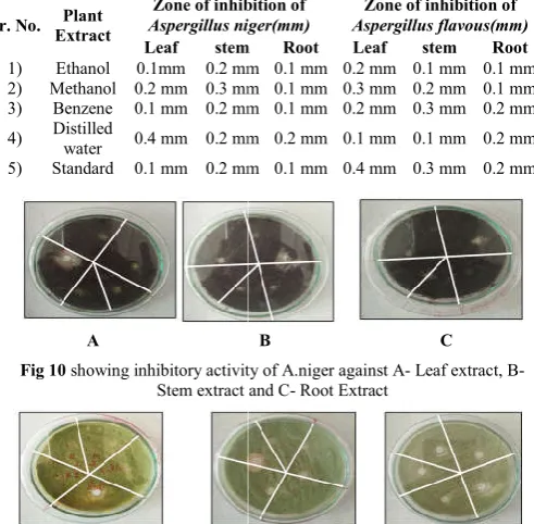
Moreover, the antifungal activity of leaf, stem and root was also checked in solvents in ethanol,
D/w extracts of Rungia repens.
observed in leaf water extract against the
mm, but in A. flavor maximum zonation was observed in leaf extract against standard antibiotics (itracunazole)
difference in the anti-microbial efficacy could be due to the presence of variable phytochemical compounds in different extracts (Hemant Kumardharma2013).
the botanical source of the drug parpataka. It is used in the treatment of fevers, fungal skin diseases and burning sensation (B.Jyoti 2010).This antimicrobial activity suggest that the plant has great antifungal activity, result noted in Table No.5.
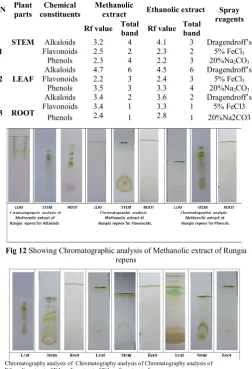
Chromatography analysis
TLC fingerprinting of methanolic and ethanolic extracts of Rungia repens was done for alkaloids, flavonoids and phenol, highest Rf value for alkaloid were recorded in leaf methanolic
0 1 2 3 4 Alkaloids Flavonoids leaf
Table No 5 Antifungal activity of A.niger and A. flavous in mm
Sr. No. Plant
Extract
Zone of inhibition of
Aspergillus niger(mm)
Leaf stem
1) Ethanol 0.1mm 0.2 mm 2) Methanol 0.2 mm 0.3 mm 3) Benzene 0.1 mm 0.2 mm
4) Distilled
water 0.4 mm 0.2 mm 5) Standard 0.1 mm 0.2 mm
A
Fig 10 showing inhibitory activity of A.niger against A
Stem extract and C
C
Fig 11 showing inhibitory activity of A. flavus against A
Stem extract and C
showing quantification of phytoconstituent in Rungia repens
A.niger and A.flavusagainst the extract
Moreover, the antifungal activity of leaf, stem and root was also checked in solvents in ethanol, methanol, benzene and Rungia repens. Maximum inhibition zone were observed in leaf water extract against the Aspergillus niger0.4 maximum zonation was observed in leaf extract against standard antibiotics (itracunazole) 0.4mm.The microbial efficacy could be due to the presence of variable phytochemical compounds in different (Hemant Kumardharma2013). Rungia repens is one of the botanical source of the drug parpataka. It is used in the tment of fevers, fungal skin diseases and burning sensation
This antimicrobial activity suggest that the plant has great antifungal activity, result noted in Table No.5.
TLC fingerprinting of methanolic and ethanolic extracts of was done for alkaloids, flavonoids and phenol, highest Rf value for alkaloid were recorded in leaf methanolic
Flavonoids Saponins
stem root
Antifungal activity of Rungia repens against A. flavous in mm
Zone of inhibition of
Aspergillus niger(mm)
Zone of inhibition of
Aspergillus flavous(mm)
stem Root Leaf stem Root
0.2 mm 0.1 mm 0.2 mm 0.1 mm 0.1 mm 0.3 mm 0.1 mm 0.3 mm 0.2 mm 0.1 mm 0.2 mm 0.1 mm 0.2 mm 0.3 mm 0.2 mm
0.2 mm 0.2 mm 0.1 mm 0.1 mm 0.2 mm
0.2 mm 0.1 mm 0.4 mm 0.3 mm 0.2 mm
B C
showing inhibitory activity of A.niger against A- Leaf extract, B- Stem extract and C- Root Extract
D E
International Journal of Recent Scientific Research Vol. 10, Issue, 09(H), pp. 35050-35055, September, 2019
35055 |
P a g e
and ethanolic extract that is 4.7 and 4.5 with 6 bands. Next to the alkaloid, flavonoid reading noted highin root methanolic and ethanolic extract with Rf value 3.4 &3.3 with single band and phenols in leaf methanolic and ethanolic extract with 3& 4 bandslikewise work also done by Pulate P.V., Wagay Nasir Aziz et.al-2015, observation noted in Table no. 6.
Acknowledgement
Authors are sincerely thankful to the Head department of botany Dr. P.G. Bansod for providing laboratory facilities and all needful help.
References
Principe, P. (2005) Monetizing The Pharmacological Benefits Of Plants. US Environmental Protection Agency, Washington DC, 1991.
Karuna Modi, Mamta Shah*Pharmacogn J. 2017; 9(2): 12repensa Complete Pharmacognostical Profile Of Rungia Repens. Vedhavathy, S. (1992). Studies On Medicinal Plants Of Tirumala
And Tirupati, Chittoor District Of Andhra Pradesh.Ph.D. Thesis, S.V. University, Tirupati.
Yoganarasimhan, S.N. (2000). Medicinal Plants Of India, Tamil Nadu, Vol.2, Cyber Media Printers, Bangalore, 472.
Trease GE, Evans WC., Pharmacognosy, 15th Edn. W.B. Saunders, Elsevier Science Limited 2002, P. 471.
J. S. Gamble, Flora Of The Presidency Of Madras, Adlard, London, UK, 1921.
Mahato SB, Sen S (1997) Advances In Triterpenoid Research, 1990-1994. Phytochemistry 44: 1185-1236.
Kokate, C.K. Purohit,A.P.And Gokhale, S.B. 2005. Pharmacognosy, Nirali Publication Pune.
Harborne, J.B. 1998. Phytochemical Method. 3rd Edition., Chapman And Hall Publication, London.
Sadashivam And Manickam, A. 2005. Biomedical Methods 2ndEdn. New Age International (P) Ltd., Publisher, New Delhi. Wallis, T.E. !990. Text Book Of Pharmacognosy. 5thEdn,CBS
Publishers And Distributors.
Prashant Tiwari, Bimlesh Kumar, Mandeep Kumar, Gurpreet Kaur, Harleen Kaur (2011) Phytochemical Screening And Extraction: A Review.
S. R. Swain*, B. N. Sinha1and P. N. Murthy 2 Pharmacologyonline 3: 58-64 (2008)Toxicological Studies Of The Hydroalcoholic Extract Of Rungia Repens Leaves.
B. Jyothi, G. Sudarsanam, Bulusu Sitaram And G. Prasada Babuaccepted : February, 2010,Pharmacognosy Of A South Indian Market Sample Of Parpatakarungia Repens(L.) Nees. Hemant Kumar Sharma1, Veerachamy Alagarsamy*2 Hemant
Kumar Sharma And Veerachamy Alagarsamy., (2013) Int. J. Res. Phytochem. Pharmacol., 3(2), 109-202In-Vitro Anti-Microbial Activity Of Various Extracts Of Rungiarepens (L) Nees.
Pharmacopoeias-- India: Indian Pharmacopoeias. India. Indian Pharmocopoeia Committee, Published By Delhi: Manager Of Publication 1966 2ndEdition.
Modi Karuna &Shah Mamta. Phytochemical Investigation And Pharmacognostic Standardization Of Polycarpaea Corymbosa Lam. Pharmacaogn J, Vol.9, Issue 6, Nov.-Dec.2017.
Bharat Gami, Farzin Parabia. Screening Of Methanol & Acetone Extract For Antimicrobial Activity Of Some Medicinal Plants Species Of Indian Folklore. Int. J. Res. Pharm. Sci., 2(1), 2011, 69-75.
Dep K, Kaur A, Ambwani S, Ambwani Tk. Preliminary Phytochemical Analyses Of Hydromethanolic Leaf Extract Of Melia Azedarach L. Journal Of Medicinal Plant Studies.2018; 6(3):4-8.
M Madhu, V Sailaja, Tnvss Satyadev, Mv Satyanarayana. Quantitative Phytochemical Analysis Of Selected Medicinal Plant Species By Using Various Organic Solvents. Journal Of Pharmacognosy And Phytochemistry 2016; 5(2): 25-29. L.P. Khalid, P.V. Pulate and N.A.Wagay. Reliminary
Phytochemical Analysis Of Miliusa Tomentosa(Roxb.) J. Sinclair By Using Various Organic Solvents. Ejbps, 2017, Volume 4, Issue 05, 276-281.
Pulate P.V., Wagay Nasir Aziz And Deshmukh V.R. Phtochemical, Ethanomedicinal And Anatomical Study Of Canthium parviflorum. World Journal Of Pharmacy And Pharmaceutical Sciences 2015, Volume 4, Issue 11, 1464-1482.
Bauer Aw, Kirby Wmm, Sherris Jc, Turck M. Antibiotic Susceptibility Testing By Standardized Single Disc Method. Am J Clin Pathol. 1966;36:493–6.
Table No 6 Chromatographic analysis of Rungia repens
SN Plant
parts
Chemical constituents
Methanolic
extract Ethanolic extract Spray
reagents
1 STEM
Rf value Total
band Rf value
Total band
Alkaloids 3.2 4 4.1 3 Dragendroff’s Flavonoids 2.5 2 2.3 2 5% FeCl3
Phenols 2.3 4 2.2 3 20%Na2CO3
2 LEAF
Alkaloids 4.7 6 4.5 6 Dragendroff’s Flavonoids 2.2 3 2.4 3 5% FeCl3
Phenols 3.5 3 3.3 4 20%Na2CO3
3 ROOT
Alkaloids 3.4 2 3.6 2 Dragendroff’s Flavonoids 3.4 1 3.3 1 5% FeCl3
Phenols 2.4 1 2.8 1 20%Na2CO3
Fig 12 Showing Chromatographic analysis of Methanolic extract of Rungia
repens
Chromatography analysis of Chromatography analysis of Chromatography analysis of Ethanolic extract of Ethanolic extract of Ethanolic extract of
Rungia repens for AlkaloidRungia repens for AlkaloidRungia repens for Alkaloid
Fig 13 Showing Chromatographic analysis of Ethanolic extract of Rungia repens.
How to cite this article: