quantification of lymphatic vessel function
Anna K. Polomska, … , Jean-Christophe Leroux, Michael
Detmar
JCI Insight.
2019;
4(4)
:e126515.
https://doi.org/10.1172/jci.insight.126515
.
Graphical abstract
Technical Advance
Dermatology
Vascular biology
Find the latest version:
Conflict of interest: The authors have declared that no conflict of interest exists.
License: Copyright 2019, American Society for Clinical Investigation. Submitted: November 29, 2018 Accepted: January 14, 2019 Published: February 21, 2019 Reference information:
JCI Insight. 2019;4(4): e126515. https://doi.org/10.1172/jci. insight.126515.
Engineering, Zürich, Switzerland.
Introduction
Lymphatic vasculature plays a vital role in maintaining tissue fluid homeostasis, immune surveillance, and the absorption of dietary fat in the intestine (1, 2). Alterations of lymphatic function have been associated with a number of diseases, including lymphedema (3), lipedema (4), venous insufficiencies (5), impaired wound healing (6), and chronic inflammatory diseases (7–9). Thus, reliable and reproducible techniques for the quan-titative assessment of lymphatic vessel functionality are essential for diagnostic and monitoring purposes.
Advanced imaging techniques, such as computer tomography and magnetic resonance, have been used to qualitatively identify structural and functional changes of the lymphatic vasculature. However, these methods are not routinely applied in the clinics due to their complexity and associated high costs of instru-mentation (10). The current “gold-standard” lymphoscintigraphy techniques are based on injection (subcu-taneous, intradermal, or intramuscular) of radiolabeled colloidal particles or proteins that are specifically taken up by the lymphatic vasculature and allow for visualization of structural and functional abnormali-ties of lymphatic vessels (11–14). A method based on the injection of a radioactive tracer that is followed by monitoring of the decrease of the signal at the injection site over time (which represents the clearance of the tracer by the lymphatic vessels) can quantitatively reflect lymphatic vessel functionality (11, 12). However, the high costs of production and disposal, the environmental risks of radionuclides, the lack of standardization of the utilized colloids (99mTc-albumin used in Europe and 99mTc-sulphur in the US), the need for correcting the results due to radioactive decay, and the use of stationary and expensive scintigra-phy cameras drastically hamper the widespread applicability of radioactivity-based methods.
Replacement of radioactive tracers with lymphatic-specific fluorescent dyes for lymphatic imaging and quantification eliminates the disadvantages of radioactivity and reduces the costs. In particular, tracers operating
in the near-infrared (NIR) range, where optical absorption and scattering in biological tissues are low, allow for the acquisition of images with high-spatial resolution and with negligible background signals (15). In analogy to the quantitative scintigraphy-based methods, we and others have previously developed techniques for the quantitative measurement of lymphatic vessel drainage function that are based on monitoring the decay of the fluorescence signal after tissue injection of lymphatic-specific NIR fluorescent tracers in animal models (16, 17). However, these approaches rely on custom designed lymphatic-specific dyes or fluorescently labeled albumin, which are not approved for clinical use. Aqueous solutions of the only currently clinically approved NIR tracer, indocyanine green (ICG), are not suitable for this application due to aggregation and change of spectral proper-ties of the dye after injection upon the monomerization caused by complexation with endogenous proteins (18, 19). This limitation might potentially be addressed by precomplexing ICG with nontoxic polymers, surfactants, or proteins that may prevent such interactions in vivo (20–23).
Another issue facing the use of NIR methods for lymphatic vessel function assessment, based on tem-poral measurements of the fluorescence signal of the locally injected probe, lies in the lack of suitable, affordable, and simple instrumentation for NIR fluorescence quantification for clinical use. Portable cam-eras (e.g., Hamamatsu PDE) would allow for quantitative imaging of ICG fluorescence in clinical settings by acquiring a series of images over time and quantifying the fluorescence signal based on image analysis (24, 25). However, the high cost of such instrumentation (ranging from several thousand up to tens of thousands of US dollars) limits their routine use outside specialized medical centers. Moreover, this type of measurement would be impractical due to the requirement for postmeasurement data processing (26–28). Therefore, the development of an affordable, compact, and portable fluorescence detector, without imaging capability but able to quantify the fluorescence signal intensity, would allow performance of the measure-ments in a routine manner outside of specialized hospital units (potentially even at a patient’s home) (29).
A major challenge concerns the reproducible delivery of lymphatic-specific tracers to defined layers of the skin (12, 30). Due to the presence of a dense lymphatic plexus in the dermal layer of human skin, deliv-ery of a NIR tracer directly to this layer would be most advantageous for the quantification of lymphatic vessel function (5, 12). However, intradermal injections, performed with conventional hypodermic needles, require special training of health professionals and are difficult to standardize (31). Potentially, this issue could be addressed by using microneedle-based injection systems that deliver molecules to a predetermined depth in the skin (31–33). Indeed, in our recent work, we developed 400- to 500-μm-long dissolving solid polymeric microneedles made of a mixture of poly(N-vinylpyrrolidone) (PVP) and ICG. After skin pen-etration, PVP dissolved and released ICG, allowing for quantitative assessment of lymphatic function in mice (20). Even though these studies identified an approach for quantitative imaging of lymphatic function, it was hampered by several limitations, such as challenging sterile manufacturing and upscale of produc-tion and the relatively the relatively low amount of tracer that could be delivered to the dermis (20).
In the present study, we pursued a three-pronged approach to develop an ICG-based tracer that can be administered intradermally via a microneedle-based injection device with hollow microneedles and a cus-tom-designed portable NIR detection device for the quantitative assessment of lymphatic vessel function based on the clearance of the NIR tracer from the skin. This quantitative imaging method was validated in a genetic mouse model with impaired lymphatic vessel function in the skin and in pig studies with distinct lymphatic drainage. The minimal invasiveness of this method, together with the use of clinically approved tracer/excipient components and the compact quantitative signal detection device, provide great potential for the rapid clinical translation for the sensitive and early detection of impaired lymphatic vessel function.
Results
Physicochemical characterization of ICG-Kolliphor HS15 formulation. In aqueous solution, ICG forms
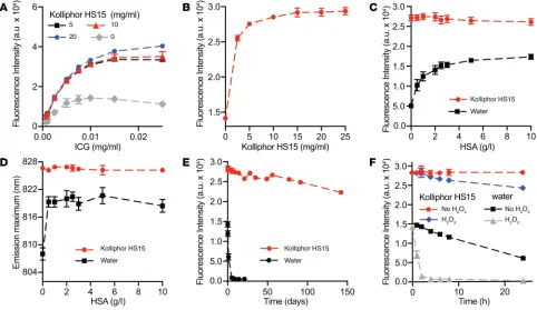
excipients, Kolliphor HS15 (nonionic FDA- and EMA-approved surfactant, polyoxyl 15 hydroxystearate) and polysorbate 80 resulted in the highest increase in ICG fluorescence intensity (approximately 4-fold) and the largest bathochromic shift of the emission maximum (20 nm) (Supplemental Figure 1), in line with previously published data (37, 38). Since Kolliphor HS15 has been shown in preclinical studies to trigger an over 100-fold lower histamine release after i.v. injection compared with polysorbate 80 (39) and has not been found to cause anaphylactic reactions after parenteral application in humans (40), it was selected for further studies. When measured as a function of Kolliphor HS 15 concentration, the fluorescence intensity increase of ICG plateaued at 10 mg/ml (Figure 1, A and B), suggesting the full monomerization of the dye.
As previously mentioned, it is desired that ICG keeps identical spectral properties regardless of the presence of serum proteins (35). Because albumin is the most abundant serum protein in the body, we investigated the in vitro spectral properties of ICG in aqueous solutions and in Kolliphor HS15 in the presence of human serum albumin (HSA). Solutions of ICG-water or ICG in Kolliphor HS15 were mixed with solutions containing HSA at increasing concentrations. As seen in Figure 1, C and D, fluorescence intensity and emission maximum of ICG in Kolliphor HS15 formulation (ICG-Kolliphor HS15) were not affected by the HSA concentration. By contrast, the fluorescence intensity of ICG-water increased with increasing HSA concentrations in the solutions, and the emission maximum shifted from 808 nm to 820 nm, indicating dye monomerization. Together, these data show that Kolliphor HS15 is a good solubilizer for ICG that keeps the tracer in the monomeric state in solution.
Increased stability of ICG in Kolliphor HS15. ICG is known to rapidly degrade in aqueous solutions,
result-ing in a loss of fluorescence intensity (41, 42). Thus, we compared the stability of ICG in a pure aqueous solution to that of ICG in the presence of 10 mg/ml Kolliphor HS15 by measuring the fluorescence intensity
of the respective dye solution over time when stored at room temperature and protected from light. The flu-orescence intensity of ICG in pure water was completely lost after 5 days, whereas for ICG-Kolliphor HS15, over 90% of the initial fluorescence intensity was retained after 60 days and 77% after 140 days (Figure 1E).
Qualitative LC-MS analysis of ICG-water incubated for 24 hours revealed the presence of a main ICG peak that eluted at 13.6 minutes (m/z = 753.42) and of a main degradation product at 12.09 minutes (m/z = 752.42) that could be assigned to an “irreversible” covalent dimer, as previously reported (42–44), as well as further smaller degradation products. In contrast, in ICG-Kolliphor HS15 solutions, the peak emerging from the plausible dimer was barely visible, even beyond 100 days after preparation (Supplemental Figures 2–4).
The increased stability of the ICG-Kolliphor HS15 may also have been due to a protective role of the encapsulation of the dye in the hydrophobic core of the surfactant micelles where the water-soluble oxidiz-ing species have limited access, as previously suggested (37, 45, 46). To further investigate this phenome-non, we treated the ICG in water (ICG-water) and ICG-Kolliphor HS15 system with an oxidizing solution containing 5 mM H2O2. After 2 hours of incubation at room temperature, the fluorescence intensity of the ICG-water solution was completely lost, while the signal intensity of ICG-Kolliphor HS15 solutions was only reduced by approximately 10% after 24 hours (Figure 1F). We also investigated the stability of ICG in solutions containing PVP, as this polymer had been previously used in one of our former studies to deliver ICG with dissolving microneedles (20). In contrast to the previous study, we selected lower PVP concentrations that would result in lower solution viscosity, thus assuring injectability of the solution. As shown in Supplemental Figure 5, ICG was rapidly degraded in low-viscosity PVP solution, presumably due to presence of peroxide impurities in the PVP (47). Preincubation of PVP solutions with an antioxidant (i.e., sodium ascorbate) to inactivate the reactive oxygen peroxides (47, 48) improved the stability of ICG (Supplemental Figure 5). These results confirm the important role of oxidizing species in the degradation of ICG and a protective role of Kolliphor HS15 against them.
Efficient lymphatic clearance of injected ICG-Kolliphor HS15 formulations in mouse ears. We and others have
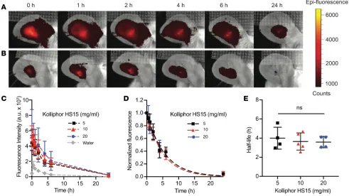
previously shown that the clearance of an intradermally delivered lymphatic-specific tracer from an injection site correlates with the lymphatic drainage efficiency and can thus be used to quantify its function (16, 17, 20). ICG-water and ICG-Kolliphor HS15 (5, 10, and 20 mg/ml) were injected intradermally (3 μl each) into the ears of WT FVB mice, and the signal at the injection site was monitored over time with an IVIS in vivo imaging system. In ears injected with ICG-water, the signal was low and exhibited an initial increase phase up to 1 hour. However, for all ICG solutions containing Kolliphor HS15, a strong initial signal (10- to 14-fold higher than ICG-water) was recorded, followed by an exponential decay (Figure 2, A–C). After normalization to the initial value directly after injection, the signals from the ears injected with ICG-Kolliphor HS15 could be fit to curves of monoexponential decay with average R2 values above 0.9 (Figure 2D). Half-lives calculated from the decay curves of ICG in Kolliphor HS15 at 5, 10, and 20 mg/ml were 4.0 ± 1.1, 3.6 ± 0.9, and 3.6 ± 0.6 hours, respectively, and were not significantly different from each other (Figure 2E).
We next investigated the perfusion of leg lymphatic vessels after intradermal administration of ICG (0.0075 mg/ml) in pure water and in the presence of Kolliphor HS15 (10 mg/ml). A bolus of 5 μl was intradermally injected in the dorsal aspect of the foot of the mice, and a stereomicroscope was used for visualization of the dye within draining lymphatic vessels of the lower hindlimb. For both solutions, the dye could be seen within the collecting lymphatic vessels. Applying external compression at the injec-tion site after injecinjec-tion of ICG-Kolliphor HS15 soluinjec-tion stimulated further uptake of the dye only into the lymphatic vessels, in contrast to ICG-water, which could also be visualized in the draining popliteal vein (Supplemental Figure 6, A–C).
Design and in vitro validation of a portable NIR fluorescence detection device. Portable NIR cameras are often
used for clinical imaging purposes and could be suitable for signal quantification. However, the measure-ments would be limited to specialized medical centers due to the high prices of such instrumentation (24, 50). Therefore, we next built a prototype system for the simple and inexpensive quantitative fluorescence detection of injected NIR tracers. This device is portable and therefore would be particularly suited for the clinical monitoring of lymphatic clearance of NIR probes outside of the hospital (e.g., at the local general practitioner or at a patient’s home) (Figure 4A, Supplemental Figure 7, and Supplemental Text).
A linear relationship between dye concentration (in 10 mg/ml Kolliphor HS15) and fluorescence inten-sity with the device was confirmed in vitro (Figure 4B). The intraday variability and repeatability of the mea-surements was assessed by measuring the ICG solutions 5 times. For interday repeatability, the same solu-tions were measured over 3 days. The results demonstrated a very good intraday and interday repeatability of the signal for all measured concentrations, as reflected by low values of coefficient of variations (%COV below 6% for intraday variability and below 15% for interday variability) (Supplemental Tables 1 and 2).
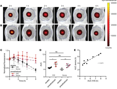
Portable device measurements in awake versus anesthetized mice. Since it is not possible to perform
measure-ments with the portable device on the small surface area of mouse ears, we initially aimed to validate the device in the back skin of mice. A bolus of 5 μl of the ICG-Kolliphor HS15 was injected intradermally, and the signal at the injection site was measured over time with both the IVIS imaging system and our device prototype. The diameter of the region of interest (ROI) analyzed by IVIS was set to 3 cm (Figure 5, A and B, blue circle) similar to the diameter of the region analyzed by the device (Figure 5, A and B). Since the lymphatic transport is reduced in anesthetized mice (51), as compared with awake mice, we first tested whether we could detect a difference in lymphatic clearance between those two conditions. Therefore, one group of mice was kept continuously anesthetized with isoflurane between the imaging sessions, and the second group was awake and anesthetized shortly only for the measurements. In contrast to our findings
Figure 2. Clearance kinetics after bolus intradermal administration of indocyanine green in water or in a solution of Kolliphor HS15 into mouse ears.
after tracer injection into mouse ears, where the signal exhibited immediate decay, the average fluorescence signal intensity in the back skin of awake mice, measured by IVIS, slightly increased in the first hour (approximately 1.1-fold). This is likely due to diffusion of the dye in the tissue and/or distribution to initial lymphatics prior to the lymphatic clearance phase (signal decrease) (5, 11–13, 16, 52). In anesthetized mice, the signal intensity slowly increased around 1.3-fold for up to 3 hours, followed by a slow decrease in signal over time. With the portable device, the decrease of the signal was observed already at 1 hour in the awake group and at 2 hours in the anesthetized group (Figure 5C).
Since fitting using a monoexponential decay function and half-life calculation were not possible, we used an AUC of the normalized clearance curves to assess the total tissue clearance of the tracer from the injection site. The AUCs calculated for curves obtained with the device were slightly lower than those obtained with the IVIS, but the values obtained with either method were not statistically different from one another. Importantly, for both measurement methods, the differences in average AUCs for awake and anes-thetized groups were highly significant (IVIS anesanes-thetized, 6.9 ± 1.1 hours; IVIS awake, 4.9 ± 0.3 hours; device anesthetized, 6.1 ± 0.8 hours; device awake, 4.0 ± 0.5 hours; P < 0.05 between IVIS anesthetized and IVIS awake; P < 0.01 between device anesthetized and device awake) (Figure 5D). Moreover, a signif-icant correlation between the AUCs calculated from the IVIS measurements and from the portable device measurements was found (Pearson’s correlation coefficient r = 0.9475, P < 0.001) (Figure 5E).
In conclusion, the portable device was able to distinguish differences in tissue clearance in vivo with close agreement to standard imaging methods.
Validation of portable device measurements in pigs. Pig skin exhibits structural and functional resemblance
to human skin, with the thickness of the pig and human epidermis varying from 30 to 140 μm and 50 to 120
μm, respectively, depending on the anatomical location and age (53–55). We used three 1-month-old female domestic pigs (10–14 kg), which are more suited for handling (i.e., easy for manual restraint under awake conditions). Injections were performed on the ventral aspect of the torso of the animals due to the large area accessible for injection as well as the thinner (56) and visually less keratinized skin compared with the rest of the body, enabling facile penetration of microneedles through the skin. The ICG-Kolliphor HS15 formulation was administered using MicronJet600 microneedles (3 pyramid-shaped microneedles, each 600 μm long), enabling standardized and uniform delivery of the tracer formulation through the epithelium directly to the dermal skin layer (57).
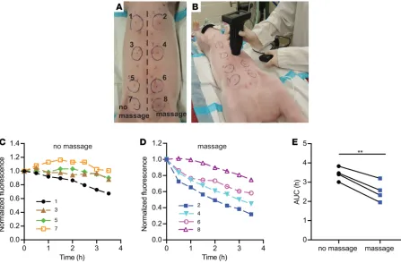
In pig 1, we investigated the effect of manual compression of the injection site on the tissue clearance from the skin under anesthesia. Similar to muscular activity, manual compression promotes the entrance of interstitial fluid into the initial lymphatic capillaries and thus increases lymphatic clearance (51, 58). Four symmetrical intradermal injections (Figure 6A) of ICG-Kolliphor HS15 (50 μl) were performed on the left and right part of the ventral aspect of the torso, and the injection sites on the right site were manually massaged with a circular motion using fingers throughout the whole duration of the experiment (except for
during the measurements). The fluorescence signal at the injection site was measured every 30 minutes with the portable device (Figure 6B). Eight to eleven measurements at the same injection site were performed by lifting the device between each measurement. Supplemental Table 3 shows the average fluorescence values obtained from all measurements, together with the exact number of readings per injection site (n), SD, and relative SD (RSD%) obtained in pig 1. There was slight variability between the measurement; however, the %RSD never exceeded 15 %. The variability arose from the difficulty in selecting the same location for each reading. Therefore, only the 3 highest values were used for further data analysis, which leads to an assump-tion that the same locaassump-tion was always selected at every time point.
Normalized clearance curves obtained for each injection site are shown in Figure 6, C and D. For 3 of 4 nonmassaged injection sites, a very slow signal decrease was observed, whereas for most of the massaged injection sites, the fluorescence intensity already decreased after 1 hour. We calculated AUCs to estimate the overall tissue clearance from the injection sites (Figure 6E). On average, applying the compression on the contralateral sites decreased the AUC from 1.2- to 1.5-fold. A paired analysis of the AUCs from the nonmassaged and massaged sites produced a highly significant statistical difference between the two groups (average AUC from nonmassaged sites, 3.4 ± 0.4 hours; average AUCs from massaged sites, 2.5 ± 0.5 hours; P < 0.01).
Validation of the portable device for tissue clearance measurements in awake pigs. In pig 1 (Figure 6), we observed
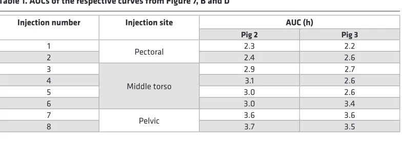
a trend of rapid tissue clearance in the pectoral region that was reduced in the pelvic area that may emerge from regional tissue properties and lymphatic vessel density differences. Thus, we next investigated the tissue clearance in two awake pigs (pigs 2 and 3) at different locations of the ventral torso (Figure 7, A and C). In pig 2, the fastest tissue clearance occurred in the pectoral region, as evidenced by the lowest AUCs (2.3 and 2.4 hours). For the injections in the middle part of the torso, the calculated AUCs increased to around 3.0 hours. The highest AUC values were found in the pelvic area of the torso (3.6 and 3.7 hours) (Figure 7B). A similar trend was found in pig 3; however, a slower tissue clearance was found in the middle part of the torso than in pig 2 (Table 1 and Figure 7D). The pooled AUCs from all 3 areas (pectoral, middle torso, and pelvic) from both pigs are shown in Figure 7E. The statistical differences between AUCs at different injection sites were highly significant. In conclusion, the portable device in combination with the ICG formulation deliv-ered via microneedles reliably distinguished different lymphatic vessel clearance patterns in pig skin.
Discussion
In this study, we established a simple method for the quantitative assessment of lymphatic vessel func-tion in the skin, based on tracking of the disappearance of the fluorescence signal of a lymphatic-spe-cific NIR tracer system containing ICG using a custom-made portable detection device after injection of the tracer with hollow microneedles into the dermis. The successful validation of this method in
mice and young pigs supports its potential utility for simple and reliable quantifications of lymphatic clearance in clinical settings.
The first component of the method is an improved ICG formulation that includes a surfactant, Kol-liphor HS15, encapsulating dye molecules in the hydrophobic micellar core. This overcomes major limita-tions associated with the use of purely aqueous solulimita-tions of this clinically approved tracer for lymphatic clearance assays. In aqueous solutions, ICG aggregates, which leads to strong fluorescence self-quenching, resulting in a nonlinear concentration-fluorescence intensity dependence as well as blue shifts in absorption and emission maxima compared with the monomeric form (41). After injection of purely aqueous ICG solutions, the dye monomerizes upon interaction with endogenous proteins (i.e., albumin and lipopro-teins), which results in an increase in fluorescence intensity and red shifts in the emission and absorption maxima (20), both of which complicate quantitative analyses over time. To address this issue, ICG should ideally be administered in a monomeric form. With the ICG-Kolliphor HS15 combination, the fluores-cence intensity and wavelength of maximum emission (826 nm) remained unchanged upon adding HSA, in contrast to purely aqueous ICG solutions. These results suggest that, after intradermal injection of ICG in Kolliphor HS15, the majority of the tracer would remain bound to the surfactant and remain in the micellar core in a monomeric state.
These findings were validated in vivo, where the injection of ICG in pure water into mouse ears result-ed in a very low initial fluorescence signal that was followresult-ed by an almost 10-fold increase after 1 hour, likely due to interactions with endogenous proteins. By contrast, the initial signal after administration of ICG-Kolliphor HS15 in a concentration range of 5–20 mg/ml was more than 20-fold higher compared with ICG-water. This is in agreement with a recent publication where a stronger signal intensity of ICG-Kol-liphor HS15 in retinal vasculature in vivo was found compared with ICG in pure water (38). Encapsulation of ICG in Kolliphor HS15 micelles also significantly improved the chemical stability of the dye compared with purely aqueous solutions, in agreement with a previous study (37).
The second important part of this work is the development of an inexpensive, compact, and portable system for NIR fluorescence detection in the skin, enabling fluorescence signal intensity measurements without the need for imaging capabilities. Due to its small size, external power source, and incorporated display, the device could be easily operated at any location, e.g., in different animal facilities and in different animal species (i.e., mice and pigs) as well as in humans.
The third component of the clearance assay is the use of a microneedle-based injection device, Micron-Jet600, that enables the painless, uniform delivery of a tracer to a controlled depth in the skin (to around 600 μm) (57). The importance of a homogenous injection depth for the reproducibility of clearance assays has been highlighted in previous studies using radioactive tracers (5, 12, 30).
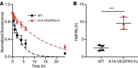
To apply a correct mathematical model for analyzing the lymphatic clearance curves, one should keep in mind that various clearance patterns can be measured in the skin in different body parts in vivo. For example, the clearance patterns of ICG in Kolliphor HS15 in the back skin of the mouse and in the pig torso differed from those obtained in mouse ears. In mouse ears, the signal always followed a monoexpo-nential-like decay over time. Therefore, the ICG removal from the injection site could be easily quantified by computing the signal half-life from normalized experimental data. The dermal elimination half-lives in
transgenic mice lacking a dermal lymphatic vasculature (K14-VEGFR3-Fc–transgenic mice) were on aver-age 4-fold higher than those in WT mice, which agrees well with the previous data (17).
In contrast, in the back skin of mice, the fluorescence signals typically started to decrease only 1 or 3 hours after injection (in awake and anesthetized mice, respectively), and this was preceded by a 10% intensity increase. Similarly, in some locations of the pig torso, the fluorescence signal decreased only after around 1 hour after injection. Such a lag phase with no or very little clearance can most likely be attributed to the tracer diffusion into the tissue, prior to entering the initial lymphatic capillaries. This phenomenon has indeed been described in several studies with both radioactive and fluorescent probes (5, 11–13, 16). Modi et al. (59) reported an initial increase of the counts of subcutaneously injected radioactive IgG prior to the decrease phase, suggesting transport of the probe to more superficial layers of the skin, which could also be the case in our experiments. The absence of the signal increase phase in ear skin is likely related to the lower tissue compliance compared with mouse back skin and to some locations in the pig torso. A low tissue compliance could create a more favorable interstitial pressure gradient after injection, thus promoting fluid (and tracer) uptake by the lymphatic vessels (60, 61). The lack of an immediate signal decrease can obviously complicate the calculation of the dermal elimination half-life of ICG. Previously, this problem has been addressed by fitting the exponential function starting from a later time point when the signal starts to decay (5, 11–13, 16, 52). However, the time point when the signal begins to decrease may vary. It could be different in healthy and in lymphedematous extremities of patients or it might change with disease progression. A longer diffusion phase could also be expected in lymphedema. To address these issues, we indirectly assessed lymphatic tracer clearance by calculating the AUC area under the normalized clearance curve, taking into account the tracer diffusion phase. Our current efforts are directed toward replacing a
simple AUC analysis with a more sophisticated mathematical model that would describe both diffusion in the skin and lymphatic clearance.
The AUC quantification method was successfully applied to analyze the results of the clearance assays in pigs. Since manual therapy is often applied as a treatment in lymphedema patients to stimulate lymphat-ic clearance, we investigated the effect of manual compression on lymphatlymphat-ic clearance of the NIR tracer from the skin in an anesthetized pig. Our findings that the clearance from the massaged injection sites was improved, as reflected by a 1.3-fold lower AUC, are in agreement with previous studies by Mortimer et al. where the half-life of clearance of 99mTc-colloid was almost 2-fold enhanced in massaged sites in pigs (12).
The fact that we observed faster lymphatic tracer clearance from the skin of awake pigs in the pectoral torso area than in the middle torso or pelvic area is likely due to the breathing motions in the pectoral area that stimulate uptake of the tracer by initial lymphatics and to differences of the skin structure, tissue compliance, and density of the lymphatic vasculature (60, 61). Our findings are in agreement with previous studies of lymph flow variations between different body parts in animals and humans (12, 59, 62) and indi-cate the importance of selecting comparable locations for tracer injections if clearance data are compared over time (e.g., during disease progression) or between healthy and lymphedematous extremities.
Our method for assessing lymphatic function presents a number of advantages over existing tech-niques. It is minimally invasive as the solution is intradermally injected with microneedles, a procedure that is simple and painless. The components of the formulation (i.e., Kolliphor HS15 and ICG) are approved for parenteral administration and the portable device can be produced at a relatively low cost and does not require any specialized facilities, potentially allowing a rapid progression in the clinic. This device must, however, be placed at the same area of the skin for each single measurement, poten-tially creating variability due to small shifts of the measured area. Therefore, our current efforts are directed toward the development of a miniaturized, wearable detection device that can be attached to the skin. This would enable the continuous monitoring of NIR fluorescence signals over several hours, shortening the measurement time period and allowing for more complex analyses that might better reflect the ratio between diffusion and clearance (e.g., by determining the time needed to reach the max-imum fluorescence signal intensity). These improvements could facilitate a more precise quantification of lymphatic function under normal and pathological conditions.
Methods
Chemicals. VERDYE, a clinical-grade ICG (Diagnostic Green GmbH) was used in this study. ICG aliquots
were prepared by dissolving the dye in anhydrous methanol, preparing small portions, and evaporating the sol-vent under a stream of nitrogen. Kolliphor HS15 and PVP (Kollidon 12 PF), injectable grades, were purchased from BASF. Sodium ascorbate (USP grade) was from AppliChem. γ-Cyclodextrin was purchased from Wacker, sodium deoxycholate was purchased from ABCR, and TPGS was purchased from BASF. HSA (≥90%, recom-binant) was obtained from MilliporeSigma, and H2O2 (35%, stabilized, for analysis) was purchased from Acros Organics. Ultrapure water from Nanopure Diamond Barnstead system (Thermo Scientific) was used.
Animals. Mice were kept under specific pathogen–free conditions until imaging. FVB mice were bred
Helsinki, Finland (17, 49). Three 4-week-old female domestic pigs (10–14 kg) were obtained from local Canadian farmers. Prior to starting any procedures, the animals were acclimatized for 1 week in the facility.
Spectroscopic measurements. ICG solutions were prepared by dissolving dye aliquots in water or in a
solution containing the respective solubilizer. Fresh solutions were always prepared and used the same day. Spectra were recorded on a plate reader (Tecan Infinite M200 Pro, Tecan GmbH). Fluorescence mea-surements were performed using Costar 96-well, black, flat-bottom plates (Corning Inc.). Briefly, 200 μl of the solutions was pipetted into the well, and the fluorescence intensity was measured using the following settings: λex/λem = 700/750–850 nm, gain: 125. Experiments were performed in triplicates.
Surfactant screening study. We investigated the influence of several FDA-approved injectable synthetic
and bioderived solubilizing excipients (Kolliphor HS15, polysorbate 80, sodium deoxycholate, Poloxam-er188, γ-cyclodextrin, d-α-tocopherol polyethylene glycol 1000 succinate [TPGS], and PVP) on the fluores-cence intensity and emission maximum of ICG. Briefly, 0.5 ml ICG dissolved in water at a concentration of 0.05 mg/ml was mixed with equal volumes of solutions of the respective excipients at 100 mg/ml, and the fluorescence intensity and emission maximum were recorded using a plate reader as described above.
Stability studies. In all stability experiments, the fluorescence intensity of the solutions was measured
over time using a plate reader as described above. The solutions were stored at room temperature and were protected from light. For H2O2-induced degradation studies, 0.75 ml of ICG solutions (0.015 mg/ml) in Ultrapure water or in Kolliphor HS15 (20 mg/ml) were diluted with equal volumes of Ultrapure water containing 10 mM H2O2. The fluorescence intensity of the resulting solutions was measured over time as described above. The solutions were stored in the dark at room temperature, and experiments were per-formed in triplicates.
Interactions with albumin. The fluorescence intensities of ICG-water and ICG-Kolliphor HS15 solutions
in the presence of HSA were measured using a plate reader. Briefly, 0.75 ml of solutions of ICG (0.015 mg/ ml) in Ultrapure water or in Kolliphor HS15 (20 mg/ml) were diluted with equal volumes of 2× PBS at pH 7.4, containing increasing concentrations of HSA. Experiments were performed in triplicates.
LC-MS analyses. All samples were analyzed using a LTQ/XL/ETD linear ion trap (Thermo Scientific)
mass spectrometer coupled to a Waters Acquity UPLC. Gradient elution was done on a Waters XBridge C18 column (3.0 × 150 mm, 3.5 μm). The mobile phase consisted of 10 mM aqueous ammonium formate plus 0.1% formic acid (eluent A) and 90% acetonitrile in ammonium formate (final 10 mM) plus 0.1% for-mic acid (eluent B). The flow rate was set to 0.3 ml/min, and the gradient was programmed as follows: 0–1 minutes 5% B, 1–20 minutes to 100% B, 20–22 minutes 100% B, 22–22.1 minutes to 5% B, 22.1–25 minutes 5% B. The column oven and the autosampler were set to 30°C and 10°C, respectively. The injection volume for all samples was 10 μl. Between each sample a blank injection (water) was programmed, accounting for possible carry over due to PVP and ICG. MS settings were used as reported by Wissenbach et al. (63). The MS was equipped with heated ESI II source was set to 200°C. All measurements were performed in the positive ionization mode.
The following samples were analyzed by LC-MS (in triplicates): fresh solutions of ICG (0.0075 mg/ml) in water and in 10 mg/ml Kolliphor HS15, the same samples stored for 24 hours, and aged solutions of ICG– Kolliphor HS15 (106 days). All solutions were stored at room temperature and were protected from light.
Lymphatic clearance assay in mouse ears. An IVIS Spectrum (Xenogen, Caliper Life Sciences) imaging
HS15 (10 mg/ml) were prepared. Briefly, 0.5 ml of the solution was placed in a well of a 6-well plate (TPP, MilliporeSigma) protected from light by black isolation tape, and the fluorescence intensity was measured with the device. The intraday repeatability of the measurements was assessed by measuring the ICG fluores-cence intensity 5 times in the same well. For interday repeatability, the same solutions were used over 3 days.
Lymphatic clearance assay in the back skin of anesthetized and awake mice. The IVIS Spectrum imaging
sys-tem and the custom portable device were used simultaneously for the in vivo clearance assay in the back skin of mice. Eight 19-week-old male FVB mice were used for the assay. Mice were anesthetized with 2% isoflurane, and fur was removed from the back skin in an area of approximately 3 cm in diameter using an electric shaver. Then, 5 μl of a freshly prepared solution containing ICG (0.0075 mg/ml) in Kolliphor HS15 solution (10 mg/ml) was injected intradermally using a 29-gauge insulin syringe. One group of mice was kept anesthetized throughout the experiment, whereas the other group was anesthetized only for the measurement and was allowed to move without any restriction between the measurements.
The fluorescence signal in the back skin was monitored at predetermined time points (0, 1, 2, 3, 4, and 6 hours) with the IVIS system using the following settings: ex/λem = 745/800 nm, exposure time: 1.5 seconds, small binning, field of view: 6.6 cm × 6.6 cm. The average fluorescence intensity in a circular ROI (3 cm in diameter) was analyzed using Living Image Software. Simultaneously, measurements were performed with the custom portable device. After tracer injection, the circular area (3 cm in diameter) around the injection site was marked. The device was moved slightly around the marked injection area until the region of highest intensity was detected. Six measurements were then recorded, and the average of the 3 highest values were used for the data analysis. After subtracting the background readout, the values obtained by each instrument were normalized to the time 0 measurement and plotted against time. The AUC (from 0–4 hours) was calculated for each mouse using a trapezoidal method.
Imaging of tracer uptake into lymphatic and blood vessels. For intravital imaging of lymphatic vessels, a
Zeiss StereoLumar.V12 stereomicroscope together with AxioVision software was used. The microscope was equipped with a cooled EMCCD camera (Evolve eXcelon, Photometrics) that has high sensitivity to NIR wavelengths. A high-powered diode system (CoolLED) with illumination at 470–770 nm served as light source. Seven 12- to 19-week-old male FVB mice were used for the imaging. Mice were anesthetized by i.p. injection of 80 mg/kg ketamine and 0.2 mg/kg medetomidine. Fur was removed from the leg with an electric shaver and topical depilation cream. Five μl ICG (0.0075 mg/ml) in Ultrapure water and 10 mg/ml Kolliphor HS15 were injected in the dorsal aspect of the foot using 29-gauge insulin syringes. The collecting lymphatic vessels were observed using a fluorescence stereomicroscope (λex/em = 770/832 nm, exposure 300 milliseconds, ×40 magnification).
Beginning at 5 minutes after injection, a sequence of 300 images was taken over 2 minutes. Manual compression with a cotton swab was applied onto the injection site for 30 seconds to stimulate uptake of the tracer into blood or lymphatic vessels.
Lymphatic clearance assays in pigs. One domestic pig (number 1, female, 14 kg) was anesthetized with
side of the medial line, starting from the pectoral region toward the pelvic part of the ventral torso. The injection sites on the right side were continuously massaged in a circular motion using fingers, starting from 30 minutes after the injection. The signal was measured immediately after each injection and then at 30-minute intervals throughout 4 hours. After the injection, the circular area (3 cm in diameter) around the injection site was marked. For each measurement, the device was moved slightly around the marked injection area in order to localize the region with the highest signal intensity. At every time point, 8–11 measurements at the same injection site were recorded by lifting the device between each measurement. From those data, the 3 highest values were selected for data analysis. Lymphatic clearance was also studied in 2 awake domestic pigs (number 2 and 3, female, 14 and 10 kg, respectively). Each animal was anesthe-tized with 2% isoflurane, and eight intradermal injections were performed as described above. After the first fluorescence measurement, the anesthesia was removed and each animal was allowed to move without any restrictions. Signal measurements were performed every hour for a total of 4 hours while the animal was manually restrained. Eight to eleven measurements were recorded, and the three highest values were used for the following data analysis.
Statistics. Data are shown as mean ± SD. Means of 2 groups were compared using the 2-tailed Student’s t test or 2-tailed paired Student’s t test. Comparisons of 3 groups were performed using 1-way ANOVA with
Tukey’s multiple comparison post hoc analysis. P < 0.01 was considered as significant. GraphPad Prism V7.0 (GraphPad Software) was used for performing the analyses.
Study approval. All mouse experiments were performed in accordance with an animal protocol
(ZH212/16) approved by the Cantonal Veterinary Office Zurich, Switzerland. Pig experiments were per-formed in the Centre National de Biologie Expérimentale, Institute National de la Recherche Scientifique (INRS), Laval, Canada. Procedures involving the care and the use of pigs in this study were reviewed and approved by the INRS Institutional Animal Care and Use Committee (protocol 1711-01) and performed according to the Canadian Council on Animal Care guidelines and policies. Throughout the experiment, the health and well-being of the pigs were closely monitored by a veterinarian.
Author contributions
AKP, MD, JCL, STP, and DB conceived and designed the study. AKP, STP, MD, and JCL wrote the manuscript. AKP and STP performed most experiments and data analyses. MM and SB designed the first prototype of the portable device, and SB built it. DF, MB, and NR designed the final version of the portable device, and DF built it. AKP, TV, and CS performed LC-MS experiments.
Acknowledgments
This work was supported by a Swiss National Science Foundation grant (310030_166490), by funding from the SKINTEGRITY flagship project of the University Medicine Zurich initiative (http://www.hochschul-medizin.uzh.ch/en/projekte/skintegrity.html, funding 2/71018/16), and by Innosuisse – Swiss Innova-tion Agency (29462.1 IP-LS). NanoPass Technologies Ltd. is acknowledged for providing MicronJet600 microneedles. Mario Filion and the animal caretakers from the Centre National de Biologie Expérimen-tale, INRS, are acknowledged for support in design and execution of the pig experiments.
Address correspondence to: Michael Detmar, Institute of Pharmaceutical Sciences, ETH Zurich, Vladimir-Prelog-Weg 3, HCI H303, CH-8093 Zurich, Switzerland. Phone: 4144.633.7361; Email: [email protected].
1. Cueni LN, Detmar M. The lymphatic system in health and disease. Lymphat Res Biol. 2008;6(3-4):109–122. 2. Alitalo K. The lymphatic vasculature in disease. Nat Med. 2011;17(11):1371–1380.
3. Sayegh HE, et al. Diagnostic methods, risk factors, prevention, and management of breast cancer-related lymphedema: past, present, and future directions. Curr Breast Cancer Rep. 2017;9(2):111–121.
4. Bilancini S, Lucchi M, Tucci S, Eleuteri P. Functional lymphatic alterations in patients suffering from lipedema. Angiology. 1995;46(4):333–339.
5. Mortimer PS. Evaluation of lymphatic function: abnormal lymph drainage in venous disease. Int Angiol. 1995;14(3 Suppl 1):32–35. 6. Saaristo A, et al. Vascular endothelial growth factor-C accelerates diabetic wound healing. Am J Pathol. 2006;169(3):1080–1087. 7. Jurisic G, Sundberg JP, Detmar M. Blockade of VEGF receptor-3 aggravates inflammatory bowel disease and lymphatic vessel
enlargement. Inflamm Bowel Dis. 2013;19(9):1983–1989.
imaging depot clearance of near-infrared albumin. Am J Physiol Heart Circ Physiol. 2012;302(2):H391–H401.
17. Karaman S, Buschle D, Luciani P, Leroux JC, Detmar M, Proulx ST. Decline of lymphatic vessel density and function in murine skin during aging. Angiogenesis. 2015;18(4):489–498.
18. Bunschoten A, et al. Targeted non-covalent self-assembled nanoparticles based on human serum albumin. Biomaterials. 2012;33(3):867–875.
19. Li X, Fu Y, Ma L, Wang Z, Zhang H. Spectrometric study on the interaction of indocyanine green with human serum albumin. Chem Res Chin Univ. 2016;32(3):343–347.
20. Brambilla D, Proulx ST, Marschalkova P, Detmar M, Leroux JC. Microneedles for the noninvasive structural and functional assessment of dermal lymphatic vessels. Small. 2016;12(8):1053–1061.
21. Gashev AA, Nagai T, Bridenbaugh EA. Indocyanine green and lymphatic imaging: current problems. Lymphat Res Biol. 2010;8(2):127–130.
22. Proulx ST, et al. Quantitative imaging of lymphatic function with liposomal indocyanine green. Cancer Res. 2010;70(18):7053–7062. 23. Weiler M, Kassis T, Dixon JB. Sensitivity analysis of near-infrared functional lymphatic imaging. J Biomed Opt.
2012;17(6):066019.
24. Unno N, et al. Quantitative lymph imaging for assessment of lymph function using indocyanine green fluorescence lymphogra-phy. Eur J Vasc Endovasc Surg. 2008;36(2):230–236.
25. Tagaya N, et al. Intraoperative identification of sentinel lymph nodes by near-infrared fluorescence imaging in patients with breast cancer. Am J Surg. 2008;195(6):850–853.
26. Reekers M, et al. Cardiovascular monitoring by pulse dye densitometry or arterial indocyanine green dilution. Anesth Analg. 2009;109(2):441–446.
27. Levesque E, et al. Plasma disappearance rate of indocyanine green: a tool to evaluate early graft outcome after liver transplanta-tion. Liver Transpl. 2009;15(10):1358–1364.
28. Werner SG, et al. Inflammation assessment in patients with arthritis using a novel in vivo fluorescence optical imaging technol-ogy. Ann Rheum Dis. 2012;71(4):504–510.
29. Göröcs Z, et al. Quantitative fluorescence sensing through highly autofluorescent, scattering, and absorbing media using mobile microscopy. ACS Nano. 2016;10(9):8989–8999.
30. Young CM, Hopewell JW. The evaluation of an isotope clearance technique in the dermis of pig skin: a correlation of function-al and morphologicfunction-al parameters. Microvasc Res. 1980;20(2):182–194.
31. Norman JJ, et al. Reliability and accuracy of intradermal injection by Mantoux technique, hypodermic needle adapter, and hol-low microneedle in pigs. Drug Deliv Transl Res. 2014;4(2):126–130.
32. Kim YC, Park JH, Prausnitz MR. Microneedles for drug and vaccine delivery. Adv Drug Deliv Rev. 2012;64(14):1547–1568. 33. Babity S, Roohnikan M, Brambilla D. Advances in the design of transdermal microneedles for diagnostic and monitoring
appli-cations. Small. 2018;14(49):e1803186.
34. Bégu S, Devoisselle JM, Mordon S. Noninvasive fluorescent study in situ and in real time of glucose effects on the pharmacoki-netic of calcein. J Biomed Opt. 2002;7(4):609–612.
35. Mordon S, Devoisselle JM, Soulie-Begu S, Desmettre T. Indocyanine green: physicochemical factors affecting its fluorescence in vivo. Microvasc Res. 1998;55(2):146–152.
36. Kraft JC, Ho RJ. Interactions of indocyanine green and lipid in enhancing near-infrared fluorescence properties: the basis for near-infrared imaging in vivo. Biochemistry. 2014;53(8):1275–1283.
37. Kirchherr AK, Briel A, Mäder K. Stabilization of indocyanine green by encapsulation within micellar systems. Mol Pharm. 2009;6(2):480–491.
38. Meyer J, et al. In vivo imaging of a new indocyanine green micelle formulation in an animal model of laser-induced choroidal neovascularization. Invest Ophthalmol Vis Sci. 2014;55(10):6204–6212.
39. Zhang H, Wang Z, Liu O. Simultaneous determination of Kolliphor HS15 and miglyol 812 in microemulsion formulation by ultra-high performance liquid chromatography coupled with nano quantity analyte detector. J Pharm Anal. 2016;6(1):11–17. 40. Lu H, Li J, Li M, Gong T, Zhang Z. Systemic delivery of alpha-asarone with Kolliphor HS 15 improves its safety and
therapeu-tic effect on asthma. Drug Deliv. 2015;22(3):266–275.
41. Saxena V, Sadoqi M, Shao J. Degradation kinetics of indocyanine green in aqueous solution. J Pharm Sci. 2003;92(10):2090– 2097.
42. Mindt S, Karampinis I, John M, Neumaier M, Nowak K. Stability and degradation of indocyanine green in plasma, aqueous solution and whole blood. Photochem Photobiol Sci. 2018;17(9):1189–1196.
44. Ott P. Hepatic elimination of indocyanine green with special reference to distribution kinetics and the influence of plasma pro-tein binding. Pharmacol Toxicol. 1998;83 Suppl 2:1–48.
45. Yan L, Qiu L. Indocyanine green targeted micelles with improved stability for near-infrared image-guided photothermal tumor therapy. Nanomedicine (Lond). 2015;10(3):361–373.
46. Rodriguez VB, Henry SM, Hoffman AS, Stayton PS, Li X, Pun SH. Encapsulation and stabilization of indocyanine green with-in poly(styrene-alt-maleic anhydride) block-poly(styrene) micelles for near-with-infrared imagwith-ing. J Biomed Opt. 2008;13(1):014025. 47. Narang AS, Rao VM, Desai DS. Effect of antioxidants and silicates on peroxides in povidone. J Pharm Sci. 2012;101(1):127–139. 48. Wu Y, Levons J, Narang AS, Raghavan K, Rao VM. Reactive impurities in excipients: profiling, identification and mitigation of
drug-excipient incompatibility. AAPS PharmSciTech. 2011;12(4):1248–1263.
49. Mäkinen T, et al. Inhibition of lymphangiogenesis with resulting lymphedema in transgenic mice expressing soluble VEGF receptor-3. Nat Med. 2001;7(2):199–205.
50. Tagaya N, et al. Intraoperative identification of sentinel lymph nodes by near-infrared fluorescence imaging in patients with breast cancer. Am J Surg. 2008;195(6):850–853.
51. Proulx ST, Ma Q, Andina D, Leroux JC, Detmar M. Quantitative measurement of lymphatic function in mice by noninvasive near-infrared imaging of a peripheral vein. JCI Insight. 2017;2(1):e90861.
52. Stanton AW, et al. Impairment of lymph drainage in subfascial compartment of forearm in breast cancer-related lymphedema. Lymphat Res Biol. 2003;1(2):121–132.
53. Swindle MM, Makin A, Herron AJ, Clubb FJ, Frazier KS. Swine as models in biomedical research and toxicology testing. Vet Pathol. 2012;49(2):344–356.
54. Meyer W, Schwarz R, Neurand K. The skin of domestic mammals as a model for the human skin, with special reference to the domestic pig. Curr Probl Dermatol. 1978;7:39–52.
55. Ito R, Suami H. Lymphatic territories (lymphosomes) in swine: an animal model for future lymphatic research. Plast Reconstr Surg. 2015;136(2):297–304.
56. Sumena KB, Lucy KM, Chungath JJ, Ashok N, Harshan KR. Morphology of the skin in large white Yorkshire pigs. Indian J Anim Res. 2010;44(1):55–57.
57. Levin Y, Kochba E, Hung I, Kenney R. Intradermal vaccination using the novel microneedle device MicronJet600: Past, pres-ent, and future. Hum Vaccin Immunother. 2015;11(4):991–997.
58. Proulx ST, et al. Use of a PEG-conjugated bright near-infrared dye for functional imaging of rerouting of tumor lymphatic drainage after sentinel lymph node metastasis. Biomaterials. 2013;34(21):5128–5137.
59. Modi S, Stanton AW, Mellor RH, Peters AM, Levick JR, Mortimer PS. Regional distribution of epifascial swelling and epifas-cial lymph drainage rate constants in breast cancer-related lymphedema. Lymphat Res Biol. 2005;3(1):3–15.
60. Negrini D, Moriondo A. Lymphatic anatomy and biomechanics. J Physiol (Lond). 2011;589(Pt 12):2927–2934.
61. Swartz MA, et al. Mechanics of interstitial-lymphatic fluid transport: theoretical foundation and experimental validation. J Bio-mech. 1999;32(12):1297–1307.
62. Ellis JP, Marks R, Perry BJ. Lymphatic function: the disappearance rate of 131-I albumin from the dermis. Br J Dermatol. 1970;82(6):593–599.
63. Wissenbach DK, Meyer MR, Remane D, Philipp AA, Weber AA, Maurer HH. Drugs of abuse screening in urine as part of a metabolite-based LC-MSn screening concept. Anal Bioanal Chem. 2011;400(10):3481–3489.