Structure be Linked to Language Acquisition?
Harvey M. Sussman
1. Introduction
Eric Lennenberg (1967) popularized the notion of a critical period for language ac-quisition, an ideal developmental time window, from approximately age two to puberty, beyond which achieving native-speaker like competence is greatly dimin-ished. The critical period hypothesis (CPH) has been and continues to be a much discussed and controversial topic, particularly in the context of second language acquisition (for a review see Birdsong, in press). My contribution to this discussion is very limited and focused on a specific issue—that is, can an enhanced, develop-mentally-based feature, empirically documented within a neuron’s dendritic ar-borization, play a role in language acquisition?
A reasonable expectation is that in a normal postnatal environment, a func-tional enrichment of neuronal circuitry interconnecting brain regions engaged in speech and language processing should parallel and underlie the emergence of a natural language in a child. From initial vocalic-like cries and squeals, to canonical and variegated babbling, to first words, to two word utterances, and culminating in the production of sentences, one would expect a concomitant maturation of the complex neural infrastructure mediating this genetically and experientially driven, but poorly understood, cognitive achievement.
What may be unreasonable, however, is an expectation of linking neuroanatom-ical features of micro-level structure to cognitive function. Fifty years ago, Lennen-berg cautioned against making such claims:
As biologists we cannot discern meaning or purpose of specific
anatom-ical developments. (Lenneberg 1967: 33)
Thus the microscopic anatomical detail does not contribute to our search for histological correlates of speech and language. (Lenneberg 1967: 56)
Broca’s area consists of large cells in the third and fifth cortical layer, but it is doubtful that this is relevant to language. (Lenneberg 1967: 62)
If Lenneberg were alive today, with our assortment of brain imaging techniques, would he still make such claims? Lenneberg readily admitted that “anatomy is a descriptive science” and as such, “[. . . ] does not imply knowledge of causality [. . . ]” (1967: 33). My guess is that Lenneberg would still hold fast to his 1967 con-victions.
Arnold Scheibel and colleagues (Scheibel et al. 1985, Scheibel 1990, 1992), employing sophisticated techniques for microscopic scrutiny of postmortem hu-man tissue samples, have cautiously suggested that the basilar dendritic branching pattern of cortical pyramidal neurons in left opercular cortex, might reveal a vi-able, underlying neuronal correlate of speech/language development during early childhood. Scheibel dubbed this dendritic branching network a “fossilized record” and an “organic autobiography” reflecting the neuro-ontogenetic development of an individual’s cognitive experiential interactions with the environment.
The goal of this paper is to (1) review Scheibel’s studies and the evidence for this claim; and (2) critically evaluate whether the neurodevelopmental processes underlying language acquisition can ever be, with confidence, causally related to specific morphological features of the cortical neuropil.
2. Neurophysiological Evidence for Age-Related Developmental Plasticity
The interaction of inherent genetic programs with environmental exposures and ex-periences directly determines the course of neurogenesis (growth and subsequent pruning of axons, dendritic arbors, and synaptic interfaces) of the brain (Tau & Pe-terson 2010). Both nonhuman and human studies have convincingly shown that the plasticity of neurosensory systems is greater during early development than in a mature organism (Harrison et al. 2004). Numerous animal studies have demon-strated the critical, time-sensitive, importance of early sensory input in brain devel-opment. Most famously, Wiesel & Hubel (1963, 1965) documented the detrimental effects of neonatal monocular deprivation on visual cortical wiring responsible for the development of ocular dominance columns in kittens. Critical periods in the human visual system have also been well documented—for example, failure to cor-rect for congenital cataracts in infants by 6 months of age or strabismus by 7 years of age produces irreversible impairments in the visual system (Levi & Li 2009, Maurer et al. 1999). Tau & Peterson in their review of normal and abnormal development of neural circuitry conclude:
Thus, certain epochs in the maturation of neural circuits for vision ap-pear to constitute critical periods of developmental vulnerability, times when experiential input is necessary for the normal development of specific neural circuits and their functional capacities, and without which the potential for development of those functional capacities is lost
for-ever. (Tau & Peterson 2010: 154)
or younger revealed the highest post-implant test scores across the battery of tests. For all age-at-implant groups, “a point in time is reached after which gains are lim-ited or absent” (Harrison et al. 2004: 255). The authors, however, were very explicit in concluding that it is highly unlikely there is one critical period. The language-related behavioral outcomes have many underlying neural mechanisms, each with their own developmental timetable. The preferred conclusion was that there is a ro-bust age- related plasticity effect, and the earlier the implant, the better the overall outcome.
The next section provides a rudimentary review of early developmental mile-stones in the infant brain that will provide a backdrop for subsequent sections.
3. Primer on Normal Development of Brain Structure in Infancy
By the end of the first year of life the human brain is approximately 70 % of its adult size, and by age 2, about 80 % of adult size, with a hierarchical progression of cerebellum growth first, followed by subcortical areas, and culminating in the cerebral cortex. Cortical synaptogenesis across the first two years of life is charac-terized by the elaboration of dendrites, spines, and synapses, within both pyrami-dal cells and GABAergic inhibitory interneurons (Mrzljak et al. 1992). Progressive and regressive developmental events that began in utero, continue in postnatal life, fine-tuning local synaptic connections spreading across long range cortical circuits (Knickmeyer et al. 2008).
The overall time course of synaptogenesis occurs at different rates across cor-tical regions (Huttenlocher & Dabholkar 1997). The time course for fine tuning neu-ral circuitry, as well as myelination, follows a path of sensory and motor cortices first, followed by association cortices and the corpus callosum, and lastly cortical areas sub-serving higher cognitive functions (Levitt 2003, Paus et al. 2001). Diffu-sion tensor imaging studies have shown that the highest rates of increase of white matter tracts occur before the age of 10, proceeding at different rates in different brain regions, and continuing well into adulthood (Ashtari et al. 2007). Gogtay et al. (2004) analyzed magnetic resonance imaging (MRI) data from 13 individuals spanning the age range of 4–21 years of age, each scanned four times at 2-year inter-vals. Cortical gray matter volume began to decline in late childhood/adolescence, proceeding in a “back-to-front” direction, occurring first in sensorimotor areas, fol-lowed by association areas, and lastly in higher-order cortical areas (superior pre-frontal cortex and posterior parietal cortex).
The consensus view of cortical thinning (pruning of synapses, axons, and dendrites) is that it is a marker of cortical maturation. Such developmental events refine connectivity within local and widely distributed networks, and enhance the efficiency and fidelity of signal transmission (Tau & Peterson 2010). Supportive evidence for this hypothesis comes from MRI studies examining overall brain meta-bolism showing that it declines at ages 9–10 years to reach adult levels by age 16–18 years (Chugani 1994). This fine-tuning of neural circuits is believed to be primarily “activity-dependent,” that is affected by environmental influences, as well as by genetic factors (Rakic et al. 1994).
pyramidal neurons, reflecting associative and commissural projections, peaks at 29 months and declines after 6 years of age. Also, of particular relevance to a critical period hypothesis, is the fact that during the “plateau phase” of brain development (i.e., brain size only grows from 80-to-90 % of adult size between the ages of 2-to-5 years), both myelination expansion and synaptic shaping are particularly active.
Travis et al. (2004) examined the basilar dendritic systems of pyramidal neu-rons across hierarchically arranged regions of human infant cortex to compare re-gional differences in neonate brains, as well as comparing infant rere-gional dendritic patterns to well established adult patterns. Tissue blocks were removed from four areas in the left hemisphere—primary (Brodmann area [BA] 4, BA 3-1-2), unimodal (BA 18), and supramodal (BA 10) from four neurologically normal neonates (aged 1 to 41 days). Their findings revealed that the relative regional pattern of dendritic complexity in the neonate was basically the inverse of adult patterns. In infants BA 4 exceeded BA 10 in total dendritic length (52 % greater) and dendritic spine num-ber (67 % greater). The inverse is true in adult brains, with BA 10>BA 4. Thus, the developmental time course of basilar dendritic systems is heterochronous and more protracted for supramodal cortex regions (BA 10) than for primary or uni-modal regions. The more complex pyramidal neurons in the adult are relatively immature at birth, develop more slowly than those in primary cortical regions, but ultimately exhibit more complex dendritic/spine systems with aging.
4. Dendritic Branching Patterns in Language Areas of Postmortem Human Brain Tissue
Arnold Scheibel was one of the earliest to speculate that the pattern and complexity of a neuron’s dendritic arborization, specifically the number and length of furcat-ing branches, could be a biological marker reflectfurcat-ing the level of computational processing of those neurons. The dendritic branching patterns emanating, in three-dimensional space, from a neuron’s soma, were thought to reflect a “fossilized record” of the ontogenetic growth pattern of that neuron, from gestation to mat-uration during the individual’s life span (Scheibel et al. 1985, Jacobs & Scheibel 1993, Jacobs et al. 1993).
In the original seminal study (Scheibel et al. 1985), human brain tissue was obtained following autopsies of 20 cadavers (10 male and 10 female) with mean ages of 52.2 and 47.8 years respectively. Tissue blocks (1–2 cm along the long axis of the gyrus) were removed from similar areas of both hemispheres (prefrontal cortex and Broca’s area). Staining and empirical analyses were restricted to the (easier to trace) basilar dendrites of supragranular pyramidal cells in lamina III of the cortex. Apical dendrite branches proved too difficult to confidently identify and measure individual branches due to the high “neighborhood density” of nearby branches from other cells (Jacobs, personal communication).
proximal, and segments 4–6 as distal branches, which appear with developing age. The lengths of the 3D dendritic segments were labeled as x1y1z1; x2y2z2; x3y3z3;
x4y4z4; etc.
The total dendritic length (tdl) of right and left hemisphere neurons (adding the cumulative lengths of all six branch segments) were comparable, but interest-ingly, the higher order branches 4–6 in the left hemisphere contributed a signifi-cantly greater percentage to the tdl relative to the right hemisphere’s higher order distal branches. Stated in another way, even though the total dendritic lengths were basically identical, the higher order distal branches were distinctively and relatively longer in left language areas relative to their homologue in the right hemisphere. A relatively longer dendritic branch allows for a greater number of dendritic spines to establish functional synapses onto that neuron, and thus provide a richer synap-tic input pattern onto that neuron. Proximal segments are known to develop early ontogenetically and appear, despite continued growth and resorptions, to be rela-tively stable across epigenetic influences in animals (Carughi et al. 1989). Distal branches develop later ontogenetically, and are remarkably sensitive to environ-mental and activity-dependent influences.
In a similar study, Jacobs et al. (1993) analyzed basilar dendrites of supra-granular pyramidal neurons from blocks of tissue extracted from left hemisphere superior temporal cortex (so-called “Wernicke’s area”). Postmortem tissue sam-ples were obtained from 20 neurologically normal individuals (10 males and 10 females ranging in age from 13 to 47). Education level (less than high school, high school, and university) was available. Significant inter-individual variation in den-dritic measurements (total denden-dritic length, mean denden-dritic length, and denden-dritic segment count) were reported that roughly reflected individuals’ personal back-grounds. The most pronounced finding was in total dendritic length of 3rd and 4thorder branches as education levels increased. Distal branches were 76 % longer than proximal branches, and also exhibited greater variability. The effect of envi-ronmental enrichment on shaping the neuronal cortical micro-structure of lab ani-mals (e.g., Greenough et al. 1973, Green & Greenough 1986) is well established. The results of Jacobs et al. (1993) seem to extend this finding to morphological growth patterns in human brain tissue.
5. Dendritic Morphological Development in Infants and Early Childhood
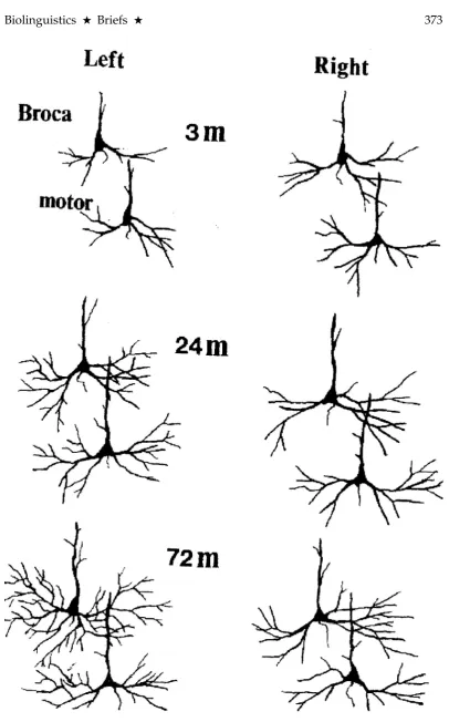
Scheibel (1992) examined postmortem tissue samples of infants (n= 17) spanning the ages of 3 to 72 months. The number of brains examined in each age group-ing was four at 3 months, three at 5–6 months, two at 12–15 months, four at 24–36 months, and four at 42–72 months. The analysis was limited to infragranular pyra-midal cells of layer V. As in all previous studies by the Scheibel group, the dendritic number and length measures were taken along the 3D branching sequence as 1st
Figure 1 presents stained images of neurons analyzed at 3 months, 24 months, and 72 months, obtained from Broca’s and oro-motor cortical regions, from right and left hemispheres. One developmental factor that can be ascertained from this image is that higher order (4thand 5th) distal dendritic branches have already formed by 24 months in both hemispheres, but appearing somewhat more dense in the left hemisphere in this isolated cross-sectional sample at 72 months.
The major conclusions from this study were: (1) histological development of the right hemisphere greatly exceeded that of the left at birth, but was gradu-ally overtaken and surpassed by the left hemisphere as language capacity devel-oped; (2) the rate of dendritic development, as measured by number and length of branches, was initially greater in motor speech areas controlling orofacial and laryngopharyngeal musculature relative to BA 44 and 45, thought to mediate more language-related activity; (3) though there was no significant difference in total dendritic length across the two hemispheres, the more proximal branches (1st, 2nd, 3rd) tended to be shorter, and the longer more distal branches (4th, 5th, 6th) were more numerous in the left hemisphere. Proximal branches develop during the pre-natal period, while distal branches emerge and grow appreciably later during the postnatal period. Furthermore, the more distal branches are the only segments likely to respond to variations of environmental sensory input (Diamond 1988). Complicating this issue, however, is the more recent findings of Travis et al. (2004), also examining basilar dendritic neuropils of pyramidal neurons, that showed the order of synaptogenesis maturation in infants was 180 degrees out of phase with adult brains when relative cortical regions were compared.
6. Caveats in Attempting to Identify Micro-Level Neural Correlates Underly-ing Language Acquisition
The basic premise of the Scheibel group (particularly Scheibel et al. 1985 and Jacobs et al. 1993), was that (1) greater relative lengths of distal dendritic branches of corti-cal pyramidal neurons (in left hemisphere language areas) provide a larger surface area, thus allowing for greater proliferation of dendritic spines, and hence higher densities of potential synaptic inputs to that neuron. This section now provides neuroanatomically-based arguments that dilute the Scheibel hypothesis.
Measuring specific features of morphological cell structure from postmortem tissue specimens has serious limitations. At the core of the limitations are sam-pling issues. After staining a small “snapshot” of brain tissue, it is microscopically examined, searching for visible and structurally intact dendritic arborizations. In the Scheibel studies ten neurons (per hemisphere) were randomly selected and an-alyzed from each individual brain, following standard criteria. To put these num-bers into proper perspective, it has recently been estimated that the human brain contains approximately 86 billion neurons (Herculano-Houzel & Lent 2005). The numerology of estimating synapses is not as precise, with a range of 1014 to 1015
life span of the deceased, and moreover, nothing can be ascertained about where afferent inputs were coming from, or where axonal outputs were going.
Another complicating issue is that neurons are likely to operate in a function-ally shared manner—neuron X may be functionfunction-ally active during a particular lan-guage function, say syntax, and also active in concert with different networks when performing mathematical operations, or accessing memory/attentional circuitry at another moment in time (Fuster 2006). The newly recognized relevance of a “tem-poral view” in speech perception, namely how different oscillating brain rhythms may be scaled for the encoding of varying, linguistic-based, information chunks (e.g., 500–2000 msec for intonational contours, 150–300 msec for syllable structure, 20–80 msec for rapidly changing featural information), suggests that the same un-derlying neural substrate can be operative for different encoding/decoding opera-tions across different temporal processing windows (Poeppel 2014, Giraud & Poep-pel, 2012, Luo & PoepPoep-pel, 2007). Thus, a specific morphological feature of the neu-ropil may play different roles relative to the changing time constants in play at the moment. However, a counter argument can also be made that the greater overall complexity of synaptic connections (due to longer distal dendritic branches) can provide the means for efficient “multi-memberships” across the various time con-stants of functional cognitive processing.
Another issue is the simple fact that language is not a single, stand alone, cognitive function. Neural circuitry underlying memory and attention must also co-develop and interactively support language-based operations. So, when a single layer III pyramidal neuron is selected to examine its dendritic/spine morphology, that neuron’s specific cognitive role is unknown and, moreover, indeed unknow-able.
Another sampling-based caveat is that all the tissue samples used in inves-tigations measuring dendritic branching/spine structure were from the cerebral cortex. The cerebral cortex receives all of its inputs directly from the thalamus, and is constantly in reciprocal communication with the thalamus, which is, un-derstandably, rarely analyzed in human postmortem anatomical studies (Sherman & Guillery 2001). In language functioning, both the cerebellum and basal ganglia have crucial roles in both planning and selecting aspects of speech motor activa-tions. These subcortical structures reach developmental maturation before synap-togenesis is fine-tuned in the cerebral cortex (Tau & Peterson 2010), but do not usu-ally get discussed when language acquisition in the context of a critical period is discussed.
Additional sampling alternatives of what and where to measure can also in-fluence outcomes. Which is more relevant — thepostsynaptic matrixor the presynap-tic matrix? The Scheibel group has settled exclusively on the postsynaptic matrix, the graded potentials arriving onto the dendritic branching arbors of the receiving neuron’s cell body. What about the presynaptic matrix? How does density of axon boutons affect emerging circuits, or stage of myelin development, or number of collateral branches off an axon?
dendrites and spines of pyramidal cells continue to grow throughout the entire life cycle, including infancy, childhood, adolescence, adult-hood and senescence, as a normal process [. . . ].
(Elston & Fujita 2014: 6)
They go on to say that
[i]n essence, though there may be a net reduction in spine density in the dendritic trees of cells from 3.5 months of age into adulthood, new spines are continually grown through this period.
(Elston & Fujita 2014: 6) Perhaps sheer numbers of dendritic spines is not really that important. Regressive neurogenesis prunes away millions of non-functional synapses throughout devel-opment. A relatively smaller number of highly functional synapses is what gets the job done, not the over abundances characterizing earlier stages of development.
Yet another problem in selecting brain tissue samples is the vast differences encountered as a function of cortex region. Jacobs et al. (2001), explored differences in dendritic/spine extent across several human cortical regions. They limited their sampling to basilar dendrites/spines of supragranular pyramidal cells across eight different Brodmann areas that spanned a functional hierarchy from primary cortex (somatosensory BA 3-1-2; motor, BA 4); unimodal cortex (Wernicke’s area, BA 22; Broca’s area (BA 44); heteromodal cortex (supplementary motor area, BA 6; angular gyrus, BA 39); and supramodal cortex (superior frontopolar zone, BA 10; inferior frontopolar zone, BA 11). Primary and unimodal cortices were designated as low integrative regions relative to heteromodal and supramodal areas which were des-ignated as high integrative areas. Tissue samples were only obtained from the left hemisphere, from 10 neurologically normal individuals, five male and five female, spanning the age interval of 13–47. Ten neurons were sampled from each region, and evaluated by total dendritic length, mean segment length, dendritic segment count, dendritic spine number, and dendritic spine density. Dendritic systems in primary and unimodal regions were consistently less complex than in heteromodal and supramodal areas. For example, total dendritic length in BA 10 (frontal cortex) was 31 % greater than in somatosensory cortex (BA 3-1-2), while dendritic spine number was 69 % greater. Jacobs et al. conclude:
These findings demonstrate that cortical regions involved in the early stages of processing (e.g. primary sensory areas) generally exhibit less complex dendritic/spine systems than those regions involved in the later stages of information processing (e.g., prefrontal cortex).
(Jacobs et al. 2001: 558) A complex cognitive function such as language spans the entire range of this cor-tical hierarchy—primary, unimodal, heteromodal, and supramodal, and in most instances, in ways not even remotely understood.
7. Conclusions
“alien” structure. The relative dendritic lengths of a miniscule number of randomly analyzed neurons cannot, unfortunately, provide the necessary insights to under-stand how a child develops language in the first decade of life. What is inescapable is the fact that a child’s self-generated oral movements and reception of speech sounds, shape their developing connectomes when it is extremely ripe for input, learning and eventual fine tuning, just like Eric Lenneberg said fifty years ago.
Perhaps the only realistic methodological approach in seeking potential neu-ral correlates of language acquisition is to (i) enlarge one’s observational lens and (ii) restrict your search to post mortem tissue samples from brains of extraordinarily (linguistically-speaking) gifted individuals.
With regard to (ii), Amunts et al. (2004) performed cytoarchitectonic scrutiny (morphometry and multivariate statistical analyses) of random cortical tissue sam-ples within BA 44 and 45, from the brain of Emil Krebs (1867–1930), a diplomat who was known to have fluently spoken more than 60 languages. Histological sections (20μm), taken from both hemispheres, were stained to see only cell bodies. Visual
area BA 18 was also included as a comparison to language-related neocortex. The fraction of cortical volume occupied by cell bodies was estimated by measuring the the gray level index (GLI), using an image analyzer. The laminar changes in the volume of cell bodies, from the pial surface to the white matter, were quantified and displayed as GLI profiles, consisting of 10 shape-describing features. The 10 features were subsequently used in a canonical principal component analysis that clearly showed the separation distances of Krebs’ cytoarchitecture features from control brains. The cytoarchitecture of Krebs’ brain differed significantly from the controls in both left and right hemisphere BA 44, but was most pronounced in right hemisphere BA 45. Of the 10 statistical features of the GLI profile, the maximal difference concerned the feature “dsk,” which is an index of the degree of lamina-tion between supra-granular layers (I to III) and infragranular layers (V and VI). Another profile feature (dmGLI), which reflects variations in volume fraction of cell bodies between cortical layers and sublayers across the whole cortex, revealed striking differences between Krebs and the control brains. Krebs’ brain had differ-ent arrangemdiffer-ents of cell bodies across the cortical lamina relative to controls, but not in the absolute amount of neuropil. There were no differences found in cy-toarchitecture laminations in visual area BA 18, and thus Krebs’ brain exhibited “a local microstructural specialization” (Aumnts et al. 2004: 350). Interestingly, Krebs’ brain showed a highly unusual pattern of hemispheric asymmetries—maximal cy-toarchitectonic symmetry for BA 44, and maximal hemispheric asymmetry for BA 45, which may have enhanced his multi-lingual talents.
So, in one respect Eric Lennenberg has been proven wrong, as he once said:
There is no cytoarchitectural peculiarity of the cortical areas involved in
language. (Lenneberg 1967: 61)
References
Amunts, Katrin, Axel Schleicher & Karl Zilles. 2004. Outstanding language com-petence and cytoarchitecture in Broca’s speech region.Brain and Language89, 346–353. doi:10.1016/S0093-934X(03)00360-2.
Ashtari, Manzar, Kelly Cervellione, Khader Hasan et al. 2007. White matter devel-opment during late adolescence in healthy males: A cross-sectional diffusion tensor imaging study. NeuroImage35(2), 501–510. doi:10.1016/j.neuroimage. 2006.10.047.
Barkat, Tania, Daniel Polley & Takao Hensch. 2011. A critical period for auditory thalamocortical connectivity. Nature Neuroscience 14(9), 1189–1196. doi:10. 1038/nn.2882.
Birdsong, David. In press. Critical periods. In M. Aronoff (ed.), Oxford Bibliogra-phies in Linguistics. New York, NY: Oxford University Press.
Carughi, Arianna, Kenneth Carpenter, & Marian Diamond. 1989. Effect of environ-mental enrichment during nutritional rehabilitation on body growth, blood parameters and cerebral cortical development of rats.Journal of Nutrition119, 2005–2016.
Chugani, Harry. 1994. Development of regional brain glucose metabolism in rela-tion to behavior and plasticity. In G. Dawson & K. W. Fischer (eds.),Human Behavior and the Developing Brain. New York, NY: Guilford,153–175.
Diamond, Marian. 1988. Enriching heredity: The Impact of the Environment on the Anatomy of the Brain. New York, NY: Free Press.
Elston, Guy & Ichiro Fujita. 2014. Pyramidal cell development: postnatal spino-genesis, dendritic growth, axon growth, and electrophysiology. Frontiers in Neuroanatomy8, 78. doi:10.3389/fnana,2014.00078.
Fuster, Joachim. 2006. The Cognit: A network model of cortical representation. International Journal of Psychophysiology60, 125–132. doi:10.1038/nn.3063. Giraud, Anne-Lise & Poeppel, David. 2012. Cortical oscillations and speech
pro-cessing: Emerging computational principles and operations. Nature Neuro-science15, 511–517. doi:10.1016/j.ijpsycho2005. 12. 015.
Gogtay, Nitin, Jay Giedd, L. Lusk et al. 2004. Dynamic mapping of human corti-cal development during childhood through early adulthood. Proceedings of the National Academy of Science of the United States of America101, 8174–8179. doi:10.1073/pnas.0402680101.
Green, Edward & William Greenough. 1986. Altered synaptic transmission in den-tate gyrus of rats reared in complex environments: Evidence from hippocam-pal slices maintained in vitro.Journal of Neurophysiology55, 739–749.
Greenough, William, A. Yuwiler & M. Dollinger. 1973. Effects of posttrial serine administration on learning in “enriched” and “impoverished” reared rats. Behavioral Biology8, 261–272.
Harrison, Robert, Karen Gordon & Richard Mount. 2004. Is there a critical period for cochlear implantation in congenitally deaf children? Analyses of hear-ing and speech performance after implantation. Developmental Psychobiology 46(3), 252–261. doi:10.1002/dev.20052.
brain. Journal of Neuroscience 25, 2518–2521. doi:10.1523/JNEUROSCI.4526-04.2005.
Hubel, David. 1979. The brain.Scientific American241, 44–65.
Huttenlocher, Peter & Arun S. Dabholkar. 1997. Regional differences in synaptoge-nesis in human cerebral cortex.Journal of Comparative Neurology387, 167–178. doi:10.1002/(SICI)1096-9861(19971020)387:2<167::AID-CNE1>3.0.CO;2-Z. Jacobs, Bob, Matthew Schall, Melissa Prather, Elisa Kapler et al. 2001. Regional
dendritic and spine variation in human cerebral cortex: A quantitative Golgi study.Cerebral Cortex11, 558–571. PMID:11375917.
Jacobs, Bob & Arnold Scheibel. 1993. A quantitative dendritic analysis of Wer-nicke’s area in humans. I. Life span changes.Journal of Comparative Neurology 327, 83–96. doi:10.1002/cne.903270107.
Jabobs, Bob, Matthew Schall, & Arnold Scheibel. 1993. A quantitative dendritic analysis of Wernicke’s area in humans. II. Gender, hemispheric, and environ-mental factors.Journal of Comparative Neurology327, 97–111. doi:10.1002/cne. 903270108.
Knickmeyer, Rebecca, Sylvain Gouttard, Chaeryon Kang et al. 2008. A structural MRI study of human brain development from birth to 2 years. Journal of Neuroscience28, 12176–12182. doi:10.1523/JNEUROSCI.3479-08.2008.
Kostovic, Ivica, Milos Judas, Zagreb Petanjek & Goran Simic. 1995. Ontogenesis of goal-directed behavior: Anatomo-functional considerations. International Journal of Psycho-physiology19, 517–527. doi:10.1016/0167-8760(94)00081-O. Lenneberg, Eric H. 1967.Biological foundations of language. New York, NY: Wiley. Levitt, Pat. 2003. Structural and functional maturation of the developing primate
brain.Journal of Pediatrics143, S35–S45. doi:10.1067/S0022-3476(03)00400-1. Levi, Dennis & Roger W. Li. 2009. Improving the performance of the amblyopic
vi-sual system.Philosophical Transactions of the Royal Society of London B: Biological Sciences364, 399–407. doi:10.1098/rstb.2008.0203.
Luo, Huan & Poeppel, David. 2007. Phase patterns of neuronal responses reli-ably discriminate speech in human auditory cortex. Neuron 54, 1001–1010. doi:10.1016/j.neuron.2007.06.004.
Maurer, Daphne, Terri Lewis, Henry Brent & Alex Levin. 1999. Rapid improvement in the acuity of infants after visual input. Science286, 108–110. doi:10.1126/ science.286.5437.108.
Mrzljak, Ladislav, Harry Uylings, Ivica Kostovic & Corbert van Eden, 1992. Prena-tal development of neurons in the human prefronPrena-tal cortex. I. A quantitative Golgi study.Journal of Comparative Neurology316, 485–496.
Paus, Tomas, Louis Collins, A. C. Evans, G. Leonard et al. 2001. Maturation of white matter in the human brain: A review of magnetic resonance studies. Brain Research Bulletin54, 255–266. doi:10.1016/S0361-9230(00)00434-2. Poeppel, David. 2014. The neuroanatomic and neurophysiological infrastructure
for speech and language. Current Opinion in Neurobiology28,142-149. doi:10. 1016/j.conb.2014.07.005.
Scheibel, Arnold. 1990. Dendritic correlates of higher cognitive function. In A. Scheibel & A. Wechsler (eds.), Neurobiology of higher cognitive function, New York, NY: Guilford, 239–270.
Scheibel, Arnold. 1992. Dendritic structure and language development. In B. de Boysson-Bardies, S. de Schonen, P. Jusczyk, P. MacNeilage, & J. Morton (eds.), Developmental neurocognition: Speech and face processing in the first year of life. Dortrecht, The Netherlands: Springer, 51–62.
Scheibel, Arnold, L. A. Paul, I. Fried et al. 1985. Dendritic organization of the anterior speech area. Experimental Neurology, 87, 109–117. PMID:3967694. Sherman, Murray & Ray Guillery. 2001. Exploring the Thalamus. San Diego, CA:
Academic Press.
Tau, Gregory & Bradley S. Peterson. 2010. Normal development of brain circuits. Neuropsychopharmacology Reviews35, 147–168. doi:10.1038/npp.2009.115. Travis, Katie, Kevin Ford & Bob Jacobs. 2004. Regional dendritic variation in
neona-tal human cortex: A quantitative Golgi study. Developmental Neuroscience 27(5), 277–287. doi:10.1159/000086707.
Wiesel, Thorton & David Hubel. 1963. Single-cell responses in striate cortex of kittens deprived of vision in one eye.Journal of Neurophysiology26, 1003–1017. PMID:14084161.
Wiesel, Thorton & David Hubel. 1965. Comparison of the effects of unilateral and bilateral eye closure on cortical unit responses in kittens.Journal of Neurophys-iology, 28, 1029–1040. PMID:5883730.
Zhou, Xiaolin & Michael Merzenich. 2008. Enduring effects of early structured noise exposure on temporal modulation in the primary auditory cortex. Pro-ceedings of the National Academy of Sciences of the United States of America105(11), 4423–4428. doi:10.1073/pnas.0800009105.
Harvey M. Sussman
University of Texas at Austin Departments of Linguistics and Communication Sciences & Disorders
305 E. 23rdSt. (B5100)
Austin, TX 78712 United States of America