ORIGINAL SCIENTIFIC ARTICLE
The Evolution of Complex Organs
T. Ryan Gregory
Published online: 10 October 2008
#Springer Science + Business Media, LLC 2008
Abstract The origin of complex biological structures has long been a subject of interest and debate. Two centuries ago, natural explanations for their occurrence were considered inconceivable. However, 150 years of scientific investigation have yielded a conceptual framework, abundant data, and a range of analytical tools capable of addressing this question. This article reviews the various direct and indirect evolution-ary processes that contribute to the origins of complex organs. The evolution of eyes is used as a case study to illustrate these concepts, and several of the most common misconceptions about complex organ evolution are discussed.
Keywords Adaptation . Constraints . Convergence . Co-option . Crystallin . Duplication . Exaptation . Eye . Functional shift . Historical contingency . Lens . Natural selection . Opsin . Photoreceptor
Introduction
As a career, science would hold very little appeal if all it entailed were the confirmation of existing knowledge or the memorization of long lists of well-established facts. Science thrives on what isnot yet known: the more vexing a problem, the more inspiring it is to investigate. With millions of species alive today (and orders of magnitude more thought to be extinct), not only describing but also explaining the diversity, history, and complexity of life is a challenge nearly without equal in all of science. Nevertheless, the diligent ac-cumulation of data punctuated by occasional empirical or theoretical breakthroughs has, over the past two centuries,
yielded tremendous advances in the understanding of life’s complexities and the historical origins thereof.
There was a time when natural processes capable of producing complex biological features were deemed incon-ceivable, leading to the conclusion that these, like human artifacts, must be the products of intelligent agency (e.g., Paley1802). Beginning with Darwin’s (1859) description of natural selection, and expanding considerably upon it in the 150 years since, the science of evolutionary biology has as-sembled a theoretical framework capable of explaining how complex features can arise naturally over time. Still, while few nonspecialists have trouble acknowledging small-scale evolutionary processes such as the evolution of antibiotic resistance within populations of bacteria, they often remain uncertain as to how similar mechanisms could account for complex structures such as eyes or wings (Ayala2007; Scott and Matzke2007).
This article provides a general overview of the various processes that play a role in the evolution of complex bio-logical systems. The classic exemplars of organ complexity, eyes, are then used as a case study to illustrate these general mechanisms. Although it is not possible to deliver a com-prehensive discussion of eye evolution within the confines of this paper, an extensive (but by no means exhaustive) ref-erence list is provided in order to facilitate further study of the subject, as well as to highlight the rich scientific literature that exists on this topic but which may be largely unknown outside professional biological circles. Finally, some common mis-conceptions regarding the evolution of complex features are discussed.
How Complex Organs Evolve
The concept of“complexity”is anything but simple. In fact, many technical definitions are in use in mathematics, T. R. Gregory (*)
Department of Integrative Biology, University of Guelph, Guelph, Ontario N1G 2W1, Canada
computer science, and other disciplines. Means of quantifying complexity have been developed in evolutionary biology as well, as have methods for assessing trends involving changes in complexity through deep time (for review, see Gregory 2008a). This paper is not about complexity per se, but about the evolution ofcomplex organs: biological structures with several intricately interacting parts that function in a sophisticated manner, the eye being the primary example dis-cussed. Complexity as defined in this intuitive sense is not restricted to organs—it also applies to biochemical, subcellu-lar, behavioral, and other biological systems. On the other hand, not all functional systems are complex, nor is com-plexity inherently advantageous or inevitable as efficiency can readily trump a convoluted arrangement.
Fundamentally, evolutionary explanations for the origin of biological features, complex or otherwise, are based on the assumption of continuity. That is to say, there is an unbroken chain of ancestry and descent linking modern organisms with earlier species that lived long ago. In order to account for the existence of complex organs as they are observed today, it is necessary to provide an account of how these could have arisen, without breaks or inexplicable leaps, from less complex antecedents. The following sections outline various processes that, taken together, are considered by evolution-ary biologists to meet this requirement.
Direct Adaptation by Natural Selection
Darwin famously noted in1859 (p.189) that “if it could be demonstrated that any complex organ existed, which could not possibly have been formed by numerous, successive, slight modifications, my theory would absolutely break down.”It should be understood that the“theory”in question is not the notion that species are related by descent; rather, it is the proposal that the mechanism responsible for evolution is natural selection acting gradually on minor heritable differ-ences(see Gregory2008b). There is absolutely no scientific disagreement as to whether natural selection occurs, as it can be observed in both experimental and natural populations; the question is whether this process alone can result in the emergence of complex organs such as eyes.
The process of gradual, stepwise adaptive change empha-sized (though not exclusively so) by Darwin has been called “direct evolution” and can be further subdivided into two major types (Thornhill and Ussery2000):
1. Serial direct evolution. The simplest form of adaptive evolution, which proceeds step by step from A1→A2→ A3→ A4 and involves gradual change along a single axis (i.e., each step serves the same function but ef-fectiveness increases from A1to A4). A second series of changes may take place in addition to the first, but in this scenario it would be only after the first series of changes ended.
2. Parallel direct evolution. A slightly more complicated form of direct adaptation in which changes occur at the same time in more than one component, as in A1/B1→ A2/B2→A3/B3→A4/B4. In this case, no single com-ponent becomes greatly modified before the others do.
Under serial direct evolution, each change that occurs is small, involves only one component of a particular system, and, in principle, is reversible. As a result, serial direct evolution does not produce organs with indivisibly integrated components. Parallel direct evolution, on the other hand, can produce a moderate interdependence of parts because these change in concert with one another. Neither serial nor parallel direct evolution necessarily leads to an increase in complexity, and in terms of highly complex organs, the most important contribution made by direct adaptive evolution probably relates to refinements of particular functions through the modification of a few components. Thus, direct evolution by itself is not sufficient to properly account for the evolution of highly complex and integrated organs, and as such, additional processes are necessary.
Indirect Evolution
Direct adaptive evolution by natural selection has, since it was first proposed, been subject to criticism by those who disagree that it is sufficient to explain the origin of complex biological features. In Darwin’s own time, opponents began listing features of organisms for which incipient or intermediate stages seem unlikely to have been functional and which there-fore could not have been be shaped incrementally by natural selection (e.g., Mivart 1871). Darwin (1872) responded by pointing to examples of organs of“intermediate”complexity that did, or indeed still do, exist in other species.1 Nevertheless, there are legitimate reasons to expect direct gradual evolution along a single axis to be incapable of pro-ducing complex adaptations composed of tightly interacting parts. As a result, evolutionary biologists (including Darwin) have long pointed out the importance of indirect routes by which complex organs and systems can evolve.
A recent analysis of the evolution of two hormone-receptor pairs provides an illustration of the basic concept of indirect evolution (Adami 2006; Bridgham et al. 2006). The close match between a hormone and the receptor to which it binds has been considered analogous to that between a“lock and key,”both of which are required for the system to function.
1Interestingly, Mivart (1871) cited, among his examples of features that he presumed would be very unlikely to evolve gradually, the appearance of two eyes on one side of the head among flounders and the feeding structures of baleen whales—both of which have been discussed recently in light of evidence indicating that intermediate forms did indeed occur (Deméré et al.2008; Friedman2008; Janvier
The challenge, as with more complex biological features, is to explain how such a system evolved through interme-diates to its current, integrated state, given that it is not rea-sonable to hypothesize that the components arose together instantaneously by mutation.
In vertebrates, the stress hormone cortisol activates the glucocorticoid receptor, which is involved in regulating metabolism and immunity whereas a related receptor, the mineralocorticoid receptor, is activated by aldosterone to regulate electrolyte homeostasis (in abstract terms A + B→X whereas C + D→Y). This specificity is important, as acti-vation of the wrong receptor would be very detrimental to the organism (it would be a problem if A + D→Y; Adami2006). Having the two receptors activated separately is also beneficial as it allows metabolism to be regulated indepen-dently of electrolytes, for example (Bridgham et al. 2006).
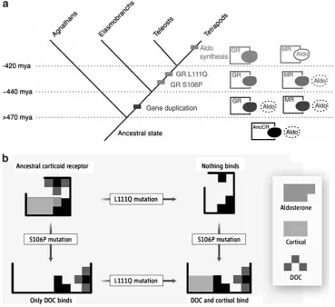
Phylogenetic analyses suggest that the two receptors are de-rived from an ancestral gene that duplicated about 450 million years ago (Mya). Aldosterone, by contrast, is found only in tetrapods and, therefore, evolved long after the origin of the two receptors (Fig. 1A). The question is, how could these receptors become specific for different hormones when one of the hormones did not yet exist?
In order to address this question, Bridgham et al. (2006) used phylogenetic approaches to reconstruct the ancestral corticoid receptor and found that it would have been sen-sitive to cortisol, aldosterone (had it existed), and another hormone known as 11-deoxycorticosterone. The difference between this early receptor and the modern glucocorticoid receptor, which does not bind aldosterone, lies in amino acid changes caused by two mutations—either one of these changes alone makes the receptor insensitive to cortisol, but both
Fig. 1 Indirect evolution of two hormone-receptor pairs. A Steps involved in the origin and evolution of the glucocorticoid receptor (GR) which is sensitive to cortisol and the mineralocorticoid receptor (MR) which is sensitive to aldosterone. The two receptors are derived from a gene for a single ancestral receptor (AncCR) which duplicated about 450 Mya. Later, two mutations occurred in the GR receptor that made it insensitive to aldosterone. Aldosterone (Aldo) did not evolve until much later (represented by a dotted circle before it arises) but was able to bind to the MR, which had retained a form close to the ancestral receptor because a third hormone similar to aldosterone formerly
activated it. From Bridgham et al. (2006), reproduced by permission of the American Association for the Advancement of Science.BThe two mutations required to make the GR insensitive to aldosterone (L111Qand
together make it sensitive to cortisol and insensitive to al-dosterone. It is unlikely that both mutations would occur simultaneously, but Bridgham et al. (2006) found that one of the mutations retained sensitivity to 11-deoxycorticosterone, meaning that this intermediate step would still be functional, though in a different way, and could still have been favored before the second mutation occurred (Fig.1B). Meanwhile, selection acting to maintain specificity for a different hor-mone that was structurally similar to aldosterone meant that once aldosterone arose, it could bind to the mineralocorticoid receptor which remained similar to the ancestral form (a process Bridgham et al.2006call“molecular exploitation”; Fig.1A).
In short, even though they are now indivisible, the two hormone-receptor pairs could have evolved through a step-wise series favored at each stage by natural selection, but the steps were not direct. The process involved gene duplication, the input of a third hormone, and shifts in function. Similar processes may operate generally in the evolution of complex proteins in a manner that is readily explained by modern evolutionary theory (see Lynch 2005 for a technical dis-cussion). These and other indirect evolutionary processes are also involved in the evolution of complex organs and their components, and are discussed in more detail in the fol-lowing sections. It is important to note that most complex organ evolution probably involves processes from various parts of the direct–indirect evolutionary continuum.
Exaptation
The notion of functional shifts is an old one in evolutionary biology, having been considered “an extremely important means of transition”by Darwin (1872, p.147). The general process has been known by many names: “co-option,” “adoption from a different function,” “recruitment” (mostly in reference to genes), or, in an outmoded term a little too suggestive of teleology,“preadaptation”(e.g., Gould and Vrba 1982; Arnold 1994; Thornhill and Ussery 2000; McLennan 2008). However, the basic concept became much more broadly appreciated when it was granted a specific name with a clearer definition:“exaptation”(Gould and Vrba1982).
The term“exaptation”derives fromex+ aptus, meaning fit (aptus) by reason of (ex) existing form, as contrasted with adaptation, which derives from ad (towards) + aptus (fit). So, whereas an adaptation is the product of natural selection favoring variants on the basis of, and gradually improving, thecurrent function, an exaptation is a feature that arose for some other reason and subsequently acquired its current function. Designating a feature as an exaptation presumes some knowledge regarding ancestral function (or lack thereof) such that it can be shown to have differed from the current role. Data bearing on this issue can be obtained using fossils and phylogenetic inference (e.g., Arnold1994). The
process-es of adaptation and exaptation are not entirely separate, however, because once a functional shift occurs natural selection may modify the feature with regard to its new role in a process of “secondary adaptation” (Gould and Vrba 1982; Fig.2). Most complex organs are likely to represent a mixture of primary adaptations, exaptations, and secondary adaptations.
The evolution of wings provides one of the classic examples of exaptation and secondary adaptation. As has been pointed out many times, a rudimentary version of a wing would not be useful in flight because it would be unable to generate sufficient lift (e.g., Mivart1871). Only when the wing reached a sufficient size and strength could it be uful for enabling powered flight, meaning that natural se-lection could not favor variants within a population on the basis of flight ability during the early stages of wing evolution. So, how then could wings have evolved to serve their current function in flight? The answer is that early wings did not function in flight but served a different function (primary adaptation). Bird feathers, for example, probably originated for thermoregulation and rudimentary wings may have been useful in capturing prey or assisting with running uphill or one or more other functions (e.g., Dial 2003). In bats, early skin flaps probably would have been functional for gliding but not in powered flight (e.g., Bishop2008). In insects, it has been hypothesized that early “wings”were used for skimming across the surface of water (Marden and Kramer 1994; Marden and Thomas 2003). Natural selection enhancing early forms of the structure— which, initially, may not have been considered a“wing”at all had biologists examined it at the time—would have, at some point, brought it to a stage that could be useful in a new function (exaptation) resembling a rudimentary version of flight (for example, controlled descent from trees in birds and bats, or skimming with less contact with the water or powered jumps in insects). Further modification for this new semiflight function (secondary adaptation) would eventually render the structure suitable for yet another functional shift, namely to weak powered flight (exaptation again), with further modifications leading to new improvements specific to flight (secondary adaptation again).
Given that exaptations are defined largely by what they are not—namely, the products of natural selection strictly for their current function—there are several possible routes by which an organ, components of an organ, or genes can become exaptations (Gould and Vrba 1982; Arnold 1994; Gould 2002; McLennan2008):
exapta-tions”in which a second function is acquired in addition to the initial function and“transfer exaptations”in which the shift to a new function involves the loss of the pre-vious function. Example: the middle ear bones of mam-mals are derived from former jaw bones (Shubin2007). 2. One organ (or gene) has an existing function but at some stage modification of the feature for the initial function makes it amenable to modification in a new role and this allows the organism to move into a new environment or adopt a new ecological lifestyle. Example: early tetrapod limbs were modified from lobe-fins and probably func-tioned in pushing through aquatic vegetation; at some point, they became sufficiently modified to allow movement on to land (Shubin et al.2006).
3. One organ (or gene) has two functions and is modified as it becomes increasingly specialized for one of them. Some-times, the organ is specialized for one of the initial functions in one lineage and for the other initial function in a different lineage. Example: an early gas bladder that served functions in both respiration and buoyancy in an early fish became specialized as the buoyancy-regulating swim bladder in ray-finned fishes but evolved into an exclusively respiratory organ in lobe-finned fishes (and eventually lungs in tetrapods; Darwin1859; McLennan 2008).
4. Two organs (or genes) perform the same function and then one becomes more specialized for the original function while the other takes on a different role. This is
particularly significant when duplication generates mul-tiple copies that subsequently diverge (see below). Ex-ample: some of the repeated limbs in lobsters are specialized for walking, some for swimming, and others for feeding.
5. A feature that had become vestigial2 in terms of its original function takes on a new function in its reduced state. Example: the vestigial hind limbs of boid snakes are now used in mating (Hall2003).
6. A feature that formerly had no function and was present for non-adaptive reasons (a “spandrel”; Gould and Lewontin1979; Gould 1997,2002) takes on a function and may become specialized for that function. This, too, can occur at both the genetic level and the organ level. Arnold (1994) considers these “first-use exaptations” because the first function they fulfill is the exaptive one. Examples: the sutures in infant mammal skulls are useful in assisting live birth but were already present in non-mammalian ancestors where they were simply byprod-ucts of skull development (Darwin 1859); some formerly parasitic transposable elements in the genome, which had no function at the organism level, have been
co-2“Vestigial” does not necessarily mean non-functional, it means reduced in form and function in a particular species relative to others in which the organ still performs the original function. Thus, finding some function for a reduced organ, which is often different from the function of the fully formed organ in other species, does not affect its status as being vestigial.
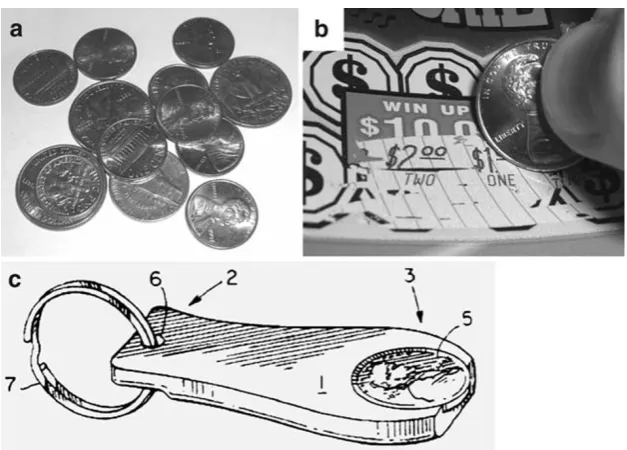
Fig. 2 A simple example of exaptation and secondary adaptation.AThe original and still primary adaptive function of coins is as currency.BA coin co-opted into a new exaptive role as an instant lottery ticket scraper. Coins would always have been capable of scraping tickets, but this function did not become apparent until an environment arose in which instant lottery tickets were abundant. Though functional as scrapers, coins
opted into a variety of other roles, such as in the vertebrate adaptive immune system (e.g., Zhou et al.2004).
The important point regarding exaptations, then, is that the current function of a feature may not reflect the reasons for its origin. Rather, the feature may only have come to occupy its current role comparatively recently.
Duplication (or Furcation)
It has long been recognized that natural selection, though capable of producing directional change, can also be a highly conservative force. If a biological feature currently serves a function vital to survival, then it is very likely that any de-viations from its current state will prove detrimental. That is to say, individuals with a different form of the feature will leave fewer offspring than those with the original form, such that there will not be change in the population from one generation to another with regard to this feature. The most widely re-cognized escape from this constraint is through duplication, a topic that has long been discussed in some detail with ref-erence to genes (e.g., Ohno1970; Taylor and Raes 2004). Ohno (1970), in particular, considered duplication and divergence of genes a critical requirement for major evolutionary diversification:“Natural selection merely mod-ified while redundancy created,”he wrote.
In general, four different outcomes are possible following a gene duplication event. One, the duplicate copy (or the ori-ginal) may simply be lost or rendered nonfunctional by mutation (to become a“pseudogene”). Two, multiple copies may prove to be beneficial such that their repetition is main-tained by selection thereafter (“isofunctionalization”; Oakley et al.2006). As an interesting example, the number of func-tional copies of the salivary amylase gene, which is involved in breaking down starch, is higher in human populations where starchy foods are common in the diet (Novembre et al.2007; Perry et al.2007). Three, mutations in different parts of the two gene copies may mean that both copies must be retained in order to fulfill the original function, or one copy may come to serve the original function in one tissue or at one time during development while the second copy is active in different places or at different times, again such that both copies are needed to serve the role of the original gene (“subfunctionalization”). Four, as per Ohno’s (1970) dis-cussion, one copy may take on a new function while the other fulfills the previous function (“neofunctionalization”).
Repeated structures are common at the organism level as well—body segments, teeth, and flower petals are among the many examples. In this case, duplicated structures may remain in repeated series (e.g., identical body segments of myriapods or annelids), one of the two repeats may retain the original function while the other takes on a new function (e.g., the reduced hind wings of true flies, called“halteres,”which now
function as a kind of gyroscope), or the repeats may become specialized for different functions that are either new or are functions that were formerly carried out by a single structure (e.g., specialized appendages in crustaceans). Darwin (1859, p.437–438), himself, recognized the importance of duplica-tion at the organism level when he wrote:
We have formerly seen that parts many times repeated are eminently liable to vary in number and structure; consequently it is quite probable that natural selection, during a long-continued course of modification, should have seized on a certain number of the primordially si-milar elements, many times repeated and have adapted them to the most diverse purposes.
Although there clearly are similarities between the gene and organism levels in this regard, it is important to note that duplication of structures (e.g., organs or components thereof) may not necessarily be the result of the duplications of genes. Organ-level multiplication can also occur with regulatory mutations that cause the feature to appear in different places, at different times, or in repeated series. To clarify this issue, Oakley et al. (2007) recently coined the term “furcation” (meaning“formation of a fork or division into branches”) to cover the multiplication of existing structures more general-ly. The important point for the present discussion is that duplications, whatever their cause and with or without divergence, can be an important mechanism for increasing complexity at both the genomic and organismal levels.
Gene Sharing
Under the process of“addition exaptation”, a feature that is functional in one capacity assumes a second function without losing its original function (Arnold1994). At the molecular level, it is becoming increasingly recognized that the same protein can carry out more than one function, though it can sometimes be difficult to determine which, if either, was the sole original function. In this sense, a descriptor other than “exaptation”or “co-option”is used in reference to the exis-tence of multifunctional proteins:“gene sharing”(Piatigorsky 2007,2008).
As Piatigorsky (2007, pp 4–5) defines it,
used in the same way wherever or whenever it is present, or that it has always done what it is doing at any given moment. The functions of genes and proteins are context dependent.
There are several ways that a single protein can serve very different functions, such as by being expressed in different tissues or at different times during development (i.e., due to changes in regulatory genes), by undergoing changes in the amino acid sequence that enable a second function but do not compromise the first, by combining with another copy of the same protein to form a“homodimer”with a different function, by combining with other proteins to form“heterodimers,”or by being subject to different patterns of folding or other chemical modifications (True and Carroll2002; Piatigorsky2007).
The process of gene sharing can be important in the evolution of complex organs because it means that functions can be enhanced or acquired without any change in the protein-coding gene itself if there is a change in the context in which it occurs—say, the emergence of a new type of tissue in which it may be expressed. Conversely, an existing gene being expressed in a new place in the body may itself lead to the evolution of a new tissue. This greatly facilitates the specialization of an organ for a new function because it does not compromise previous functions for the gene, does not require gene duplication and divergence (though this remains an important process in its own right), and may involve little more than a quantitative change in the amount or localization of the gene’s protein product.
Bricolage (Tinkering) and Collage
In light of the processes described above, it may seem an obvious point that the evolution of complex organs does not involve redesign from scratch at each stage; whether by direct adaptation or shifts in function, the process builds upon and modifies what is already present. This was recognized by early evolutionists including Darwin (see Jacob1977,1982; Laubichler2007) but has often been overlooked when authors characterize natural selection as an optimization process. The clear exposition by Jacob (1977, 1982) was therefore an important reminder of this point, from which it is worth quoting at length (Jacob1982, pp 33, 34):
The action of natural selection has often been compared to that of an engineer. This comparison, however, does not seem suitable. First, in contrast to what occurs during evolution, the engineer works according to a preconceived plan. Second, an engineer who prepares a new structure does not necessarily work from older ones. The electric bulb does not derive from the candle….To produce something new, the engineer has at his disposal original blueprints drawn for that particular occasion, materials and machines specially prepared for that task. Finally, the objects thus producedde novoby the engineer, at least by
the good engineer, reach the level of perfection made possible by the technology of the time.
...
In contrast to the engineer, evolution does not produce innovations from scratch. It works on what already exists, either transforming a system to give it a new function or combining several systems to produce a more complex one. Natural selection has no analogy with any aspect of human behavior. If one wanted to use a comparison, however, one would have to say that this process resembles not engineering but tinkering, brico-lagewe say in French. While the engineer’s work relies on his having the raw materials and the tools that exactly fit his project, the tinkerer manages with odds and ends. Often without knowing what he is going to produce, he uses whatever he finds around him…none of the materials at the tinkerer’s disposal has a precise and definite function. Each can be used in different ways. What the tinkerer ultimately produces is often related to no special project. It merely results from a series of contingent events, from all the opportunities he has had to enrich his stock with leftovers. In contrast with the engineer’s tools, those of the tinkerer cannot be defined by a project. What can be said about any of these objects is just that“it could be of some use.”For what? That depends on the circumstances.
Though they describe more a principle than a process, the terms“tinkering”and“bricolage”include, and are now most often used as substitutes for, “co-option of a gene or other feature into a new function”or simply“mutation and natural selection leading to an alteration of preexisting traits”(e.g., Bock and Goode2007). As Jacob (2001) noted, one must be cautious to avoid a potentially confusing anthropomorphism in which an actual tinkerer or bricoleur is imagined who invents through trial and error.
What is perhaps missing, and for the purposes of this discussion is useful to emphasize, is a particular process mentioned by Jacob (1977, 1982) that differs from both direct adaptation and exaptation, in which existing compo-nents, be they functional for something else or nonfunctional initially, arebrought together or rearranged to form a new, more complex combination with a novel function. Rather than “bricolage,” the term “collage” may more effectively encapsulate this concept.3Because a new function emerges
through the combination of existing components, and es-pecially once further modified by natural selection for this new function, a feature produced through“collage”becomes much more than the sum of its parts. As noted by Jacob (1977,1982), the feature is not assembled with a predefined outcome in mind, rather its function depends on circum-stances and on which components are available and happen to become linked. Two important points bear mentioning about the process of indirect evolution through“collage”: (1) the linking of components is not an“all or nothing”process in which two or more already complex structures suddenly are joined—individual parts, which themselves may vari-ously be simple or relatively complex and functional for something else or nonfunctional, can be added in series, with each new addition leading to a different function for the combined structure and (2) the newly combined structure may carry out its new function rather poorly at first, with subsequent direct adaptation leading to improvement along
this novel axis, for example by enhancing the integration of the newly combined components (Fig.3).
Scaffolding
Many organs, having been built up in overall complexity by direct adaptation, exaptation, and collage, and further specialized through secondary adaptation, exhibit a level of integration to the point that their components are interde-pendent on one another. In these cases, the removal of one or more components may render the organ nonfunctional—at least with regard to its current integrated function (after all, exaptation can also occur following a loss of parts). Shifts in function help to explain how such a system could be assembled through less complex intermediate steps, but another process known as “scaffolding” is sometimes involved in the evolution of such functionally indivisible organs. In this case, a component of the organ that is present
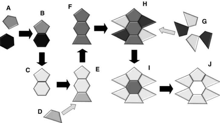
Fig. 3 A diagram showing the processes of exaptation (shifts in function), collage (assembling existing elements into new functional combinations), scaffolding (loss of a component that was formerly required for the assembly of a complex arrangements of parts), and direct adaptation by natural selection (including secondary adaptation) in the evolution of a complex feature. The complex organ (J) includes many parts, all of which must be present for the organ to carry out its current function. Although it can now carry out this function only when all of its components are present, an organ such as this can evolve through intermediates, all of which havesomefunction—though not necessarily the function of the final complex organ (J). At an early stage, two simple structures, each already present and performing its own distinct function (A), come together into a combined structure (B) that is capable of carrying out a function that neither component could before. At first, the combined structure (B) may perform the new function rather poorly, but if it nonetheless confers some advantage over alternatives lacking the structure, then the components may be modified by natural selection favoring improvements in this new function (C). Later, a third
through some early stages has the effect of supporting the assembly of other components and, when lost in later stages, leaves behind a complex structure that by all appearances could not have been assembled one piece at a time (Fig.3). Draper (2002) has provided an abstract description that is readily applied to biological systems (including organs or biochemical pathways). In this case, a complex system with two required parts (AB) that performs a function (F) evolves through a complicated but essentially direct path involving both the addition and loss of parts:
Originally, Z performs F, though perhaps not very well (this is possible because, from the fact that AB cannot perform F without A or B, it does not follow that Z cannot perform F by itself). Then, A is added to Z because it improves the function, though it is not necessary. B is also added for this reason. One now has a reducibly complex system composed of three parts, Z, A, and B [i.e., the system could still function if the number of parts is reduced]. Then Z drops out, leaving only A and B [perhaps only after A and B have become modified to work in a more integrated fashion in their new joint arrangement]. And without Z, both A and B are required for the system to function.
This can perhaps be illustrated even more simply with a straightfoward architectural analogy involving the construc-tion of a stone arch (e.g., Cairns-Smith1985; Dawkins1986; Schneider 2000; Thornhill and Ussery 2000). As Dawkins (1986, p.149) put it:
An arch of stones…is a stable structure capable of standing for many years even if there is no cement to bind it. Building a complex structure by evolution is like trying to build a mortarless arch if you are allowed to touch only one stone at a time. Think about the task naïvely, and it can’t be done. The arch will stand once the last stone is in place, but the intermediate stages are unstable. It’s quite easy to build the arch, however, if you are allowed to subtract stones as well as add them. Start by building a solid heap of stones, then build the arch resting on top of this solid foundation. Then, when the arch is all in position, including the vital keystone at the top, carefully remove the supporting stones and, with a modicum of luck, the arch will remain standing.
A second issue is that a stone does not become a keystone until it is added to the rest of the assembled arch, and serving this role cannot be the reason it is maintained until that point. This is analogous to a component of a complex organ that can be incorporated only relatively late in the process, and therefore cannot be maintained for this function earlier in the process. One manner in which such components may remain present is if they serve a different function and are preserved
more or less in their current form on that basis. In this regard, dual functionality may itself be a form of scaffolding that, in retrospect, will have played a role in facilitating the production of a complex feature.
Viewing a complex structure—be it an arch (or, for that matter, the Great Pyramids or Stonehenge), an organ, or a biochemical pathway—only as it appears in the present, with no consideration of the scaffolding that may have been involved in its construction, can lead to undue pessimism regarding the plausibility of its assembly by comparatively unremarkable processes.
Non-adaptation: Constraints, Trade-offs, and Historical Contingency
In the sixth and final edition of The Origin of Species, Darwin (1872, p 421) expressed the following frustration:
[As] it has been stated that I attribute the modification of species exclusively to natural selection, I may be permitted to remark that in the first edition of this work, and subsequently, I placed in a most conspicuous position—namely, at the close of the Introduction—the following words:“I am convinced that natural selection has been the main but not the exclusive means of modification.”This has been of no avail. Great is the power of steady misrepresentation.
Case Study: The Evolution of Eyes
The eye has long held a special place in discussions regarding the origin of complex organs. Paley (1802) famously compared the intricacies of an eye to those of a finely crafted watch and concluded that both were the work of an intelligent designer.4Darwin (1859) offered a different explanation for the origin of biological complexity and again used the eye as a prominent example. Thus, in a passage that is often (partially) quoted, Darwin (1859, pp 186, 187) remarked:
To suppose that the eye, with all its inimitable con-trivances for adjusting the focus to different distances,
for admitting different amounts of light, and for the correction of spherical and chromatic aberration, could have been formed by natural selection, seems, I freely confess, absurd in the highest possible degree. Yet reason tells me, that if numerous gradations from a perfect and complex eye to one very imperfect and simple, each grade being useful to its possessor, can be shown to exist; if further, the eye does vary ever so slightly, and the variations be inherited, which is certainly the case; and if any variation or modification in the organ be ever useful to an animal under changing conditions of life, then the difficulty of believing that a perfect and complex eye could be formed by natural selection, though insuperable by our imagination, can hardly be considered real.
A considerable amount of research has illuminated many details of eye evolution in different groups of animals since Darwin penned these words. The comparatively recent rise of
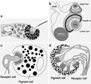
Fig. 4 Very simple, but nonetheless functional, light-sensing systems.
AA single light-sensitive cell (ocellus) as found in the larva of the box jellyfishTripedalia cystophora. In this case, the light-sensitive cell is not connected to a nervous system of any kind but instead includes a cilium that can be stimulated to move the larva in response to light. The pigments (dark spots) within the cell are arranged in a simple cup, meaning that some measure of the directionality of light is provided to the cell. From Nordström et al. (2003), reprinted by permission of The Royal Society.BIn stark contrast, the adult of the same box jellyfish species, T. cystophora, has complex upper and lower eyes with retinas, lenses, and irises at the end of a sensory club called a rhopalium. From Nilsson et al. (2005), reproduced by permission of Nature Publishing Group.CA simple eye spot found
in the larva of the trematode flatworm Multicotyle purvisi, which consists of one pigment cell and one photoreceptor cell. This organ itself provides no information about the direction of a light source, but this can be achieved by comparing the input from two of these organs. Redrawn by Land and Nilsson (2002) based on Rohde and Watson (1991), reproduced by permission of Oxford University Press.DA slightly more complex visual organ involving a single pigment cell but multiple receptor cells found in the turbellarian flatwormBdellocephala brunnea. In this species, the pigment cell is cup-shaped, such that information about the direction of light can be obtained by comparing input from the different receptors. Redrawn by Land and Nilsson (2002) based on Kuchiiwa et al. (1991), reproduced by permission of Oxford University Press. Note that these images are not drawn to the same scale
4
disciplines including molecular biology, phylogenetics, and evolutionary developmental biology (“evo–devo”), in particu-lar, has generated a great many insights regarding this subject. So much, in fact, that any more than a cursory review of the available information must be considered well beyond the scope of this article (however, references to papers containing this information are provided whenever possible). Instead, the eye is used as a case study to illustrate the various general principles of complex organ evolution outlined above, and in particular to demonstrate that the multifaceted nature of the topic requires that it be examined from a variety of perspectives.
Eyes: Definition and Diversity
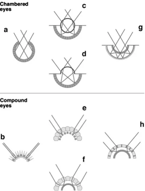
According to some authors, an eye is defined at minimum as a photoreceptor shielded on one side by nearby pigment which allows the detection of the direction of a source of light. The simplest eyes, then, may consist of just one photoreceptor and one pigment cell or even a single cell that includes both photo-and shading pigments as found in some flatworms photo-and algae (e.g., Arendt and Wittbrodt 2001; Oakley 2003). Some examples of relatively simple “eyes” are shown in Fig. 4. Others argue for a more restrictive definition under which an eye is an organ that can produce an image, however crude, and not simply detect light (e.g., Land and Nilsson 2002; Piatigorsky2008; Serb and Eernisse2008). Even under this stricter definition, there are at least eight different types of eyes,5prominent examples of which variously employ cups, pinholes, camera-type lenses, arrays of lenses, concave mirrors, or telescope-like arrangements for image formation (Land and Fernald1992; Land and Nilsson2002; Serb and Eernisse2008). These are illustrated in Figs.5and6. To the extent that the eyes of each species are at least slightly different from each other, and given that many species have more than one type of eyes, there are probably millions of different kinds of eyes peering at the world around them at this very moment.
There is no doubt that access to visual information has been important in a great many groups of organisms. Eyes can be found in about one third of the world’s animal phyla while another one third has light-sensing organs but not eyes.
Roughly one third of the world’s animal phyla have no light sensitive organs, but these tend to be groups exhibiting low diversity; by contrast, the phyla with eyes include more than 95% of all animal species (Land and Nilsson2002; Fernald 2004a; but see de Quieroz1999). The extraordinary benefits provided by the ability to see are also shown by the fact that eyes appeared very early in animal evolution. In fact, most of the major types of eyes are recognizable in fossils from the Cambrian some 530 Mya (Land and Nilsson2002; Nilsson 5
For more detailed discussions of the form, function, and evolution of diverse animal eyes, see Land (1988), Nilsson (1989), Land and Fernald (1992), Arendt and Wittbrodt (2001), and Land and Nilsson (2002), or consult recent reviews on the eyes of specific groups including annelids (Purschke et al.2006), crustaceans (Gaten1998; Elofsson 2006; Reimann and Richter 2007; Marshall et al. 2007; Cronin and Porter2008), tardigrades (Greven 2007), insects (Land
1997; Buschbeck and Friedrich 2008), velvet worms (Mayer2006), millipedes (Müller et al. 2007), jellyfishes (Nilsson et al. 2005; Kozmik et al.2008a,b), mollusks (Serb and Eernisse2008), trilobites (Clarkson et al. 2006), horseshoe crabs (Battelle 2006), and vertebrates (Lamb et al.2007,2008).
Fig. 5 Eight major types of complex eyes found in living animals, divided into two major categories: chambered eyes (top) and
2004). Interestingly, the phylum Chordata (of which humans are members) may have been among the last groups to evolve discernable eyes, as these were not present in the known Cambrian chordates (e.g.,Pikaia); chordate eyes first appear in the fossil record of conodonts from 30 million years later (Land and Nilsson2002). On the other hand, the extensive physical and molecular similarities between the eyes of lampreys and other vertebrates indicate that complex camera-type eyes were already present in their last common ancestor 500 Mya (Lamb et al.2007).
There are several ways of categorizing complex image-forming eyes, such as by the type of photoreceptor cells, the arrangement of photoreceptors relative to pigment cells (i.e., inverted or everted), or by the mechanism of image formation (via shadows, refraction, or reflection; e.g., Land and Nilsson 2002; Fernald2004a,b). Perhaps the best-known distinction is between chambered (or simple) and compound eyes (Nilsson 1989; Land and Nilsson2002; Fernald2006). Although based on a very narrow sampling of animal diversity, it is clear that most references to the evolution of “the” eye relate to the chambered, camera-type lens eyes found in humans and other vertebrates, as well as in cephalopod mollusks, some annelid worms, and various arthropods including spiders. Certainly, these are the most effective at image formation and are the most familiar, and they will form the basis of most of the remaining discussion. However, the evolution of compound eyes is no less interesting than that of chambered eyes—and, given the extraordinary diversity of groups exhibiting them
(most notably arthropods), this is an important question in biology (Land 1997; Nilsson and Kelber 2007; Buschbeck and Friedrich2008; Cronin and Porter2008).
Direct Adaptive Evolution: From Eyespot to Eyeball?
The simplest hypothesis for how a complex feature arose is one involving direct adaptive evolution, with incremental improvements in function favored at each stage by natural selection. Not surprisingly, this has been the starting point for many discussions of eye evolution, which is often depicted as a linear series of small changes, each of which adds very slightly to the organism’s ability to process visual information (e.g., Salvini-Plawen and Mayr1977; Miller 1994; Nilsson and Pelger1994; Osorio1994; Dawkins1996; Bahar2002; Kutschera and Niklas2004).
The major question under these linear scenarios is whether indeed each step along the path not only is functional but in fact increases some aspect of visual ability. In order to test this, and moreover to investigate how much time such a process might require, Nilsson and Pelger (1994) created a theoretical model that began with nothing more than “a flat patch of light-sensitive cells sandwiched between a transparent pro-tective layer and a layer of dark pigment”. In the model, they used incremental changes of 1% in one parameter at a time (length, width, or protein density) that improved visual acuity as calculated based on established optical principles. Their model proceeded through a series of changes including
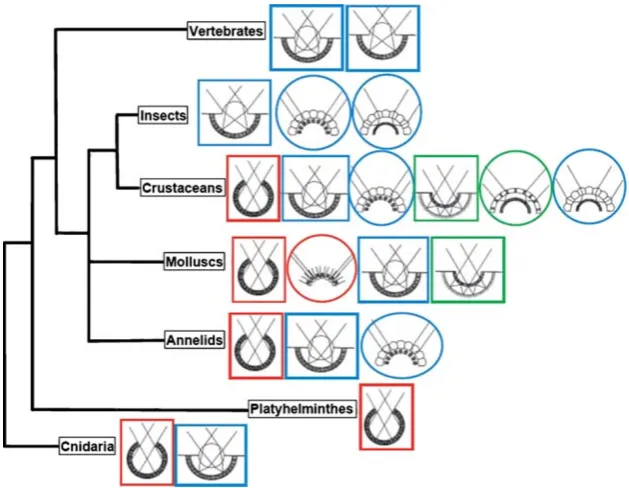
Fig. 6 The distribution of eye types among major taxa of animals. Single-chambered eyes are outlined withrectangles and compound eyes are outlined withovals. See Fig.5for more information about the
an inward folding of the flat patch to form a pit and then a cup, and when resolution could no longer be improved along this trajectory, a very simple lens was added (as they note “even the weakest lens is better than no lens at all”) which then changed incrementally to become spherical and then to develop a gradient of refractive indices (Fig. 7), with the
visual organ finally becoming similar in basic form to the eye of an aquatic animal like a fish or octopus (Fig. 8).
Overall, Nilsson and Pelger (1994) found that small, incremental changes that improve vision by a quantifiable degree could connect both ends of the continuum, from a simple patch of cells to a complex camera-type eye.
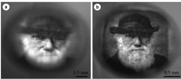
More-Fig. 7 The impacts of refinements in lens organization. (a) Image formed by a spherical glass bead with a single refractive index, showing the blurring resulting from spherical aberration. (b) The same image through the lens from a fish eye, which has a graded refractive index, resulting from a higher crystallin protein concentration in the centre than at the edges. Although the first“lens”may have functioned relatively
poorly, it is only a matter of incremental adaptive changes to improve its functioning to the level seen in modern camera-type lens eyes. For images of eye lenses focusing light from a laser, see Piatigorsky (2007,
2008). From Sweeney et al. (2007), reproduced by permission of The Royal Society and A.M. Sweeney
Fig. 8 The results of a theoretical model (not, as it is sometimes described, a“simulation”) developed by Nilsson and Pelger (1994) to test the time required for a complex camera-type eye to evolve through a series of gradual steps from a simple patch of light-sensitive tissue consisting of an outer protective layer, a layer of receptor cells, and a bottom layer of pigment cells. The number of generations passing between each step is indicated, based on a change of only 0.005% in some parameter (length, width, or protein density) per generation with changes resulting in an improved calculated image formation retained
over, only 1,829 steps of 1% improvements were needed to complete this transition. Even assuming a change of only 0.005% per generation, the model suggests that the entire sequence could be completed in about 360,000 generations (Fig. 8). Given that many fishes and aquatic invertebrates have at least one generation per year, this would mean that the entire sequence in the model could be completed, to invoke an appropriate cliché, in an evolutionary blink of an eye and well within the tens of millions of years available during the Cambrian.
However, while the intermediate stages used by Nilsson and Pelger (1994) are functional in an abstract model, the most important question is whether organisms possessing them could actually survive in nature. It is in this regard that the approach of comparing living species becomes useful even though they are not ancestors and descendants of one another. In this way, it can be shown that each of the hypothetical intermediates depicted in Fig.8does still exist and clearly is functional for the organism in which it occurs. According to Land and Nilsson (2002, p.4), “nearly every imaginable intermediate exists between the acute vision of an eagle and the simple light sensitivity of an earthworm.”
In fact, it has been recognized for over a century that such a diversity of eye types still exists (Darwin 1859; Conn 1900), as seen in Fig.9.
It is important not to take linear models and comparisons of living species too far. Although they demonstrate how direct adaptive evolution could play a major role in the evolution of visual organs, their emphasis on linear transformation tends to obscure the complexity of the actual process. In the simplest terms, it is clear that a single, linear path of eye evolution is too simplistic even for comparisons among mollusks, which appear to have followed several distinct routes leading to divergent eye types (Fig. 10). In vertebrates there is ample evidence for the gradual evolution of eyes, but this does not follow the linear model given in Fig.8. Indeed, the current hypothesis of vertebrate eye evolution involves at least one functional shift even at the organ level, from an early photoreceptive organ performing nonvisual circadian (day–night cycle) functions to a primitive eye capable of sight (Lamb et al. 2007, 2008; Table 1). As will be shown in the following sections, the influence of indirect evolutionary processes is even more pronounced at the level of the components of
Fig. 9 Varying levels of complexity in the visual organs of living species as illustrated by H. W. Conn in1900:Aa simple, flat patch of pigmented cells connected to nerve fibers (see also Fig. 4C); B a slightly more complex pigment cup as found in the limpetPatella, which does not form an image but provides information about the direction of incoming light;Ca pinhole camera-type eye filled with water as found inNautilus; D a camera-type eye with a large lens filling the cavity;Ea camera-type eye with a basic lens and cornea as found in the marine snailMurex;Fa complex camera-type eye with a cornea, lens, iris, and retina as in a cuttlefish. This shows visual organs
camera-type eyes specifically involved in image formation, either in receiving light (photopigments and photorecep-tors) or focusing it (lenses and corneas; Table2).
Photopigments and Photoreceptors6
Vision is not the only function for molecules capable of reacting with light, and it should be no surprise that photosensitive molecules can be found not only in eyes but in animal tissues unrelated to vision as well as in plants, bacteria, and other types of organisms that do not see. The photopigments involved in vision, in particular, are made up of two components: (1) a light-sensitive molecule (chromophore) which changes physical conformation when it interacts with light; in all eyes studied to date this consists of a photosensitive molecule known as retinal that is derived
from vitamin A, (2) a membrane-bound opsin protein that is involved in the chemical cascade that transduces the incoming light to an electrical signal. Opsins are members of a broad category known as G protein-coupled receptor proteins that also serve a range of nonvisual functions including chemore-ception (Nilsson2004). Both component molecules predate the origin of vision, and their merger and subsequent specialization in visual systems represents an important example of evolution through collage, exaptation, and secondary adaptation.
More than 1,000 distinct opsin molecules have been iden-tified since the first example (bovine rhodopsin) was se-quenced in 1982 (Terakita2005). Those that occur in animals are divided into seven subfamilies, all of which appear to have originated before the split between the protostomes (most invertebrates) and deuterostomes (chordates and rela-tives including echinoderms) (Terakita 2005; Larusso et al. 2008). The extraordinary diversity of opsin molecules is likely a product of extensive gene duplication and subse-quent divergence (Arendt2003; Plachetzki and Oakley2007; Oakley and Pankey 2008). Importantly, the duplication of opsin genes and their divergence in becoming reactive to different wavelengths of light forms the basis of color vision (e.g., Dulai et al.1999; Spady et al.2006; Briscoe2008; Gerl and Morris2008).
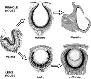
Fig. 10 Two of the several paths of eye evolution followed in mollusks. Contrary to some representations of the eyes in Fig.9, there is no simple linear series from eye patch to complex camera type eye. Rather, eyes may evolve in a variety of ways, becoming specialized as pinhole-type, lens-type, or other types of eyes from an early beginning. These are examples of eyes from modern species, and not actual ancestor–
descendant transitions. Note that these images are not drawn to the same scale (Nautiluseyes are about 10 mm across whereas the others are about 1 mm). From Land and Nilsson (2002), based on drawings by Hesse (1908;Patella,Haliotis, andHelix), Young (1964;Nautilus), and Newell (1965; Littorina), reproduced by permission of Oxford University Press and Blackwell
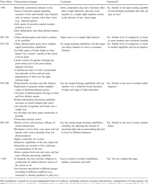
Table 1 Summary of the steps in the evolution of vertebrate eyes as proposed by Lamb et al. (2007)
Time (Mya) Characteristics/changes Comments Functional?
>580 Bilaterally symmetrical animals evolve Some components may have functions other than in light detection, and may work together in a simple light response system in the absence of any visual organ
Yes. Similar to the light sensing capability of soil-dwelling nematodes that lack any type of eyesa
Various G-protein-coupled signaling cascades evolve and initially may function only in sensory systems other than vision (e.g., chemoreception)
Early opsins (G-protein-coupled receptor proteins) evolve
Early rhabdomeric and ciliary photoreceptors evolve
580–550 Ciliary photoreceptors and opsins continue to be modified
Organ serves as a simple light detector Yes. Similar level of complexity as found in some modern non-vertebrate chordates 550–530 Ciliary photoreceptor gains more complex
signal transmission capabilities
No image-forming capabilities, but the organ can detect shadows or serve a circadian function
Yes. Similar level of complexity as found in modern hagfishes and larval lampreys Eye-field region of brain bulges to form
lateral“eye vesicles”outside of the newly evolved skull
Lateral vesicles invaginate, bringing the proto-retina next to the proto-retinal pigment cell layer
A transluscent layer of cells (a primordial lens placode) evolves and prevents pigmentation of skin over the light-sensing organ
530–500 Photoreceptors develop cone-like features Eye has image-forming capabilities and can operate over a relatively broad spectrum of light and range of light intensities
Yes. Similar to the eyes of modern adult lampreys
Duplication of genome creates multiple copies of phototransduction genes Cell types of photoreceptors diverge in form
and have distinct opsins
Retinal information processing capability increases as neural changes take place Lens placode invaginates and forms into a
simple lens
Iris develops and basic pupil constriction is possible
Extraocular muscles evolve
500–430 Myelin evolves and increases efficacy of neural transmission
Eye has strong image-forming capabilities, including for adjusting the amount of incoming light and accommodating the lens to focus at different distances
Yes. Similar to the eyes of many modern fishes
Rhodopsin evolves from cone opsin and rod bipolar cells evolve (possibly from rod photoreceptors)
Highly contractile iris evolves
Refractive capabilities of the lens improved Intraocular eye muscles evolve, allowing
accommodation of the lens
Retina contains both rods and cones and has more efficient processing capability < 430 In tetrapods, the lens becomes elliptical to
compensate for added refractive power of the cornea in air
Eyes as found in modern amphibians, reptiles, mammals, and birds
Yes. You are reading this page
Eyes become specialized in different groups according to different conditions (e.g., nocturnal vs. diurnal, predators vs. prey, etc.)
This hypothesis is based on several independent lines of evidence, including analyses of genes and proteins, comparisons of living species with differing degrees of eye complexity, and information regarding the development of eyes in embryonic vertebrates. For a detailed discussion, see Lamb et al. (2007) and for a less technical review see Lamb et al. (2008).
As noted, photopigments are membrane-bound proteins, which means that maximizing the number of molecules that can be contained within specialized light-sensitive cells involves increasing the surface area of the cell membranes. This has generally been accomplished in two ways, thereby defining two distinct categories of photoreceptors: (1) in rhabdomeric photoreceptor cells, membrane area is increased through the growth of projections of the upper end of the membrane (apical microvilli), (2) in ciliary photoreceptor cells,
a fold of the ciliary membrane is used for increasing the amount of photopigment that can be held. These two cell types differ in other important ways, including in the type of opsin they contain (rhabdomeric or r-opsin, and ciliary or c-opsin, respectively) and the mechanism by which interactions with photons are transduced into electrical information (Arendt and Wittbrodt2001; Arendt2003; Nilsson2004; Fig.11).
It was traditionally thought that rhabdomeric photorecep-tors were found only in protostomes whereas the ciliary type
Table 2 Examples of some of the direct and indirect evolutionary processes that may be involved in the evolution of eyes
Process Examples from eye evolution
Direct adaptive evolution Gradual evolution of lens crystallin concentrations resulting in evolution of graded refractive index lenses in aquatic animals
Exaptation
One structure has one function and takes on or switches to a new function in a new environment
The cornea, which has no refractive capacity in water, became the primary focusing structure after tetrapods moved onto land
The lens became far less important in image formation in terrestrial vertebrates and became specialized for accommodation instead
One structure has one function but becomes modified enough to allow a shift in function
Circadian organ in early chordates became modified sufficiently that it became capable of visual functions
An early protective, transparent layer of cells became sufficiently thickened and invaginated that it could begin serving as an early lens
Two structures perform the same function but become differently specialized
Though both cell types were probably found in the distant bilaterian ancestor, ciliary photoreceptors became the dominant type in vertebrates whereas rhabdomeric photoreceptors came to predominate in most other animals (see also duplication and divergence)
A vestigial structure takes on new function In vertebrates, rhabdomeric photoreceptors lost their microvilli and became retinal ganglion cells that function in circadian entrainment rather than in vision
Duplication and maintenance of repetition The compound eyes of arthropods are composed hundreds or thousands of repeated lens eyes called ommatidia
Duplication and divergence Opsin genes duplicated and diverged to become r-opsins and c-opsins, along with specialization of rhabdomeric cells with r-opsins and ciliary cells with c-opsins
In certain taxa, duplications and diversification of opsins to respond to different wavelengths of light allowed the evolution of color vision
The rod cells of vertebrates are derived from cone cells, both of which are derived from a single ancestral ciliary photoreceptor
Gene sharing Some lens crystallin proteins function both in the eye in light refraction and elsewhere in the body for other functions (e.g., cellular stress response)
Collage The first photopigment was formed by the combination of a preexisting light sensitive molecule derived from vitamin A (which became retinal) with a preexisting G protein-coupled receptor protein (which became the ancestral opsin)
The first“eye”arose by the combination of a photoreceptor cell with a pigment cell During the evolution of complex camera-type eyes, various types of tissue that already
existed (e.g., blood vessels, nerves, muscles) were incorporated
Scaffolding May apply to the evolution of phototransduction pathways or other relevant biochemical systems, but more data are required
Constraints, trade-offs, and historical contingency Trade-off between resolution versus brightness in pinhole camera eyes Trade-off between visual acuity versus size of compound eyes Inverted retina in vertebrates
A narrow range of available wavelengths of sunlight is perceived in most animals, probably because eyes first evolved in water which filters most wavelengths
Convergence Lenses, irises, and various other components of camera-type eyes emerged independently in vertebrates and cephalopods
was restricted to deuterostomes. However, further research has identified species containing both types. The polychaete wormPlatynereis dumerilii, for example, has rhabdomeric photoreceptors in both its larval eyes and in its two pairs of adult eyes as would be expected for a protostome. However, it was also recently discovered to have ciliary photoreceptors in its brain, complete with c-opsins and regulatory genes more similar to those of vertebrates than to those associated with its rhabdomeric photoreceptors. These ciliary cells are not in-volved in vision but apparently serve a circadian function (Arendt et al.2004).
The box jellyfishTripedalia cystophoraprovides a second example. It has 24 eyes, including two pit-shaped and two slit-shaped pigment cups and two camera-type lens eyes posi-tioned at right angles to one another on each of four spe-cialized sensory clubs called rhopalia (Nilsson et al.2005; Kozmik et al.2008a, b; Fig. 4). The jellyfish camera-type eyes appear to use ciliary opsin as the photopigment and melanin as the shielding pigment—both as in vertebrates— though its lens protein is distinct from those of other animals (Kozmik et al.2008a, b; as an aside, it is somewhat enig-matic that complex camera-type eyes complete with tiny, spherical, graded index lenses and, in the lower eye, an iris would occur in these animals, as they are not connected to a brain and are not arranged in such a way as to generate a sharp image in any case; Nilsson et al.2005; Wehner2005). Humans, like other vertebrates, make use of ciliary photo-receptors with c-opsins in their retinas for image processing
(see Kolb2003for a review of retina morphology and func-tion). However, a second category of photoreceptor was discovered in 1991, in the form of a subset of retinal ganglion cells containing a photopigment dubbed melanopsin. It is now recognized that these cells are remarkably similar to the rhabdomeric cells of invertebrates and that melanopsin is similar to r-opsin (see Van Gelder2007for review). Though they are located within the retina, these cells do not function in image formation; instead, they appear to serve a circadian function. It is for this reason that some blind people lacking rods and cones can nonetheless respond to day–night cycles (Van Gelder2007; Zaidi et al. 2007).
The cases ofPlatynereis,Tripedalia, and humans suggest that most animals will turn out to exhibit both types of pho-toreceptor cells, or at least that they had both at some stage in their ancestry (Plachetzki et al. 2005). They also suggest that both cell types were present in the common ancestor of all bilaterally symmetrical animals (the Urbilaterian; Arendt and Wittbrodt2001; Arendt et al. 2004). In particular, these cells are thought to derive from a common ancestral cell via duplication (or furcationsensuOakley et al.2007) and diver-gence (making them“sister cell types”; Arendt2003), with protostomes eventually using the rhabdomeric type in their eyes and deuterostomes using the ciliary type. The reason for this distinction is not clear, and may represent little more than a quirk of history.
There is also evidence that duplication and divergence have occurred within cell types. Not only are the rod cells
Fig. 11 The two photoreceptor types found in animals.ARhabdomeric photoreceptors, which use extensions of the membrane (apical microvilli) to increase the amount of photopigment (r-opsin) that they can contain. Rhabdomeric photoreceptors are the predominant type found in proto-stomes (most invertebrates).BCiliary photoreceptors, which make use of a modified ciliary membrane to increase the surface area available for storing photopigment (c-opsin). Ciliary photoreceptors are the main type
of the vertebrate retina thought to be derived from cone cells within the ciliary type (Okano et al.1992; Kawamura and Tachibanaki 2008), but so too are the bipolar cells in the retina (Lamb et al. 2008). Similarly, it has been suggested that the descendants of rhabdomeric photo-receptors include not only the retinal ganglion cells but also the horizontal and amacrine cells (Arendt 2003). If correct, then this would mean that the retina at large is derived from two ancestral types of photoreceptors, which themselves are derived from a single type that existed long before the evolution of vision.
Lenses7
Sharp image formation requires that incoming light rays be redirected so that they converge on the photoreceptors of the eye. There are various ways by which this can be accom-plished, for example, by either reflecting or refracting light. Animals with camera-type eyes use the latter mechanism by placing material of a refractive index different from the external medium in between the source of light and the retina (Fig.5).
It is not difficult to see how gradual changes (i.e., direct adaptive evolution) could refine the function of lenses once they appeared in very rudimentary form. As Land and Nilsson (2002, p 58) explained,
a small blob of jelly or mucus, with a refractive index somewhat higher than the surrounding water, placed in the pupil of the eye, will converge entering rays slightly, and this in turn will reduce the width of the blur circle on the retina without requiring a decrease in pupil diameter. The process of improvement could continue until the ‘lens’ converged the light to a point on the retina, at which stage the transformation to an image-forming eye would be complete.
Any substance with an appropriately different refractive index can improve focus in an eye lacking a lens: as Dawkins (1996) showed, even a bag of water or glass of white wine can enhance the image formed by a pinhole camera. That a “blob of jelly”could be functional is demonstrated by the fact that such a simple lens, which converges light but is not sufficiently effective to form an image, exists in snails of the genusHelix(Land and Nilsson2002; Fig.10). The lenses of cephalopod mollusks or vertebrates are far more sophisticat-ed and are capable of focusing light sharply. They also ex-hibit graded refractive indices, lower at the edges and higher
in the centre, which correct for spherical aberration (Land and Nilsson2002; Fig. 7). Nevertheless, the difference be-tween the lens ofHelixand that ofOctopusis one of degrees, easily connected through incremental steps. The origin of the earliest lenses, on the other hand, can only be understood as a result of indirect evolution.
As Lamb et al. (2007,2008) explain, the tissues that form the lens in vertebrate eyes did not always have this function. At first, they probably served a developmental role by trig-gering the invagination of the eye vesicle, later thickening and becoming transparent, and eventually becoming suffi-ciently modified in this regard to provide a small amount of focusing power. Certainly this series of changes does occur within the early development of vertebrate embryos (Cvekl and Piatigorsky 1996; see Fig.12), though of course one must be very cautious not to overstate any similarities between ontogeny (development) and phylogeny (evolution-ary history). One of the major ways that such rudiment(evolution-ary lenses became specialized for their new imaging function was by the accumulation of refractive proteins known as crystal-lins within the cells of which they are composed. All crystallins have in common the fact that they are globular, water-soluble proteins capable of refracting light either alone (monomers) or in combinations of various numbers of molecules (dimers, tetramers, or more complex aggregates), can be densely packed, and remain stable for the lifetime of the organism (Piatigorsky2007).
Unlike opsins in the retina, crystallins are not descended from a common ancestral protein. Rather, these have been co-opted from a wide range of preexisting proteins in different lineages, especially from common stress proteins or metabolic enzymes (Fig.13). For this reason, True and Carroll (2002) suggest that crystallins represent“by far, the classic and best studied cases of co-option in animal evolution.” Notably, many lens crystallins are not only similar but identical to proteins that serve other functions in the eye and elsewhere in the body. That is, they provide not just an example of co-option but of gene sharing, such that only comparatively minor changes—such as in the amount and location of gene expression of existing proteins—was required to produce the first simple layers of tissue capable of refracting light (Piatigorsky 2007, 2008). Once this occurred, gradual natural selection could have refined them and the tissues in which they reside to produce increasingly efficacious lenses. Vertebrate lens crystallins consist predominantly of repre-sentatives from two protein groups, theα-family and theβγ -superfamily. Theα-family crystallins in the lens are derived from chaperone proteins and include two major forms that have emerged through duplication and divergence: αA is largely lens-specific whileαB is expressed elsewhere in the body, such as in the heart, muscles, and brain. Both still function in a chaperone capacity in addition to their role in 7