The Genomic Standards Consortium
Non-contiguous finished genome sequence and
description of
sp. nov.
Mamadou Bhoye Keita1, Seydina M. Diene1, Catherine Robert1, Didier Raoult1,2, Pierre-Edouard Fournier1, and Fadi Bittar1*
1URMITE, Aix-Marseille Université, Faculté de Médecine, Marseille, France
2King Fahad Medical Research Center, King Abdul Aziz University, Jeddah, Saudi Arabia *Correspondence: Fadi Bittar ([email protected])
Keywords:, genome, culturomics, taxonogenomics
Strain G2T sp. nov. is the type strain of B. massiliogorillae, a proposed new species within the
genus
fecal sample of a wild western lowland gorilla from Cameroon. B. massiliogorillae is a facul-tative anaerobic, Gram-variable, rod-shaped bacterium. Here we describe the features of this organism, together with the complete genome sequence and annotation. The 5,431,633 bp long genome (1 chromosome but no plasmid) contains 5,179 protein-coding and 98 RNA genes, including 91 tRNA genes.
Introduction
Strain G2T (= CSUR P206 = DSM 26159) is the type
strain of B. massiliogorillae sp. nov. This bacterium
is a Gram-variable, facultatively anaerobic, indole-negative bacillus having rounded-ends. It was iso-lated from the stool sample of Gorilla gorilla goril-la as part of a “culturomics” study aiming at
culti-vating bacterial species within gorilla feces.
The genus
140 years ago [1]. To date this genus, comprised mostly of Gram-positive, motile, and spore-forming bacteria, includes 276 species with validly published names [2]. Members of the genus
environments including soil, fresh and sea water, food, and occasionally from humans and animals in which they are either pathogens, such as
[4], or saprophytes [5].
rarely be involved in a variety of human infec-tions, including pneumonia, bacteremia, meningi-tis, endocardimeningi-tis, endophthalmimeningi-tis, osteomyelitis and skin/soft tissue infection [5]. However, in great apes, few data are available about the
pres-ence of the genus
described the isolation of atypical
gorillas from Africa [6-8].
Here we present a summary classification and a set of features for B. massiliogorillae sp. nov. strain
G2T together with the description of the complete
genome sequence and annotation. These charac-teristics support the circumscription of the spe-cies B. massiliogorillae [9].
Classification and features
In July 2011, a fecal sample was collected from a wild western lowland gorilla near Messok, a vil-lage in the south-eastern part of the DJA FAUNAL Park (Cameroon). The collection of the stool sam-ple was approved by the Ministry of Scientific Re-search and Innovation of Cameroon. No experi-mentation was conducted on this gorilla. The fecal specimen was preserved at -80°C after collection and sent to Marseille. Strain G2T (Table 1) was
iso-lated in January 2012 by cultivation
agar medium (Oxoid, Dardilly, France). This strain exhibited a 97.3% 16S rRNA nucleotide sequence
similarity with
phylogenetically closest validly publishe
species (Figure 1). This value was lower than the 98.7% 16S rRNA gene sequence threshold
rec-ommended
Table 1. Classification and general features o strain G2T
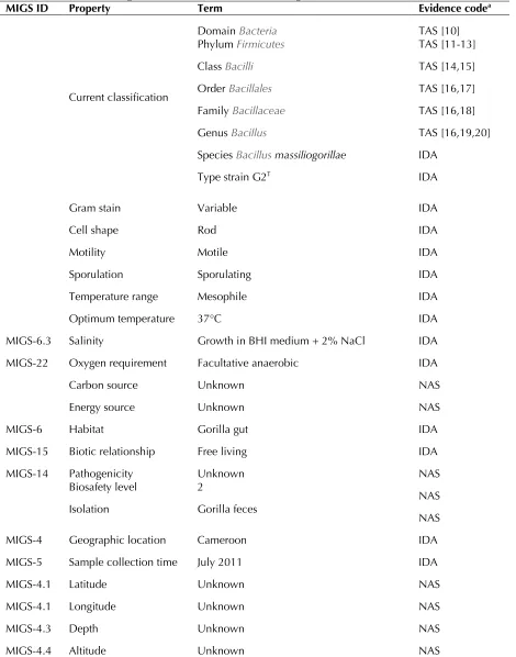
MIGS ID Property Term Evidence codea
Domai TAS [10]
Phylum TAS [11-13]
Cla TAS [14,15]
Current classification Orde TAS [16,17]
Family TAS [16,18]
Genu TAS [16,19,20]
Species IDA
Type strain G2T IDA
Gram stain Variable IDA
Cell shape Rod IDA
Motility Motile IDA
Sporulation Sporulating IDA
Temperature range Mesophile IDA
Optimum temperature 37°C IDA
MIGS-6.3 Salinity Growth in BHI medium + 2% NaCl IDA MIGS-22 Oxygen requirement Facultative anaerobic IDA
Carbon source Unknown NAS
Energy source Unknown NAS
MIGS-6 Habitat Gorilla gut IDA
MIGS-15 Biotic relationship Free living IDA
MIGS-14 Pathogenicity Unknown NAS
Biosafety level 2
NAS Isolation Gorilla feces
NAS
MIGS-4 Geographic location Cameroon IDA
MIGS-5 Sample collection time July 2011 IDA
MIGS-4.1 Latitude Unknown NAS
MIGS-4.1 Longitude Unknown NAS
MIGS-4.3 Depth Unknown NAS
MIGS-4.4 Altitude Unknown NAS
Bacillus infernus (NR027227) Bacillus methanolicus (NR 040985) Bacillus fumarioli (FJ973527) Bacillus jeotgali (JX094165)
Bacillus lentus (NR040792) Bacillus smithii (NR036987)
Bacillus badius (EU717967) Bacillus firmus (JX428993)
Bacillus benzoevorans (Y14693) Bacillus nealsoniis (NR044546) Bacillus circulans (JF833093)
Bacillus massiliogorillae (JX650055)
Bacillus simplex (HQ327114)
Bacillus psychrosaccharolyticus (JX429005) Bacillus flexus (JN033557)
Bacillus megaterium (FJ685764) Bacillus thuringiensis (HF545324)
Clostridium botulinum (AM412317)
100 100
87 97
54
74
56
52 78
39
40 62
23 15
18
Figure 1. Phylogenetic tree highlighting the position o strain G2T relative to other
type strains within the
quences were aligned using CLUSTAL X (V2), and phylogenetic inferences obtained using the maximum-likelihood method within the MEGA 5 software [22]. Numbers at the nodes are percentages of bootstrap values obtained by repeating the analysis 1,000 times to generate a majority consensus tree
Different growth temperatures (25, 30, 37, 45°C) were tested. Growth occurred at all tested tem-peratures, and the optimal growth was observed at 37°C. Colonies were 2-5 mm in diameter on Co-lumbia agar, grey opaque in color. Growth of the strain was tested under anaerobic and microaerophilic conditions using GENbag anaer and GENbag microaer systems, respectively (BioMérieux), and in aerobic conditions, with or without 5% CO2. Growth was achieved under
aer-obic (with and without CO2), microaerophilic and
anaerobic conditions. Gram staining showed Gram variable bacilli (Figure 2). A motility test was posi-tive. Cells grown on agar sporulate and the rods have a length ranging from 3.2 to 7.5 µm (mean 5.4 µm) and a diameter ranging from 0.8 to 1.2 µm (mean 1 µm) as determined by negative staining transmission electron microscopy (Figure 3).
Strain G2T exhibited catalase activity but not
oxi-dase activity. Using the API 50CH system (BioMerieux), a positive reaction was observed for D-glucose, D-fructose, D-ribose, N-acetylglucosamine, amygdalin, arbutin, aesculin, salicin, cellobiose, maltose, D-lactose, D-trehalose, D-saccharose, and hydrolysis of starch. Using the API ZYM system, positive reactions were observed for esterase (C4), esterase lipase (C8), phospha-tase acid, α- glucosidase and N-acetyl-β-glucosaminidase. The urease reaction was also positive, but nitrate reduction and indole produc-tion were negative. B. massiliogorillae is
Figure 2. Gram staining of B. massiliogorillae strain G2T
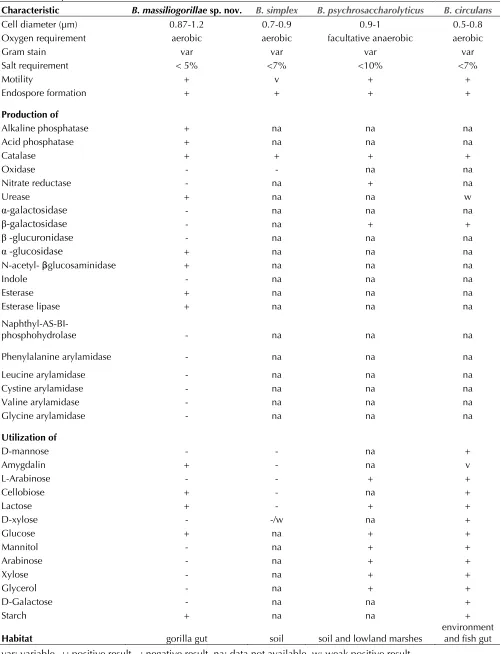
Table 2. Differential phenotypic characteristics between B. massiliogorillae sp. nov. strain G2T and phylogenetically
clos
Characteristic B. massiliogorillae sp. nov.
Cell diameter (µm) 0.87-1.2 0.7-0.9 0.9-1 0.5-0.8 Oxygen requirement aerobic aerobic facultative anaerobic aerobic
Gram stain var var var var
Salt requirement < 5% <7% <10% <7%
Motility + v + +
Endospore formation + + + +
Production of
Alkaline phosphatase + na na na
Acid phosphatase + na na na
Catalase + + + +
Oxidase - - na na
Nitrate reductase - na + na
Urease + na na w
α-galactosidase - na na na
β-galactosidase - na + +
β -glucuronidase - na na na
α -glucosidase + na na na
N-acetyl- β -glucosaminidase + na na na
Indole - na na na
Esterase + na na na
Esterase lipase + na na na
Naphthyl-AS-BI-phosphohydrolase - na na na
Phenylalanine arylamidase - na na na
Leucine arylamidase - na na na
Cystine arylamidase - na na na
Valine arylamidase - na na na
Glycine arylamidase - na na na
Utilization of
D-mannose - - na +
Amygdalin + - na v
L-Arabinose - - + +
Cellobiose + - na +
Lactose + - + +
D-xylose - -/w na +
Glucose + na + +
Mannitol - na + +
Arabinose - na + +
Xylose - na + +
Glycerol - na + +
D-Galactose - na na +
Starch + na na +
Habitat gorilla gut soil soil and lowland marshes environment and fish gut
When compared to other B.
massiliogorillae differed fr
the utilization of amygdalin, cellobiose, lactose and glucose (Table 2). It also differed from
and β-galactosidase production, and in the utiliza-tion of L-arabinose, mannitol, xylose and glycerol (Table 2). Differences were also observed with
the utilization of mannose, L-arabinose, D-xylose, mannitol, arabinose, D-xylose, glycerol and D-galactose (Table 2).
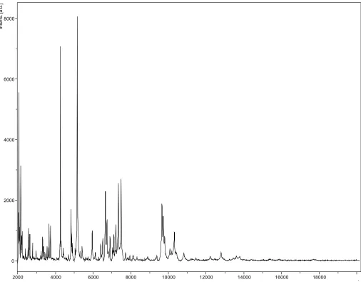
Matrix-assisted laser-desorption/ionization time-of-flight (MALDI-TOF) MS protein analysis was carried out as previously described [27,28]. De-posits were done for strain G2T from 12 isolated
colonies. Each smear was overlaid with 2µL of ma-trix solution (saturated solution of alpha-cyano-4-hydroxycinnamic acid) in 50% acetonitrile, 2.5% tri-fluoracetic-acid, and allowed to dry for five minutes. Measurements were performed with a Microflex spectrometer (Bruker Daltonics, Leipzig, Germany). Spectra were recorded in the positive linear mode for the mass range of 2,000 to 20,000 Da (parameter settings: ion source 1 (IS1), 20 kV; IS2, 18.5 kV; lens, 7 kV). A spectrum was obtained after 675 shots at a variable laser power. The time of acquisition was between 30 seconds and 1 mi-nute per spot. The 12 G2T spectra were imported
into the MALDI BioTyper software (version 2.0, Bruker) and analyzed by standard pattern match-ing (with default parameter settmatch-ings) against 6,252 bacterial spectra including 199 spectra from
10
BioTyper database. The method of identification included the m/z from 3,000 to 15,000 Da. For every spectrum, 100 peaks at most were taken into account and compared with spectra in the database. A score enabled the identification, or not, from the tested species: a score > 2 with a val-idated species enabled the identification at the species level, a score > 1.7 but < 2 enabled the identification at the genus level; and a score < 1.7 did not enable any identification. For strain G2T,
the scores obtained ranged from 1.177 to 1.343, thus suggesting that our isolate was not a member of a known species. We incremented our database with the spectrum from strain G2T (Figure 4).
Spectrum differences with other of
cies are shown in Figure 5.
Genome sequencing information
Genome project history
The organism was selected for sequencing on the basis of its phylogenetic position and 16S rRNA simi-larity to other members of the genus
part of a “culturomics” study of the gorilla flora aim-ing at isolataim-ing all bacterial species within gorilla feces. It was the 61st genome of
the first genome of sp. nov.
A summary of the project information is shown in Table 2. The Genbank accession number is CAVL000000000 and consists of 66 large contigs. Table 3 shows the project information and its asso-ciation with MIGS version 2.0 compliance [29].
Growth conditions and DNA isolation
B. massiliogorillae sp. nov. strain G2T, CSUR P206,
DSM 26159, was grown aerobically on 5% sheep blood-enriched Columbia agar at 37°C. Four petri dishes were spread and resuspended in 3x500µl of TE buffer and stored at 80°C. Then, 500µl of this suspension were thawed, centrifuged 3 minutes at 10,000 rpm and resuspended in 3x100µL of G2 buffer (EZ1 DNA Tissue kit, Qiagen). A first me-chanical lysis was performed by glass powder on the Fastprep-24 device (Sample Preparation sys-tem, MP Biomedicals, USA) using 2x20 seconds cy-cles. DNA was then treated with 2.5µg/µL lysozyme (30 minutes at 37°C) and extracted using the BioRobot EZ1 Advanced XL (Qiagen). The DNA was then concentrated and purified using the Qiamp kit (Qiagen). The yield and the concentration was measured by the Quant-it Picogreen kit (Invitro-gen) on the Genios Tecan fluorometer at 50ng/µl.
Genome sequencing and assembly
0 2000 4000 6000 8000
In
te
ns
. [a
.u
.]
2000 4000 6000 8000 10000 12000 14000 16000 18000
/ Figure 4. Reference mass spectrum from B. massiliogorillae strain G2T. Spectra from 12 individual colonies were compared and a reference spectrum was generated.
The paired-end library was clonally amplified with 0.5 cpb and 1 cpb in 2 emPCR reactions with the GS Titanium SV emPCR Kit (Lib-L) v2 (Roche). The yield of the emPCR was 19.4%, slightly above the expected yield ranging from 5 to 20% recom-mended by the Roche procedure.
Approximately 790,000 beads for a ¼ region were loaded on the GS Titanium PicoTiterPlate PTP Kit 70x75 and sequenced with the GS FLX Titanium Sequencing Kit XLR70 (Roche). The run was per-formed overnight and then analyzed on the cluster through the gsRunBrowser and Newbler assem-bler (Roche). A total of 322,962 passed filter wells were obtained and generated 64.2 Mb of sequenc-es with a length average of 310 bp. The passed filter sequences were assembled using Newbler with 90% identity and 40 bp as overlap. The final
assembly identified 60 scaffolds generating a ge-nome size of 4.6 Mb.
Genome annotation
Open Reading Frames (ORFs) were predicted us-ing Prodigal [30] with default parameters but the predicted ORFs were excluded if they spanned a sequencing gap region. The predicted bacterial protein sequences were searched against the GenBank database [31] and the Clusters of Orthol-ogous Groups (COG) databases using BLASTP. The tRNAScanSE tool [32] was used to find tRNA genes, whereas ribosomal RNAs were found by using RNAmmer [33] and BLASTn against the GenBank database. ORFans were identified if their BLASTP E-value was lower than 1e-03 for
To estimate the mean level of nucleotide sequence similarity at the genome level between B. massiliogorillae sp nov. strain G2T and another 3
pairwise and determined the mean percentage of nucleotide sequence identity among orthologous ORFs using BLASTn. Orthologous genes were de-tected using the Proteinortho software [34].
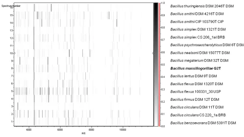
Figure 5. Gel view comparing
massiliogorillae G2T spectra with other members
of the
and
raw spectra of all loaded spectrum files arranged in a pseudo-gel like look. The x-axis records the m/z value. The left y-axis displays the running spectrum number originating from subsequent spectra loading. The peak intensity is expressed by a Gray scale scheme code. The color bar and the right y-axis indicate the relation between the color a peak is displayed with and the peak intensity in arbitrary units.
Table 3. Project information
MIGS ID Property Term
MIGS-31 Finishing quality High-quality draft
MIGS-28 Libraries used 454 paired-end 3- kb libraries MIGS-29 Sequencing platform 454 GS FLX Titanium
MIGS-31.2 Sequencing coverage 13×
MIGS-30 Assemblers Newbler version 2.5.3 MIGS-32 Gene calling method Prodigal
EMBL Date of Release April 18, 2013 EMBL ID CAVL000000000
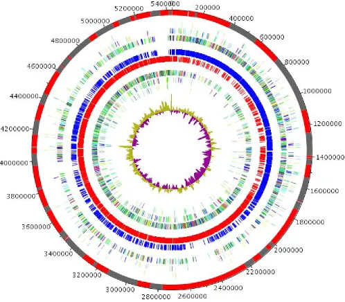
Genome properties
The genome is 5,431,633 bp long (1 chromosome, but no plasmid) with a 34.95% G+C content (Fig-ure 6 and Table 5). It is composed of 66 large contigs. Of the 5,276 predicted genes, 5,179 were protein-coding genes and 98 were RNAs (1 16S rRNA, 1 23S rRNA gene, 5 5S rRNA genes and 91 tRNA genes). A total of 3,801 genes (73.39%) were assigned a putative function (by COGS or by
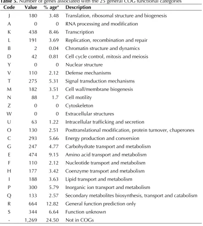
NR BLAST) and 368 genes were identified as ORFans (7.11%). The remaining genes were anno-tated as hypothetical proteins (666 genes, 12.86%). The distribution of genes into COGs functional categories is presented in Table 6. The properties and statistics of the genome are sum-marized in Tables 4 and 5.
Table 4. Nucleotide content and gene count levels of the genome
Attribute Value % of totala
Genome size (bp) 5,431,633 100
Coding region (bp) 4,561,287 83.98
G+C content (bp) 1,898,498 34.95
Total genes 5,276 100
RNA genes 98 1.84
Protein-coding genes 5,179 98.63
Genes with function prediction 3,801 73.39
Genes assigned to COGs 3,910 75.49
Genes with peptide signals 610 11.78
Genes with transmembrane helices 1,347 26.01 a The total is based on either the size of the genome in base pairs or the total number of protein coding genes in the annotated genome
Table 5. Number of genes associated with the 25 general COG functional categories Code Value % agea Description
J 180 3.48 Translation, ribosomal structure and biogenesis A 0 0 RNA processing and modification
K 438 8.46 Transcription
L 191 3.69 Replication, recombination and repair B 2 0.04 Chromatin structure and dynamics D 42 0.81 Cell cycle control, mitosis and meiosis Y 0 0 Nuclear structure
V 110 2.12 Defense mechanisms
T 275 5.31 Signal transduction mechanisms M 182 3.51 Cell wall/membrane biogenesis N 88 1.7 Cell motility
Z 0 0 Cytoskeleton
W 0 0 Extracellular structures
U 63 1.22 Intracellular trafficking and secretion
O 130 2.51 Posttranslational modification, protein turnover, chaperones C 293 5.66 Energy production and conversion
G 247 4.77 Carbohydrate transport and metabolism E 474 9.15 Amino acid transport and metabolism F 110 2.12 Nucleotide transport and metabolism H 177 3.42 Coenzyme transport and metabolism
I 188 3.63 Lipid transport and metabolism
P 300 5.79 Inorganic ion transport and metabolism
Q 133 2.57 Secondary metabolites biosynthesis, transport and catabolism R 664 12.82 General function prediction only
S 344 6.64 Function unknown - 1,269 24.50 Not in COGs
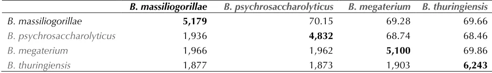
Comparison with other
genomes
Here, we compared the genome of B. massiliogorillae
strain G2T with those of
strain ATCC 23296,
and
The draft genome of B. massiliogorillae is larger in
size than those of
and smaller in size than that of
vs 6.26 Mb). B. massiliogorillae has a lower G+C
con-tent than
38.8%) and
slightly higher than tha
34.8%). The protein content of B. massiliogorillae is
higher than those of
and fewer than that of
6,243) (Table 6). In addition, B. massiliogorillae
shares 1,936, 1,966 and 1,877 orthologous genes
with
sequence identity of orthologous genes ranges from 68.46 to 70.15% among
69.28 to 70.15% between B. massiliogorillae and
other
new species status. Table 6 summarizes the number of orthologous genes and the average percentage of nucleotide sequence identity between the different genomes studied.
Table 6. The number of orthologous proteins shared between genomes†
B. massiliogorillae
B. massiliogorillae 5,179 70.15 69.28 69.66
1,936 4,832 68.74 68.46
1,966 1,962 5,100 69.86
1,877 1,873 1,903 6,243
†Lower left triangle- shared orthologous, upper right triangle- average percentage similarity of nucleotides correspond-ing to orthologous proteins shared between genomes, bold- number of proteins per genome
Conclusion
On the basis of phenotypic (Table 2), phylogenetic and genomic analyses (taxonogenomics) (Table 6), we formally propose the creation of massiliogorillae sp. nov. that contains the strain
G2T. This strain has been found in a stool sample
collected from gorilla in Cameroon.
Description of sp. nov.
(ma.sil.io.go.ril’ae. L. gen.
masc. n. massiliogorillae, combination of
the Latin name of Marseille, where strain G2T was
isolated, and of Gorilla, the Latin name of the goril-la, from which the stool sample was obtained).
B. massiliogorillae is an aerobic Gram-variable
bacte-rium. Optimal growth is achieved aerobically. No growth is observed in microaerophilic or anaerobic conditions. Growth occurs on axenic media between 25 and 45°C, with optimal growth observed at 37°C. Cells stain Gram-positive or negative, are rod-shaped, endospore-forming, motile and have a mean diameter of 1 µm (range 0.8 to 1.2 µm) and a mean length of 5.4 µm (range 3.2 to 7.5 µm). Colonies are grey opaque and 2-5 mm in diameter on blood-enriched BHI agar.
Catalase positive but oxidase negative. Using the API 50CH system (BioMerieux), a positive reaction is obtained for D-glucose, D-fructose, D-ribose, N-acetylglucosamin, amygdalin, arbutin, aesculin, salicin, cellobiose, maltose, lactose, trehalose, D-saccharose, and hydrolysis of starch. Using the API ZYM system, positive reactions are obtained for es-terase (C4), eses-terase lipase (C8), phosphatase acid, α- glucosidase and N-acetyl-β-glucosaminidase. Us-ing API 20NE, there are neither nitrate reduction nor indole production but urease reaction was positive. Susceptible to amoxicillin, nitrofurantoin, erythro-mycin, doxycycline, rifampin, vancoerythro-mycin, gentamy-cin and imipenem but resistant to trimethoprim- sulfamethoxazole, ciprofloxacin, ceftriaxon and amoxicillin-clavulanic acid.
The G+C content of the genome is 34.95%. The 16S rRNA and genome sequences are deposited in GenBank under accession numbers JX650055 and CAVL00000000, respectively. The type strain G2T (=
Acknowledgements
Fadi Bittar was supported by a Chair of Excellence IRD provided by the Institut de Recherche pour le Développement / Méditerranée-Infection foundation.
The authors thank the Xegen company for automating the genome annotation process.
References
1. Cohn F. Untersuchungen über Bakterien. Beitrage zur Biologie der Pflanzen Heft 1872; 1:127-224. 2. Abstract for the genus
LLC. Retrieved September 22, 2013.
http://doi.namesforlife.com/10.1601/nm.4857 3. Jernigan JA, Stephens DS, Ashford DA, Omenaca
C, Topiel MS, Galbraith M, Tapper M, Fisk TL, Zaki S, Popovic T, et al. Bioterrorism-related inha-lational anthrax: the first 10 cases reported in the United States. Emerg Infect Dis 2001; 7:933-944
4. Bottone EJ
pathogen. Clin Microbiol Rev 2010; 23:382-398
5. Mandell GL, Bennett JE, Dolin R. (2010) Princi-ples and Practice of Infectious Diseases. Elsevier. 4320 p.
6. Klee SR, Ozel M, Appel B, Boesch C, Ellerbrok H, Jacob D, Holland G, Leendertz FH, Pauli G, Grunow R, Nattermann H. Characterization of
great apes from Cote d'Ivoire and Cameroon. J Bacteriol 2006; 188:5333-5344
7. Leendertz FH, Yumlu S, Pauli G, Boesch C, Couacy-Hymann E, Vigilant L, Junglen S, Schenk
S, Ellerbrok H. A new
wild chimpanzees and a gorilla from West and Central Africa. PLoS Pathog 2006; 2:e8
8. Leendertz FH, Lankester F, Guislain P, Néel C, Drori O, Dupain J, Speede S, Reed P, Wolfe N, Loul S, et al. Anthrax in Western and Central Afri-can great apes. Am J Primatol 2006; 68:928-933
9. Sentausa E, Fournier PE. Advantages and limita-tions of genomics in prokaryotic taxonomy. Clin Microbiol Infect 2013
10. Woese CR, Kandler O, Wheelis ML. Towards a natural system of organisms: proposal for the do-mains ArchaEukarya.Proc Natl
Acad Sci USA 1990; 87:4576-4579
11. Gibbons NE, Murray RGE. Proposals concerning the higher taxa oInt J Syst Bacteriol 1978; 28:1-6.
12. Garrity GM, Holt JG. The Road Map to the Man-ual. In: Garrity GM, Boone DR, Castenholz RW (eds), Bergey's Manual of Systematic Bacteriolo-gy, Second Edition, Volume 1, Springer, New York, 2001, p. 119-169.
13. Murray RGE. The Higher Taxa, or, a Place for Everything...? In: Holt JG (ed), Bergey's Manual of Systematic Bacteriology, First Edition, Volume 1, The Williams and Wilkins Co., Baltimore, 1984, p. 31-34.
14. Ludwig W, Schleifer KH, Whitman WB. Class I. In: De Vos P, Garrity G, Jones D, Krieg NR, Ludwig W, Rainey FA, Schleifer KH, Whitman WB (eds), Bergey's Manual of Systemat-ic Bacteriology, Second Edition, Volume 3, Springer-Verlag, New York, 2009, p. 19-20. 15. List of new names and new combinations
previ-ously effectively, but not validly, published. List no. 132. Int J Syst Evol Microbiol 2010; 60
:469-472
16. Skerman VBD, McGowan V, Sneath PHA. Ap-proved Lists of Bacterial Names. Int J Syst Bacteriol 1980; 30:225-420.
17. Prévot AR. In: Hauderoy P, Ehringer G, Guillot G, Magrou. J., Prévot AR, Rosset D, Urbain A (eds), Dictionnaire des Bactéries Pathogènes, Second Edition, Masson et Cie, Paris, 1953, p. 1-692. 18. Fischer A. Untersuchungen über bakterien.
Jahrbücher für Wissenschaftliche Botanik 1895; 27:1-163.
20. Cohn F. Untersuchungen über Bakterien. Beitr Biol Pflanz 1872; 1:127-224.
21. Ashburner M, Ball CA, Blake JA, Botstein D, But-ler H, Cherry JM, Davis AP, Dolinski K, Dwight SS, Eppig JT, et al. Gene ontology: tool for the unification of biology. The Gene Ontology Con-sortium. Nat Genet 2000; 25:25-29
22. Tamura K, Peterson D, Peterson N, Stecher G, Nei M, Kumar S. MEGA5: Molecular Evolutionary Genetics Analysis using Maximum Likelihood, Evolutionary Distance, and Maximum Parsimony Methods. Mol Biol Evol 2011; 28:2731-2739
23. Stackebrandt E, Ebers J. Taxonomic parameters revisited: tarnished gold standards. Microbiol To-day 2006; 33:152-155.
24. Heyrman J, Logan NA, Rodríguez-Díaz M, Scheldeman P, Lebbe L, Swings J, Heyndrickx M, De Vos P. Study of mural painting isolates, lead-ing to the transfer o' and
' to
description of
the strains previously attributed to
nov. Int J Syst Evol Microbiol 2005; 55:119-131
25. Larkin JM, Stokes JL. Taxonomy of psychrophilic strains oJ Bacteriol 1967; 94:889-895
26. De Paolis MR, Lippi D. Use of metabolic and mo-lecular methods for the identification of a strain isolated from paper affected by foxing. Microbiol Res 2008; 163:121-131
27. Roux V, El Karkouri K, Lagier JC, Robert C, Raoult D. Non-contiguous finished genome sequence and description of sp. nov. Stand Genomic Sci 2012; 7:221-232
28. Seng P, Drancourt M, Gouriet F, La Scola B, Fournier PE, Rolain JM, Raoult D. Ongoing revo-lution in bacteriology: routine identification of bacteria by matrix-assisted laser desorption ioni-zation time-of-flight mass spectrometry. Clin In-fect Dis 2009; 49:543-551
29. Field D, Garrity G, Gray T, Morrison N, Selengut J, Sterk P, Tatusova T, Thomson N, Allen MJ, Angiuoli SV, et al. The minimum information about a genome sequence (MIGS) specification. Nat Biotechnol 2008; 26:541-547
30. Prodiga
31. GenBank database.
32. Lowe TM, Eddy SR. t-RNAscan-SE: a program for improved detection of transfer RNA gene in ge-nomic sequence. Nucleic Acids Res 1997; 25:955-964
33. Lagesen K, Hallin P, Rodland EA, Staerfeldt HH, Rognes T, Ussery DW. RNAmmer: consistent and rapid annotation of ribosomal RNA genes. Nucle-ic Acids Res 2007; 35:3100-3108
34. Lechner M, Findeib S, Steiner L, Marz M, Stadler PF, Prohaska SJ. Proteinortho: Detection of (Co-)orthologs in large-scale analysis. BMC Bioinfor-matics 2011; 12:124
![Figure 1. Phylogenetic tree highlighting the position of quences were aligned using CLUSTAL X (V2), and phylogenetic inferences obtained using the maximum-type strains within the likelihood method within the MEGA 5 software [22]](https://thumb-us.123doks.com/thumbv2/123dok_us/697033.2067640/3.612.77.517.51.389/phylogenetic-highlighting-position-clustal-phylogenetic-inferences-likelihood-software.webp)