R E V I E W
Open Access
The march of pluripotent stem cells in
cardiovascular regenerative medicine
Haissam Abou-Saleh
1, Fouad A. Zouein
2†, Ahmed El-Yazbi
2,4†, Despina Sanoudou
5, Christophe Raynaud
6,
Christopher Rao
7, Gianfranco Pintus
3, Hassan Dehaini
2and Ali H. Eid
1,2,3*Abstract
Cardiovascular disease (CVD) continues to be the leading cause of global morbidity and mortality. Heart failure remains a major contributor to this mortality. Despite major therapeutic advances over the past decades, a better understanding of molecular and cellular mechanisms of CVD as well as improved therapeutic strategies for the management or treatment of heart failure are increasingly needed. Loss of myocardium is a major driver of heart failure. An attractive approach that appears to provide promising results in reducing cardiac degeneration is stem cell therapy (SCT). In this review, we describe different types of stem cells, including embryonic and adult stem cells, and we provide a detailed discussion of the properties of induced pluripotent stem cells (iPSCs). We also present and critically discuss the key methods used for converting somatic cells to pluripotent cells and iPSCs to cardiomyocytes (CMs), along with their advantages and limitations. Integrating and non-integrating reprogramming methods as well as characterization of iPSCs and iPSC-derived CMs are discussed. Furthermore, we critically present various methods of differentiating iPSCs to CMs. The value of iPSC-CMs in regenerative medicine as well as myocardial disease modeling and cardiac regeneration are emphasized.
Keywords:Cardiovascular disease, Stem cell therapy, iPSCs, Heart failure, Cardiomyocytes, Regenerative medicine
Background
Cardiovascular disease (CVD) remains the leading cause of death worldwide, killing 17 million people each year. The World Health Organization (WHO) estimates that by 2020 this number will reach 24 million. With com-plex multifactorial pathologies, including both genetic and environmental factors, CVD continues to be difficult to prevent. Current strategies against CVD include preven-tion (i.e., lifestyle changes) and pharmacological and/or surgical intervention. However, the effectiveness of drug treatment varies among individuals, while surgical inter-ventions may not be applicable to all patients. New ap-proaches need to be established to better understand the mechanisms of CVD and improve diagnostic and thera-peutic strategies, particularly in the context of heart failure.
Loss of myocardium results in the clinical syndrome of heart failure [1]. The long-term prognosis of heart failure is poor and current therapies are largely palliative [2, 3]. The only treatment for end-stage heart failure with established long-term efficacy is transplantation. However, the increasing prevalence of heart failure and existing shortage of donor organs are frequent chal-lenges [4,5].
Stem cell therapy (SCT) aims to reduce cardiac degen-eration by regenerating cardiomyocytes (CMs) and is currently considered one of the most promising thera-peutic strategies [6, 7]. Stem cells are undifferentiated cells theoretically capable of renewing themselves indef-initely under appropriate conditions through mitotic cell division, and can maintain, generate, or replace damaged tissue by differentiating into specialized cell types [8]. This review describes different types of stem cells, in-cluding embryonic stem cells (ESCs) and adult stem cells (ASCs), and focuses primarily on induced pluripotent stem cells (iPSCs). The key methods used for converting somatic cells to iPSCs and then to CMs are presented, along with their advantages and limitations. Emphasis is
* Correspondence:ae81@aub.edu.lb
†Fouad A. Zouein and Ahmed El-Yazbi contributed equally to this work. 1
Department of Biological and Environmental Sciences, Qatar University, Doha, Qatar
2Department of Pharmacology and Toxicology, Faculty of Medicine,
American University of Beirut, Beirut, Lebanon
Full list of author information is available at the end of the article
given to the value of iPSC-derived CMs (iPSC-CMs) in regenerative medicine and myocardial disease modeling.
Stem cell potency
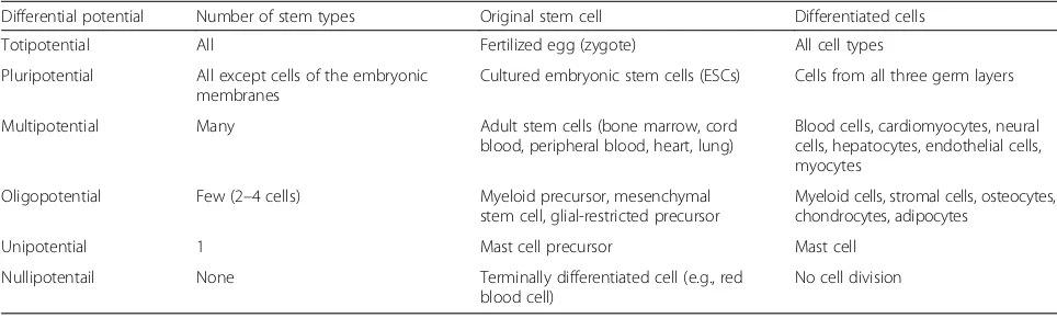
Stem cells can be classified according to their“potency” or “differentiation potential” (Table 1). Importantly, newer cell types, such as iPSC-CMs, directly transdiffer-entiated CMs, and endogenous cardiac stem cell derived CMs (CSC-CMs), could be easily obtained from any in-dividual and used to create patient- and disease-specific models, enabling the elucidation of molecular and gen-etic mechanisms that underlie inherited diseases pheno-types and unveiling novel therapeutic and personalized therapeutic targets [9–14].
Multipotent stem cells for SCT
Adult or somatic stem cells (ASCs) are non-embryonic multipotent stem cells found in the adult organism after embryonic development and residing in an area in tis-sues called the “stem cell niche” [15, 16]. ASCs exist in various tissues, such as the bone marrow [17, 18], cord blood [19,20], skeletal muscles [21,22], peripheral blood [23, 24], adipose tissue [25, 26], lung [27, 28], and the heart [29, 30]. Unlike ESCs, ASC origins are not well defined and their multipotency is very limited. Their primary functions are to maintain the homeostasis of mature cell tissues and, with limitations, to regenerate damaged organs. However, ASCs are rare in mature tis-sues, have limited capacity to differentiate into multiple cell lineages, and behave differently depending on envir-onmental stimuli. In addition, their isolation from adult tissues is challenging, and methods of culture have not yet been optimized. For example, bone marrow-derived hematopoietic stem cells (HSCs) have been studied in multiple diseases, including bone-marrow failure [31], vas-culogenesis [32, 33], and cardiac regeneration [17, 34]. However, HSCs represent a very small fraction (only 0.01–0.015%) of the total bone marrow cells and their therapeutic and differentiation potential is highly controver-sial [35,36]. Consequently, although ASCs would represent
a valuable and promising source of stem cells and SCT, their use is still hindered by a series of biological and tech-nical limitations that require further investigation.
Pluripotent stem cells for SCT: shift from ESCs to iPSCs
ESCs are isolated from embryos and can be classified as totipotent or pluripotent depending on their temporal existence during fetal development. Totipotent ESCs are present in the earliest eight-cell stage embryo, whereas pluripotent ESCs are found throughout the remainder of embryonic development. In this review, ESCs refer to the pluripotent type of ESCs, obtained from a 4- or 5-day-old embryo, also known as the blastocyst phase of development. ESCs are extracted from the inner cell mass of blastocysts and placed in a controlled culture that allows them to divide indefinitely without further cell differentiation. These ex vivo expanded cells serve as a paramount source of stem cells for transplantation therapies for many diseases, including cardiomyopathies, neurological disorders, and diabetes (Fig. 1). However, a series of ethical and technical issues restricts ESC use [37]. Technically, the use of ESCs for cell transplantation requires a differentiation step to the target cell lineage with formation of undifferentiated cells amongst the cel-lular product [38]. This can induce spontaneous tera-toma formation in host tissue, raising safety concerns that must be carefully addressed [39, 40]. Moreover, the allogeneic nature of ESCs may induce immune responses with a prominent risk of rejection.
Ethically, the use of human ESCs (hESCs) is contro-versial, with many pro-life advocates being concerned about the isolation of hESCs from “living” embryos. In 2001, the USA government banned stem cell research by restricting federal funding for research on hESCs. To allow responsible scientific research involving human stem cells, the National Institutes of Health (NIH) estab-lished the “Human Embryonic Stem Cell Registry”, which lists 177 stem cell lines that are suitable for em-ployment in federally funded research. Unfortunately,
Table 1Differential potential of stem cells
Differential potential Number of stem types Original stem cell Differentiated cells
Totipotential All Fertilized egg (zygote) All cell types
Pluripotential All except cells of the embryonic membranes
Cultured embryonic stem cells (ESCs) Cells from all three germ layers
Multipotential Many Adult stem cells (bone marrow, cord
blood, peripheral blood, heart, lung)
Blood cells, cardiomyocytes, neural cells, hepatocytes, endothelial cells, myocytes
Oligopotential Few (2–4 cells) Myeloid precursor, mesenchymal
stem cell, glial-restricted precursor
Myeloid cells, stromal cells, osteocytes, chondrocytes, adipocytes
Unipotential 1 Mast cell precursor Mast cell
Nullipotentail None Terminally differentiated cell (e.g., red
blood cell)
not all of these stem cell lines are readily available, and scientists have concerns about the quality and the lon-gevity of these stem cell lines. To bypass these chal-lenges, an increasing number of laboratories around the world are currently using iPSCs to limit the use of hESCs and the destruction of living human embryos.
iPSCs: the promising era of SCT
Practical considerations such as the availability of em-bryonic tissues and the isolation of relatively rare cell types have limited the large-scale production of pure stem cells for industrial and clinical applications. As such, the stem cell research field has explored other op-tions, such as transforming fully differentiated adult somatic cells into pluripotent stem cell (PSCs). The reacquisition of a pluripotent state, known as“cell repro-gramming”, represents a paradigm shift in our under-standing of cellular differentiation and of the plasticity of the differentiated state.
Historical overview
The concept of cell reprogramming is not novel (Fig.2). It was first proposed in 1950 by Robert Briggs and Thomas King, who successfully achieved nuclear transfer
adult cell-derived animal (a sheep known as Dolly) was achieved using the SCNT method [51]. In 2006, Shinya Yamanaka (Nobel Prize in Medicine, 2012) from Kyoto University established the first iPSCs by insertion of de-fined“stemness” genes into the nucleus of somatic cells [52]. These genes were retrovirally introduced into adult mouse fibroblasts and encoded four transcription factors (Oct3/4, Sox2, Klf4, and c-Myc (OSKM)) known to be involved in the maintenance of pluripotency. Yamanaka’s work transformed our understanding of epigenetic re-programming of somatic cells to a pluripotent state and set the ground for the development of human iPSCs (hiPSCs). This can now be achieved using either the original four genes [53] or a different combination of Oct3/4, Sox2, Nanog, and Lin28 [54,55].
Nanog: the ever-young player in the iPSC orchestra
To date, the transcription factor Oct3/4 is thought to be indispensable for inducing pluripotency in somatic cells whereas Sox2, Klf4, and c-Myc are alternative supporting factors [56]. In 2003, Ian Chambers from the University of Edinburgh isolated a mouse gene, named Nanog, after the mythological Celtic land of the ever young, Tir nan Og. The Nanog gene is specifically expressed in PSCs and thought to be a key factor in maintaining the pluripotency state [57, 58]. Thus, it has been shown that the overex-pression of Nanog in mESCs causes them to self-renew in the absence of cytokines and growth factors. Similar
results were obtained with hESCs; Nanog overexpression enabled their propagation for multiple passages during which the cells remained pluripotent [59]. Conversely, the knockdown of Nanog promotes the differentiation of ESCs into other cell types, thereby demonstrating the cap-ability of this gene to preserve the stemness state [60,61]. Further, Nanog has been used in concert with other tran-scription factors to reprogram human somatic cells to iPSCs, in which it can serve as a selective marker of pluri-potency [53–55,62].
Inducing PSCs
iPSCs are reprogrammed adult somatic cells, originally produced by retrovirus-mediated transduction of four transcription factors—Oct3/4, Sox2, Klf4, and c-Myc— known subsequently as OSKM factors [52]. The newly created iPSCs display phenotypic and functional proper-ties of ESCs and contribute to embryonic development when injected into mouse blastocysts. Since then, mouse iPSCs (miPSCs) have been generated from embryonic fi-broblasts [62], adult tail-tip fibroblasts [55], hepatocytes and gastric epithelial cells [63], pancreatic cells [64], neural stem cells [65], and B lymphocytes [66]. Add-itionally, researchers have reported generating iPSCs from somatic tissues of monkey [67] and rat [68]. In humans, many tissue sources have been used for suc-cessful generation of iPSCs. These include peripheral blood cells [24], cord blood cells [69, 70], keratinocytes Fig. 2Stem cell research: key dates. Genetic reprogramming started as early as 1958 with the first somatic nuclear cell transfer, demonstrating that the nucleus was responsible for the function of a cell. The derivation of the first embryonic stem cell from mice was only achieved in the early 1980s. The major breakthrough that turned world attention toward cloning and genetic manipulation happened in 1997 with the first animal cloning of the famous sheep Dolly. Soon after, in 1998, the first human embryonic stem cell was derived. Those cells remained the only pluripotent stem cells at the disposal of researchers until 2006, when Shinya Yamanaka identified the reprogramming factors capable of inducing pluripotency in adult cells. Somatic nuclear cell transfer image is courtesy of Howard Hughes Medical Institute (HHMI). Mouse ESC image is courtesy ofemouseatlas.org. Dolly the sheep, human ESC, and mouse iPSC images are courtesy ofwikipedia.org.ESCembryonic stem cell,
[71], skin fibroblasts [53, 72–74], melanocytes [75], adi-pocytes [76], and neural stem cells [77]. Consequently, the development of hiPSCs has rapidly emerged as a promising source of PSCs, a tremendously valuable source of cells for tissue engineering, cell-based therap-ies, novel drug screening, as well as the molecular and cellular characterization of disease pathogenesis. Several approaches towards the generation of iPSCs have emerged. The methods used to reprogram adult cells to iPSCs can be grouped into two major categories, inte-grating and non-inteinte-grating methods [78].
Integrating reprogramming methods
Viral integration method
The viral integration method represents the first success-ful approach for somatic cell reprogramming to iPSCs and uses viral delivery (retrovirus or lentivirus) of four repro-gramming factors (OSKM) into the host genome [79]. In this method the transgenes carried by the viral vectors are randomly inserted into the host genome and iPSC col-onies appear in culture within 3–4 weeks (Fig.3). Expres-sion of the transgenes is normally silenced in iPSCs, although a low level of expression or spontaneous reacti-vation may be observed. This may in turn affect other aspects of gene expression, DNA methylation, or pluripo-tency potential [72, 80–83]. As a result, such iPSCs may affect the phenotypes of their derived cells, rendering them refractory to differentiation in vitro or in vivo
following transplantation. For example, c-Myc is a well-known proto-oncogene whose reactivation following retroviral gene transduction resulted in tumor formation in almost 50% of chimeric mice generated from iPSCs [62, 84, 85]. Therefore, other reprogramming factors have been screened and c-Myc-free iPSCs were generated using a combination of four or three of the Oct3/4, Sox2, Nanog, and Lin28 factors [54,55,85–87]. These alterna-tive approaches were successful in the production of iPSCs without transgenic insertion of c-Myc, albeit with reduced efficiency [55,84]. Other studies have further re-duced the number of genes required for reprogramming to one or two factors using Oct3/4 alone [77, 88] or in combination with Sox2 or Klf4 [65,89–91]. Of note, the omission of one or more of the reprogramming factors is largely dependent on the endogenous expression of these factors in the donor cell type. For example, hiPSC deriv-ation using the lentiviral system takes several weeks with skin fibroblasts but only 10 days with keratinocytes, in which the expression levels of Klf4 and c-Myc are much higher [92]. Therefore, the best combination of reprogram-ming factors is partly dependent on the hosting cell type.
Viral integration followed by excision: the Cre-Lox system
The problem of permanent integration of transgenes in a host genome was partially solved by viral integration of OSKM factors into the host genome followed by their excision using the Cre-Lox recombinase system (Fig.4).
In mammalian cells, Cre-Lox recombination is widely used to control gene expression, induce chromosomal re-arrangement, or delete undesired DNA segments (Fig. 5) [93,94]. In the context of hiPSCs, LoxP-lentiviral vectors containing either four (Oct3/4, Sox2, Klf4, c-Myc) or three (Oct3/4, Sox2, Klf4) reprogramming factors flanked be-tween two unidirectional LoxP sites have been employed [95]. The hiPSCs are then transiently transfected with an expression vector encoding Cre-recombinase that medi-ates the excision of the integrated transgene (Fig.5). This has the advantage of inducing the generation of transgene-free hiPSCs, favoring the translation of iPSC technology into clinical applications. Despite the efficiency of Cre-recombinase-driven excision and the advantages of this approach, residual viral vector sequences can remain at the sites of integration, which may in turn trigger un-desirable downstream effects, while the overall reported reprogramming efficiency remains very low.
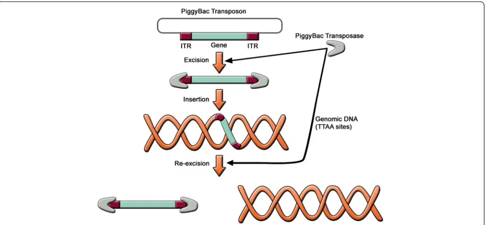
Non-viral integration followed by removal: the PiggyBac transposition
In order to avoid viral integration altogether, transposon-based non-viral integration methods have been developed using the PiggyBac (PB) transposon system. The PB trans-posons are mobile genetic elements used to transpose target sequences between vectors and chromosomal DNA via a“cut and paste”mechanism (Fig.6) [96]. The procedure consists of co-transfecting cells with PB transposon vectors (containing target sequence) and PB transposase expression plasmids. The PB transposase recognizes specific inverted terminal repeat (ITR) sequences located on both ends of the transposon vector, efficiently removes the contents from the transposon sites, and integrates them into TTAA chromo-somal sites. Cells harboring an inserted PB vector are transi-ently re-transfected with the PB transposase expression vector. The PB transposase substantially re-excises the trans-posons from the genome,“footprint”-free.
Fig. 4Lox site. The 8-bp core sequence is flanked by two 13-bp inverted repeats
Transgene-free iPSC lines were generated from human embryonic fibroblasts (hEFs), human embryonic kidney 293 (HEK293) cells, and adult skin fibroblasts using the PB transposon-based system [97]. This approach has several advantages over the traditional viral integrating methods for reprogramming. First, the plasmid DNA and the transfection protocol used for cell delivery of PB transposon vectors are innocuous and offer the oppor-tunity to reprogram cell types that are prone to viral in-fection. Second, the feasibility of the protocol and the reliability of the PB transposase-mediated excision en-hance the establishment of transgene-free hiPSC lines. However, this approach results in low yields (< 2%) of bona fide iPSCs. Of note, it has been shown that the effi-ciency of iPSC derivation from human adult fibroblasts using PB transposon vectors is enhanced by 15- to 51-fold after addition of butyrate, a small-chain fatty acid [98]. The mechanism of butyrate action includes histone acetylation, DNA demethylation, and the expres-sion of endogenous pluripotency associated genes.
Although remarkable progress has been made towards safe and efficient reprogramming, the aforementioned methods involve integration of transgenes into the host genome with unpredictable interruptions to the host cell genome and downstream consequences. In order to avoid any permanent or transient genomic modifications a safer approach for iPSC derivation is to avoid both permanent and transient genomic modification. There-fore, non-integrating methods for cell reprogramming have been developed and considered.
Non-integrating reprogramming methods
Viral non-integrating method
The viral non-integrating method involves the gener-ation of iPSCs using non-integrating viruses such as ade-noviruses and sendai viruses for the delivery of OSKM factors (Fig. 7). As opposed to retroviruses and lentivi-ruses, these expression vectors do not integrate into the host genome and show high-level expression of exogen-ous genes [99–101]. So far, the adenoviral/sendaiviral iPSCs display features of reprogrammed cells, express endogenous pluripotency genes, and contribute to tissue development in chimeric mice. Furthermore, viral gen-ome and viral proteins were totally absent in iPSC clones generated by adenoviral or sendaiviral transduction. However, major issues are hindering the long-term suc-cess of this method. For example, in most cases, iPSC lines generated by adenoviral/sendiviral transduction formed teratomas when injected into immunodeficient mice [99–101]. Furthermore, Stadtfeld and colleagues found that almost 25% of the adenoviral iPSC lines were tetraploid, which is not seen in iPSCs produced with retro- or lentiviral vectors [99]. The authors postulate that adenoviral reprogramming either induces cell fusion or, alternatively, selects for rare tetraploid cells pre-exist-ing in the startpre-exist-ing cell populations. In addition, the effi-ciency of deriving iPSCs was ~ 100-fold lower than that obtained with integrating viruses. This is probably due to the fact that many cells do not maintain gene expres-sion of OSKM factors long enough to trigger entry into a pluripotent state.
Non-viral non-integrating methods
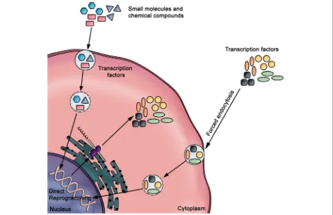
Non-viral non-integrating methods consist of the deriv-ation of iPSCs through virus-free and transgene-free techniques. This relies on the induction of iPSCs by transient transfection of plasmid DNA, minicircle DNA, or synthetic RNA encoding OSKM factors, as well as the direct delivery of recombinant proteins of OSKM factors into the cells.
Plasmid DNA
When transfected into cells, plasmid DNA replicates in-dependently of the genomic DNA without incorporating into the genome of the host cells. Transgene-free iPSCs have been produced from mouse [102] and human [100, 103] fibroblasts by transient transfection with plasmid vectors. In particular, hiPSCs were generated by repeated transient transfection with three plasmids
expressing seven reprogramming factors. These factors include Oct3/4, Sox2, c-Myc, Klf4, Nanog, and Lin 28, along with Epstein-Barr nuclear antigen-1 (EBNA-1), and SV40 large T antigen (SVLT), which allow stable extra-chromosomal replication of the plasmid vectors [100]. Interestingly, the omission of the later factor resulted in cell toxicity and disappearance of iPSC colonies. Although the isolated hiPSCs were devoid of vector or transgene ex-pression, the differentiation process remained extremely low and required repetitive transfections.
Minicircle DNA
Minicircle DNA are small supercoiled derivatives of plasmids that are free of all prokaryotic vector sequences and are composed essentially of a small eukaryotic expression cassette (~ 4 kb). The absence of bacterial DNA backbone makes them powerful tools for genetic Fig. 7Non-integrative methods using plasmids, sendaiviruses, or RNA delivery. Non-integrative methods (DNA- or RNA-based) have been
manipulation of mammalian cells. In addition, their small size enhances their transfection capacity and con-fers a long ectopic expression pattern compared to standard plasmids [104,105]. Minicircle vectors carrying a cassette of the transcription factors Oct3/4, Sox2, Lin28, and Nanog have been employed for derivation of hiPSCs from adipose stromal cells [106] and neonatal fi-broblasts [107]. No genomic integration of the minicircle transgene has been detected in hiPSC subclones as con-firmed by Southern blot analysis. However, the reprogram-ming efficiency remains extremely low (0.0005–0.005%) compared to viral integration techniques used for the ex-pression of the same transcription factors [54,55].
RNA delivery
The RNA-based method for somatic cell reprogramming consists of delivering OSKM factors by repeated adminis-tration of synthetic messenger RNA (mRNA), an approach that overcomes viral genome integration or immune re-sponses to foreign DNA. Multiple human cell types have been reprogrammed using synthetic modified messenger RNA [108]. Furthermore, the same technology has been employed to differentiate the mRNA-induced iPSCs into
myogenic cells. Recently, the use of selected microRNAs (miRNAs) with or without OSKM factors has been shown to be an efficient method of producing iPSCs [109–111]. The mechanism by which miRNAs enhance iPSCs repro-gramming is unclear, but it could be related to their ability to regulate the cell cycle [111]. Of note, several miRNAs used in the reprogramming process are usually expressed in ESCs and are thought to maintain the ESC phenotype [112, 113]. The RNA-based method represents a promis-ing strategy to reprogram somatic cells with less or no genetic modifications, qualifying mRNA-reprogrammed cells for clinical applications. Nonetheless, this approach entails a small risk of genetic modification due to the introduction of nucleic acids into the cell.
Protein delivery
The protein delivery method involves the direct delivery of reprogramming factors (i.e., proteins) into the cell (Fig. 8). Through this approach, hiPSCs have been successfully generated from mouse [114] and human neonatal fibroblasts [115] by direct delivery of the OSKM factors conjugated with a cell-penetrating polyar-ginine peptide. Of note, this method has an attractive
advantage of being virus-free and does not include gen-etic modification or DNA transfection. However, the low reprogramming efficiency and the need for repeated treatments represent the major limitations.
Improving iPSC reprogramming efficiency
Numerous chemicals and small molecules have been shown to improve the efficiency of iPSC generation or enable the reduction of the reprogramming factors re-quired for pluripotency induction [116]. These mole-cules and compounds can be divided into two groups: 1) chromatin modifiers and 2) regulators of cell signaling pathway [117]. For instance, valproic acid (VPA) is a small molecule histone deacetylase inhibitor which has been used to successfully reprogram foreskin fibroblasts with only two factors: Oct3/4 and Sox2 [89]. The repro-gramming efficiency was significantly improved when VPA was applied to cells expressing high endogenous levels of c-Myc and Klf4, such as keratinocytes or adi-pose stromal cells [92, 118]. Other studies optimized the reprogramming efficiency by combining two or three small molecules with transcription factors. For example, neonatal epidermal fibroblasts have been reprogrammed by using Oct3/4 and Klf4 supplemented with CHIR99021 (Wnt signaling pathway activator) and Parnate (histone demethylase inhibitor) [119]. Similarly, the combination of SB431542 (transforming growth factor, TGF-β inhibitor), PD0325901 (MEK inhibitor), and thiazovinin (cell-survival enhancer) significantly promotes the reprogramming effi-ciency of fibroblasts [119]. Also, the addition of vitamin C together with VPA to serum-containing culture media im-proved reprogramming efficiency by threefold compared with VPA alone [120]. Despite the tremendous efforts invested to achieve a high reprogramming efficiency, the yields of bona fide hiPSCs have rarely exceeded 1%. Two conflicting models have been proposed to explain the renitence to pluripotency induction, namely the “elite” and“stochastic”models [121,122]. The elite model postu-lates that only a small fraction of somatic cells, most likely the tissue-resident stem cells, are subjected to reprogram-ming. The stochastic model argues that under specific cul-ture conditions, either tissue-resident stem cells or fully differentiated cells can be successfully reprogrammed to a pluripotent state in a stochastic fashion [64, 66, 123]. Further investigation is needed to establish a consensus model that allows a better understanding of the mechanisms of reprogramming at the multicellular and single-cell levels.
Characterization of iPSC lines
Reprogramming of somatic cells is hindered by the het-erogeneity of the derived iPSC lines, which affects their differentiation potential into specific cell lineages. Even a single reprogramming experiment could generate multiple
iPSC lines which exhibit distinct molecular and functional characteristics [124–126]. This problem is largely due to the differential propensity to pluripotency induction among cells and our limited understanding of the under-lying reprogramming mechanisms. In this context, several methods have been employed to evaluate the characteris-tics of established iPSC clones. Whole genome expression or quantitative reverse-transcription polymerase chain reac-tion (qRT-PCR) can be used to assess the gene expression signatures of the iPSC clones, while immunocytochemistry and western blots are employed to examine protein expres-sion. The differentiation potential of iPSC clones can be assessed in vitro by embryoid body formation and in vivo by teratoma formation after transplantation in animals. In another exciting approach, Chan and colleagues attempted to define the molecular signature of the fully repro-grammed hiPSCs using in situ live cell imaging [127]. They found that transgene silencing and expression of the pluripotency markers TRA-1-60, DNA (cytosine-5-)-meth-yltransferase 3 beta(DNMT3B), and REX1 marked the fully reprogrammed state whilst alkaline phosphatase, SSEA-4, growth differentiation factor 3 (GDF3), human telomerase reverse transcriptase (hTERT), and Nanog are insufficient as markers. Recently, Burridge and colleagues claimed to have established culture conditions that circumvent the interline variability of iPSC lines, which could significantly facilitate the downstream characterization of the repro-grammed iPSCs and increase the number of suitable iPSCs for the needs of each project [128].
Host cells used for iPSC reprogramming
Fibroblasts
The vast majority of studies on hiPSC derivation from somatic cells have employed dermal fibroblasts as the start-ing population for reprogrammstart-ing [129–131]. Fibroblasts play an important role within the dermis and are respon-sible for the synthesis of connective tissues and remodeling of the extracellular matrix. They can be obtained from a single skin biopsy followed by 3–4 weeks of in vitro incu-bation to generate a sufficient amount of starting cell population [132]. Their easy isolation and expansion ren-ders them the best source of iPSCs. However, the efficiency of reprogramming is very low, ranging from 0.0001% (when using reprogramming factors without c-Myc) to 0.01% (in the presence of c-Myc) [53, 55, 89, 132]. In addition, the time required for the formation of iPSCs is relatively long and colonies usually take up to 2 months to appear in culture [133]. However, recent reports suggest approaches that increase efficiency of reprogramming of primary fibroblasts [129,130].
Keratinocytes
provide strength to the hair and nails. One study has re-ported the generation of iPSCs from keratinocytes ob-tained from human foreskin biopsies and plucked hair [71]. These cells showed a significant improvement in re-programming efficiency and speed compared to skin fi-broblasts. However, the keratinocytes used in this study were derived from neonatal and juvenile individuals. In yet another study, iPSCs were established from human hair follicle keratinocytes, suggesting that some microen-vironmental cues of hair follicles may allow for efficient hair follicle re-differentiation [134]. Recently, integration-free iPSCs have also been established from keratinocytes of healthy donors [135].
Melanocytes
Melanocytes are skin-specialized cells responsible for the production of melanin, the darkening pigment of the skin. Similar to fibroblasts and keratinocytes, melano-cytes have been derived from skin biopsies and expanded in vitro [75]. When compared to fibroblasts, these cells showed a higher reprogramming efficiency and speed using the four OSKM factors. Interestingly, melanocytes express high endogenous levels of Sox2 and can be repro-grammed with only three factors (Oct3/4, Klf4, and c-Myc). Unfortunately, the age of the melanocyte donor was not indicated in this study, thus limiting the compari-son with other cell types. More recently, a new protocol for deriving iPSCs from melanocytes in serum-free culture has been described [136], making their application in re-generative medicine potentially more feasible.
Fetal neural stem cells
The major advantage of fetal neural stem cells is their ability to be reprogrammed using only the Oct3/4 factor [77]. However, their fetal origin makes the comparison to other cell types difficult, while the invasive procedures required for their isolation limits their potential usage.
Cord blood cells
Cord blood cells (CBCs) have also been used to derive iPSCs. In fact, CD133+ cells isolated from freshly iso-lated or cryopreserved cord blood units have been repro-grammed to iPSCs using Oct3/4 and Sox2 [69]. Another study has reported the generation of iPSCs from cord blood-derived endothelial cells using Oct3/4, Sox2, Nanog, and Lin28 [70]. CBCs can be readily collected from the umbilical cord at birth without invasive proce-dures. Unlike ASCs, CBCs are neonatal stem cells which have a reduced risk of acquiring and transmitting somatic mutations onto the derived iPSCs and retain the immuno-logical immaturity of neonatal cells. However, CBCs com-prise different populations of cells, including HSCs [137], mesenchymal stem cells [19], and endothelial progenitor cells [138]. This mixing of cells could generate a
heterogeneous population of derived iPSCs with low re-programming efficiency [69, 70]. Of note, patient-specific CBC-derived iPSCs would be available for patients who had their cord blood banked at childbirth. Thus, the long cryopreservation time may alter the reprogramming effi-ciency and the regenerative therapy of these cells.
Peripheral blood CD34+cells
CD34+ cells are a subset of stem cells with a therapeutic potential against multiple hematologic malignancies and immunodeficiency disorders. Cells expressing CD34+ are normally found in the bone marrow; however, the adminis-tration of some cytokines, such as the granulocyte colony-stimulating factor (G-CSF) and the granulocyte-macrophage colony-stimulating factor (GM-CSF) enhance their trafficking to the peripheral blood [139]. This process, known as stem cell mobilization, can markedly increase the number of circulating CD34+to ~ 1% of the total cell count, offering an abundant source of progenitor cells for repro-gramming [140]. Peripheral blood CD34+ cells have been used as a starting population for iPSC derivation using the OSKM factors [24]. So far, the reprogramming efficiency of these cells is comparable to skin fibroblasts. However, in vitro expansion of CD34+cells is challenging. Furthermore, the intake of G-CSF for mobilization may lead to undesir-able effects, especially in patients with cardiovascular dis-eases such as headache, nausea, and bone pain [141].
Adipose-derived stem cells
Adipose tissue is a specialized connective tissue derived from embryonic mesenchyme that contains a mixture of multipotent stem cells that have the potential to differenti-ate into multiple cell lineages, including bone, cartilage, and muscle [26,142,143]. Adipose-derived stem cells (ADSCs) are derived by aspiration of adipose tissue (lipoaspiration) and can be directly reprogrammed to iPSCs using the four OSKM factors [76]. A high amount of ADSCs could be col-lected from a small amount of lipoaspirates following a short culture period (~ 48 h). In addition, the reprogram-ming of ADSCs does not require the support of mouse feeder cells for the reprogramming, thereby avoiding the possibility of contaminating the derived iPSCs with animal pathogens. In comparison with human fibroblasts, the reprogramming efficiency of human ADSCs was 20-fold higher and twofold faster and the expression levels of Klf4 and c-Myc are relatively high [76,118]. The abundance of adipose tissue, the ease of harvesting of ADSCs, the pluri-potency capacity, and the low morbidity put ADSCs at the top of the somatic cell list for use in reprogramming.
Saving the failing heart: iPSC differentiation into cardiomyocytes
therapeutic option for end-stage heart failure. Alterna-tive approaches may include the refurbishment of the CM population to rescue the failing myocardium and re-store heart function. The derivation of CMs from hiPSCs is a novel therapeutic strategy that could transform the future of cardiovascular medicine. However, the estab-lishment of differentiated CMs that fulfill this purpose requires a substantial improvement of hiPSC culture methods and CM differentiation. Various methods have been described to induce the differentiation of iPSCs into CMs. These methods are closely related to those traditionally employed for the derivation of CMs from hESCs, since hiPSCs and hESCs share similar character-istics and differentiation potential.
Small-scale protocols of differentiation
In general, three small-scale PSC-to-CM differentiation strategies have been implemented: 1) embryoid body (EB) formation assays; 2) co-culture of undifferentiated PSCs with a visceral endodermal cell line (END-2); and 3) a confluent PSC monolayer in the presence of defined cardiogenic growth factors (Fig.9).
Embryoid body formation assays
EB formation assays are the most common method to generate CMs from iPSCs. EB assays involve the growth of undifferentiated iPSCs as aggregates in suspension,
causing them to form structures called EBs [144–147]. Formation of EBs has been reported with different ap-proaches, including static suspension culture, hanging drops, and forced aggregation, followed by stage-specific application of cardiogenic factors. Under serum-free con-ditions that do not support pluripotency and with the sup-plementation of several cytokines, such as activin A and BMP4, EBs can efficiently differentiate into beating CMs [147–152]. Zhang and colleagues reported the derivation of functional CMs from the EBs of hiPSCs that were lenti-virally transduced with Oct3/4, Sox2, Nanog, and Lin28 [153]. These hiPSC/EB-derived CMs were comparable to those generated from hESCs and expressed similar pheno-typical, structural, and functional characteristics. More specifically, cultures of hiPSC-derived CMs have shown down-regulation of Oct3/4 and Nanog as well as upregu-lation of cardiac genes, contractile protein expression, and sarcomeric organization. Moreover, the cells generated atrial, nodal, and ventricular action potentials (APs) and responded adequately to electrical stimulation and pharmacological activation of the β-adrenergic signaling pathway. The authors noted that the contractility of hiPSC-CMs was less than that of hESC-CMs and the si-lencing of the transgenes Oct3/4 and Nanog was not as ef-ficient. However, these differences normally occur among cell lines derived from the same PSC population and are shared between all differentiation methods.
Co-culture of PSCs with visceral endoderm-like cells
Visceral endoderm is an extraembryonic cell layer formed in the early stage of embryonic development that secretes critical factors involved in embryonic development. Mum-mery and colleagues reported that co-culture of human and mouse PSCs with visceral END-2, derived from mouse P19 embryonal carcinoma cells, can efficiently induce their differentiation into CMs [150, 154, 155]. Although the cardio-inductive mechanism of END-2 is unclear, the tran-scriptome and secretome profiles have been determined [156, 157]. Analysis of the serum-free media conditioned by END-2 revealed that SB203580, a specific p38 MAP kinase inhibitor, and prostaglandin E are potent promoters of cardiac differentiation [158], whereas insulin or insulin growth-factor-1, activators of the PI3/Akt signaling path-way, act as potent inhibitors [158,159].
Confluent PSC monolayer differentiation by specific cardiogenic growth factors
This method consists of direct differentiation of iPSCs towards the cardiac lineage by sequential addition of de-fined growth factors known to induce cardiac develop-ment in various animal models. This sequential addition of specific growth factors aims to recapitulate, in vitro, the embryonic development of heart tissue. Nodal sig-naling in the ectoderm evokes mesoderm induction, thus marking the onset of gastrulation. The role of Nodal in the development of germ layers and the primitive streak is crucial. Indeed, loss of Nodal function has been shown to lead to loss of mesoderm and excessive ectoderm, as well as embryonic lethality during early gastrulation [160, 161]. When gastrulation ensues, mesodermal cells start to emerge from the primitive streak. Among the earliest cell lineages to emerge are cardiac progenitor cells. These cells express a myriad of mesoderm genes, including Wnt3, Brachyury T, BMP4, and MESP-1 [162– 164]. As a major determinant of cardiovascular lineage commitment, MESP-1 orchestrates the increased expres-sion of several transcription factors involved in cardiac differentiation and maturation, such as GATA4, NKx2.5, Mef2c, and Tbx5 [165, 166]. Moreover, by virtue of its ability to directly inhibit Wnt and Nodal via DKK1 and CER1, MESP-1 imparts a strong repressing effect on early mesoderm induction [167, 168]. Based on the above, approaches that stimulate human PSCs with suc-cessive rounds of recombinant growth factors such as basic fibroblast growth factor (bFGF), BMP4, Wnt3, and Activin A, followed by addition of DKK1 or other Wnt inhibitors, have been employed to induce cardiac differ-entiation [148, 149, 169]. In addition, other modulators such as Noggin [170], VEGF [148], CHIR and IWR-1/ IWP-2 [171, 172], TGF-β signaling inhibitor [173], and SHH signaling activation [173] have been shown to increase the differentiation efficiency.
Large-scale protocols
Although small-scale protocols are successful in producing a high percentage of iPSC-derived CM, they suffer from limited scalability, limited reproducibility, and heterogen-eity. Moreover, large animal models, high-throughput assays, and tissue engineering need a constant supply of bil-lions of CMs, which require, more advanced and scalable strategies. Large-scale production using 2D culture was successfully achieved by scaling out the culture surfaces. This approach, however, is not cost- or space-efficient. Therefore, mass production of PSC-derived CMs was suc-cessfully implemented using 3D industry-compatible plat-forms. Such platforms include matrix-dependent cultures, such as microcarrier suspension cultures and sphere cul-ture with gellan gum polymer, and matrix-independent sus-pension cultures, including spinner flasks and bioreactors. Transition to 3D cultures require the generation of suspen-sion aggregates from dissociated clumps, microcarriers, self-assembling aggregates, or forced aggregation by micro-patterning. Maintenance of aggregates in homogenous conditions is achieved by rocking, agitating, or stirring the culture depending on the platform format being used. Multiple studies have successfully produced high yields of ventricular-like CMs in a large scale and from different hPSC lines using multiple chemical modula-tors and different bioreacmodula-tors [172, 173]. Excellent re-views describing large-scale techniques in detail can be found elsewhere [174,175].
Improving CM differentiation
Several approaches geared towards improving the differen-tiation and maturation of iPSC-derived CMs have been suggested. Some of these promising strategies include knockdown of certain genes [176], bioreactors [177], hyp-oxic culture conditions [178–181], controlled feeding strat-egies and variation in chemical supplementation [172,173], as well as aggregation of iPSC-derived EBs in chemically pre-defined medium [126]. Combining hypoxia and bio-reactor hydrodynamics to boost iPSC differentiation into CMs has been established [177]. Correia and colleagues ex-plored the impact of dissolved oxygen (DO) at 4% tension and mechanical forces using two distinct bioreactor sys-tems, namely WAVE (high mechanical loading frequency) and stirred tank (low mechanical loading frequency) biore-actors [177]. They found that intermittent agitation with changes of stirring direction in stirred bioreactors led to high cell lysis and low CM numbers, but higher yields when compared to normoxic conditions (20% O2tension) [177].
This is in line with other bioengineering technologies that are geared to transform the discipline of regenerative medi-cine [182].
with higher CM yields and faster kinetics compared to stirred tanks. Additionally, 97% CM purity by puromycin selection was achieved in 2 days (total 11 days of differ-entiation) with WAVE bioreactors versus 7 days (total 16 days of differentiation) with stirred tank cultures [177]. These findings are interesting since it is been shown that CMs isolated at day 11 of differentiation sur-vived cardiac engraftment following intramyocardial transplantation better compared to CMs differentiated for 16–18 days [183]. Ting and colleagues also demonstrated that an intermittent rocking platform (Wave type) to inte-grate micro-carrier suspension resulted in much higher CM yields than stirring platforms, which showed reduced CM yields compared to static microcarrier cultures [184].
Another strategy consists of replacing the mouse em-bryo fibroblast (MEFs) feeder layer with human cells. Current methods of hiPSC culture involve the utilization of a feeder layer of MEFs. These inactivated MEFs are known to promote the proliferation of hiPSCs as well as to maintain them in an undifferentiated state. This, how-ever, is not without the risk of exposing the cultured hiPSCs to animal contaminants. Attractively, however, and as has been published with hESCs, MEFs can be ef-ficiently replaced by culturing autologous skin fibroblasts obtained from the same donor/patient [185,186]. Alter-natively, matrigel-coated surfaces have also been utilized with promising results [187]. Application of a layer of synthetic matrices over the monolayer culture (sandwich method) in addition to the sequential application of growth factors further promotes hPSC-CM differentiation [188]. Burridge and colleagues developed an optimized cardiac differentiation that produced contractile sheets of up to 95% troponin-positive cardiomyocytes in 11 hiPSC lines. Their strategy was based on using synthetic matrices and a chemically defined medium consisting mainly of RPMI 1640, L-ascorbic acid 2-phosphate, and rice-derived recombinant human albumin along with other small mol-ecules [189].
In an attempt to define the various molecules that could promote differentiation of iPSCs to CMs, a high-through-put screening system has been developed. Some of the identified molecules include resveratrol [190], vitamin C [120], cyclosporine A [191], and triiodothryonine [192]. Moreover, it was reported that differentiation and matur-ation of hESCs and hiPSCs may be potentiated by activa-tion of Wnt/β-catenin signaling [193] or by exogenously expressing human apolipoprotein-A1 [194]. These cardio-genic effects are thought to be mediated by the BMP4/ SMAD signaling pathway. Of note, manipulation of differ-entiation protocols using different protein factor concen-trations and treatment strategies, matrix components, or SMs resulted in large variations in CM differentiation effi-cacy among different cell types and lines, suggesting the importance of optimization procedures [173, 174, 195,
196]. In addition to different protocols, a key player that influences the differentiation potential is the cellular origin of iPSCs [197]. This is not surprising given the notion of “epigenetic memory” of iPSCs, which dictates various as-pects of gene expression and differentiation potential [197–199]. iPSCs derived from cardiac lineage cells are be-lieved to be more effective for transplantation and engraft-ment than non-cardiac lineage-derived iPSCs [190, 200,
201]. Sanchez-Freire and colleagues compared the effect of human donor cell source on CM differentiation and function of derived iPSCs [200]. They found that human cardiac progenitor cells (CPCs) have higher CM differenti-ation efficiency than human skin fibroblasts of the same donor due to epigenetic differences. However, iPSC-CMs derived from both cell types have similar therapeutic cap-abilities after implantation in an animal MI model [200]. Chun and colleagues studied the impact of different types of anisotropic mechanical strain on iPSC-CMs derived from skin fibroblasts of healthy versus dilated cardiomy-opathy (DCM) patients [202]. They revealed that genetic backgrounds carried from healthy and DCM patients highly influence responses to different types of strain con-ditions [202].
In summary, many factors play critical roles in influen-cing the differentiation of iPSCs to CMs. Some of these include the starting cell population, cardio-inductive molecules and growth factors, as well as culturing condi-tions. Empirically determined optimum employment of these factors is key for successful and efficient differenti-ation of iPSCs to CMs.
[212]. To overcome this problem, genetically modified hESC lines that allow for selection of terminally differenti-ated CMs have been developed. This approach is based on the expression of a reporter gene (such as green fluores-cent protein (GFP)) that has been fused to the regulatory sequence of a cardiac-specific gene like MYH6 [213], Nkx2.5 [211], myosin light chain 2 V (MLC2V) [214], or insulin gene enhancer protein 1 (ISL1) [215]. Mitochon-drial labeling with a fluorescent dye has also been postulated to be a good selective marker of hESC/hiPSC-derived CMs [204]. Indeed, this approach, combined with FACS, has been shown to generate very highly enriched (> 99% pure) CMs [204]. It is important to note that although more homogenous EBs can be established via massive suspension culture systems, a significant number of iPSCs did not differentiate and thus still carried a strong potential for teratoma formation [205]. Interest-ingly, in this very study, metabolic purification of CMs using a glucose-depleted and lactate-enriched medium proved to be powerful in eliminating undifferentiated iPSCs, thus generating purer iPSC-derived CMs [205].
It is important to note that a major limiting step for SCT in cardiac regeneration is the purification and enrich-ment of stem cell-derived CMs. While several approaches for this goal have been employed, their efficiency remains somewhat debatable. There is an agreement, however, that for any such method of purification to be efficient, it ought to be fast, specific, and scalable with no genetic modifications. It is then that such a method can be viewed as a potential therapeutic approach for the use of iPSC-derived CMs in the cardiology clinic.
Characterization of iPSC-derived CMs: structural and functional properties
Following purification, the iPSC-derived CMs need to be characterized to ensure they have the expected characteris-tics. The study of structure and function of iPSC-derived CMs is complicated by the fact that the differentiation method [216] and culture conditions [217] may strongly influence phenotype. It is also unclear whether hESC-CMs and iPSC-CMs have different phenotypes. Such method-or cell type-related variation would have significant impli-cations for CM use in both cell therapy and disease model-ing. Structure and function in CMs are intimately related and could be assessed using different techniques, including live cell imaging, molecular biology, electrophysiology, and HPLC mass spectrometry (HPLC-MS).
Live cell imaging
Live cell imaging yields a large number of cellular mea-surements that can be used to monitor multiple aspects of cell structure and function. Ultrastructural analysis shows that hESC-CMs develop in vitro from spheroidal cells to elongated cells with a more organized sarcomeric
pattern [218] (Fig. 10). Transmission electron micros-copy (TEM) of the hESC-CMs at varying developmental stages shows progressive ultrastructural maturation from an irregular myofilament distribution with parallel nas-cent Z-bands containing myofibrils to a more mature sarcomeric organization containing well-defined sarco-meres with recognizable A, I, and M-bands in older hESC-CMs [217–219]. iPSC-CMs also have functional, albeit immature, sarcomeric structures [220] and com-parative studies between hESC-CMs and iPSC-CMs have not shown any difference in ultrastructural phenotype [153]. EM revealed abundant myofibrillar bundles and developed mitochondrial structure in both neonatal mouse CMs and iPSC-CMs. However, iPSC-CMs contained fewer mitochondria with lower density cristae [221]. In addition, Ca2+ fluorescent dyes and confocal laser scanning microscopy are commonly used to detect the presence of intact Ca2+handling proteins and assess Ca2+ signaling in differentiated CMs [11, 133, 222]. Higher resolution microscopy like two-photon excitation has also been employed to assess the functional coupling
Fig. 10Myosin heavy chain (MHC,green) and nuclear (DAPI,blue) staining of hESC-CMs without (a) and with (b) characteristic sarcomeric striation patterns, compared withcadult rat ventricular myocyte.
(synchronous Ca2+ transients) between host and differ-entiated CMs [223–225]. Recently, Vondriska and col-leagues used super resolution stimulated emission depletion (STED) microscopy to investigate chromatin rearrangements in CMs following the induction of cellu-lar hypertrophy [226]. STED imaging techniques can provide spatial resolution that is below the diffraction limit, approaching virtually molecular resolution [227]. They are valuable for the characterization of iPSC-de-rived CMs and provide novel insights into the structural organization of the differentiated CMs and the dynamics of molecular interactions and cell coupling. Liu and col-leagues studied the immunogenicity and rejection of iPSC and iPSC-CM allogenic transplants in murine is-chemic myocardium using bioluminescent imaging (BLI) [228]. Their findings revealed that unlike iPSCs, iPSC-CMs and iPSCs differentiated in vivo possess high immunogenicity and are immediately recognized and rejected by the immune system. Immunosuppression stopped this but increased the risk of teratoma formation [228]. Of note, in a separate study, iPSC-CMs efficiently in-tegrated into the healthy myocardium 2 weeks following their transplantation into nude rat hearts [221]. These con-tradicting findings suggest that iPSC-CM viability and inte-gration into the myocardium might be disease-dependent.
Molecular biology
Among the different evaluation approaches, molecular biol-ogy, immunocytochemistry, qRT-PCR, and phosphoproteo-mic assays are also used to characterize the functional properties of iPSC-derived CMs. Immunocytochemistry uses antibodies that target specific peptides or protein anti-gens in the cell via specific epitopes. It is a valuable tool to detect the presence of gap junction proteins (e.g., connex-ins) at the borders of differentiated CMs where they mediate functional coupling with host CMs [220,229,230]. Immunocytochemistry revealed that the major contractile protein in neo-CMs, β-MHC, was similarly expressed in both neonatal mouse CMs and iPSC-CMs, although adhe-sion molecules such as N-cadherin, α-dystroglycan, and laminin-α2 were less expressed in iPSC-CMs compared to neonatal mouse CMs [221]. Interestingly, transplantation of iPSC-CMs into adult nude rats increased their α-MHC expression, an adult CM-specific molecule, but to a lesser extent than the adult and fetal murine heart [221]. Adhe-sion molecule protein expresAdhe-sion was also detected in iPSC-CMs post-transplantation. These findings strongly support the integration efficiency of iPSC-CMs into the adult myocardium and their capacity to potentially restore myocardial function [221]. In addition, immunocytochem-istry has been used to detect the presence of various structural and functional cardiac proteins, such as sarcomeric α-actinin, CTNT, connexin43, α-sarcoglycan, tropomyosin, potassium/sodium hyperpolarization-activated
cyclic nucleotide-gated channel 4 (HCN4), Nkx2.5, GATA4, and ANP [153, 221, 230–232]. Quantitative RT-PCR is the next best option to assess the cardiogenic potential of iPSC-derived CMs. It enables reliable detection and quantification of pluripotency and cardiac gene ex-pression levels in differentiated CMs. These data are crit-ical for demonstrating the pluripotency of iPSC lines and for assessing the functionality of CMs. qRT-PCR has been employed in multiple studies to determine the expression levels of stemness genes like OSKM, Nanog,GDF3,REX1, andTERTin iPSCs during their differentiation into func-tional CMs [11,12,153,233], and similarly in other studies by measuring the expression levels of cardiac genes like Nkx2.5, GATA-4, MEF2C, Tbx5, CTNT, MYH6,α-actinin, myosin light chains (MLCs), myosin heavy chains (MHCs), phospholamban (PLN), ANP, and natriuretic peptide precursor type A (NPPA) [153, 231, 234]. In addition to immunohistochemistry and qRT-PCR, quantitative phos-phoproteomic assays have been used to characterize ESC/ iPSC functional properties in physiological and patho-logical conditions [12,206,235,236]. Phosphoproteomics is a branch of proteomics that assesses the phosphorylation of proteins as one of the most important post-translational modifications. Protein phosphorylation acts as a molecular switch to activate or inactivate different proteins. It is a critical event for regulating cellular processes, including cell cycle, growth, differentiation, and signal transduction pathways [237, 238]. During the differentiation process of PSCs, the emerging phosphoproteomic methods enable the deciphering of the cellular signaling that drives cells from pluripotency to specific fates [236,239,240].
Electrophysiology
“ventricular” AP, for example, often have a high degree of automaticity and an upwards sloping phase 4, which is more typical of nodal cells in adult myocardium. Interestingly, some groups report that the iPSC differen-tiation method seems to affect the electrophysiological phenotype (Fig.11). For instance, differentiation protocols based on EBs lead to equal numbers of ventricular- and atrial-like cells whereas the END-2 co-culture method re-sults in homogeneous populations of ventricular-like cells [154]. Pharmacologically, several groups reported that iPSC-and ESC-derived CMs have similar responses to pharmaco-logical agents as adult CMs, suggesting expression of ion channels and key receptors resembling adult CMs. In par-ticular, pharmacological blockade of the rapid delayed recti-fier potassium (Ikr) channels results in the prolongation of
the AP duration in ESCs and iPSC-derived CMs, whilst blockade of calcium channels results in the shortening of the AP duration [10,216,217,220,232,241]. Chronotropic responses to adrenergic stimulation have also been re-corded [10,147,151,216,220,232].
HPLC-mass spectrometry
Structural changes following cellular differentiation can be assessed using analytical chemistry methods. Kawamura and colleagues analyzed N-glycan transition during iPSC-CM differentiation using HPLC-MS methods [242]. Cell surface glycans are functional proteins with multiple roles, including cell–cell adhesion, cell activation, and cellular
response to growth and arrest. Expression patterns of cell surface glycans change during differentiation as shown in ESCs [243]. In their study, Kawamura and colleagues iso-lated 68 different N-glycans and identified the structures of 60 of these proteins. Isolated N-glycans were analyzed based on their HPLC elution positions and MALDI-TF/ MS. Findings showed structural differences between iPSCs, iPSC-CMs, and mouse myocardium. Decreases in high mannose and neutral N-glycans versus increases in focusylated, monosialyl, and disialyl N-glycans were ob-served when comparing iPSCs to iPSC-CMs to mouse myocardium sequentially. Additionally, some structural differences were detected between iPSC-CMs and mouse myocardium. The murine myocardium was especially rich in NeuGc-type sialyl structures, which corresponded to cytidine monophosphate-N-acetylneuraminic acid hydro-xykase (CMAH) expression that was relatively limited in the heart. iPSC-CMs also expressed several unique glycans with Galα1-6Gal structure [242]. The pattern of N-glycan distribution revealed in this study could be used as a platform for future investigations in order to define markers of maturity following iPSC-CM transplantation into the myocardium.
In summary, iPSC- and ESC-derived CMs appear to re-semble the biochemical and molecular signatures of adult CMs, along with some of their structural and functional properties. However, some iPSC- and ESC-derived CMs retain the phenotype of immature myocytes. Whether this
Fig. 11aDifferent action potential phenotypes recorded from hESC-CMs. Figure reproduced with permission of Rao and colleagues. Phenotype and developmental potential of cardiomyocytes from induced pluripotent stem cells and human embryonic stem cells. In: Ainscough J. et al. eds. Nuclear reprogramming and stem cells. Humana Press, 2011 (159).bDiagram of an idealized adult human ventricular action potential. The phases of the action potential are labeled (phases 0–4). The predominant cardiac ion currents at each point in the action potential are labeled (INa= sodium current,Ito= transient outward potassium current,ICa= calcium current,IKr= rapidly activating delayed rectifier potassium current,
IKs= slowly activating delayed rectifier potassium current,IK1= inward rectifier potassium current). Figure reproduced with permission of Rao and
colleagues. Phenotype and developmental potential of cardiomyocytes from induced pluripotent stem cells and human embryonic stem cells. In: Ainscough J. et al. eds. Nuclear reprogramming and stem cells. Humana Press, 2011 (159).CMcardiomyocyte,hESChuman embryonic stem cell,
affects their utility in regenerative medicine or as disease models is not apparent and will be further discussed in the following sections.
Cardiovascular disease modeling
Disease modeling is an integral component of research efforts to understand the pathogenic mechanisms of CVDs and unveil promising therapeutic targets. Al-though large and small animal models have been extensively used for modeling human CVDs, they are expensive, ethically problematic, and their contribution to understanding human disease is arguably limited by their fundamental biological differences. hESCs, whilst also ethically problematic, have also been explored as CVD models by introducing causative human gene mu-tations into hESCs and inducing their differentiation into functional CMs in vitro. iPSCs have significant ad-vantages over ESCs as they are derived from somatic cells, circumventing most ethical objections to ESC tech-nology, and carry genetic mutations as well as any other modifier genes and contributing genetic factors, poten-tially facilitating recreation of patient-specific disease phenotypes in vitro.
However, literature shows that iPSC-derived CMs are structurally, functionally, and genetically similar to early embryonic CMs [244]. Despite maturation in culture, these cells are arrested at a phase corresponding to the late embryonic/early neonatal stage [245]. This could po-tentially mask a disease phenotype due to differential ex-pression of proteins with interfering or modulatory functions [246]. As well, with most cardiomyopathic manifestations appearing in adulthood, it becomes im-perative to direct iPSC-derived CMs to complete matur-ation. To this end, studies have used mechanical and electrical stimulation approaches in vitro to promote structural and functional maturation [247–249]. Others used varied culture substrate/arrangement to enhance maturation [250,251]. In addition, work with native car-diac extracellular matrix in a 3D culture system im-proved iPSC-derived CM maturation [252]. A recent study used a different approach to obtain mature human iPSC-derived CMs [253]. Human iPSCs were differenti-ated in vitro into cardiac progenitor cells that were later transplanted into rat neonatal hearts. Within one month of transplantation, these cells developed into adult CMs and revealed patient-specific disease phenotype.
Modeling long-QT syndromes
iPSCs were first used to replicate a cardiovascular dis-ease phenotype in vitro by Moretti and colleagues [10]. They compared wild-type cells with patient-specific iPSC-CMs containing an inherited autosomal dominant [596G→A] missense mutation in the KCNQ1 gene asso-ciated with LQTS1. Using single-cell patch clamp assays,
the authors found that patient-specific iPSC-CMs dis-played prolonged atrial and ventricular APs and reduced repolarization velocity compared to wild-type cells. Fur-thermore, electrophysiological analysis showed reduction in the slow outward potassium current (Iks) in
ventricu-lar patient-specific iPSC-CMs compared with controls (Table 2). In contrast to wild-type cells, β-adrenergic stimulation of ventricular patient-specific iPSC-CMs with isoproterenol had no significant effect on the repolarization and Iks currents (Table 2). Additionally,
immunocytochemical analysis of patient-specific CMs showed impaired protein trafficking and membrane de-livery which correlated with the disease phenotype. Two other studies modeled LQTS2 using similar methods. Itzhaki and colleagues derived iPSC-CMs from a patient with LQTS2 containing a missense mutation in the KCNH2 gene, which affected the delayed rectifier potassium channel (Ikr) [229]. The derived iPSC-CMs
displayed the electrophysiological abnormalities of the disease, including prolonged AP duration and reduced repolarization velocity. As expected, the Ikr was
signifi-cantly reduced in patient-specific cells in which an in-creased sensitivity to arrhythmogenic agents was detected. The authors further tested the therapeutic effect of nifedi-pine (antihypertensive), pinacidil (vasodilator), and ranola-zine (antianginal) on the electrophysiological properties of diseased iPSC-CMs. The drugs were found to shorten AP duration and abolish abnormal depolarization (early after depolarization (EAD)). Similarly, Matsa and colleagues successfully derived CMs from related LQTS2 patients with KCNH2 mutation [13]. Using patch clamp and mi-croelectrode array mapping, the authors demonstrated that LQTS2 iPSC-CMs displayed prolonged APs and cor-rected field potential duration (cFPD). The authors tested the effect of E-4031 (antiarrhythmic) on patient-specific iPSC-CMs and found an elongation of AP duration and induction of EAD. In addition, application (individually or together) of nicorandil (vasodilator) and PD-118057 (antiarrhythmic) was found to shorten AP duration and abolish EAD (nicorandil).
Yazawa et al. [11] have also successfully recreated the LQTS phenotype in iPSC-derived CMs generated from patients with Timothy syndrome (LQTS8). This disease, characterized by a mutation in the CACNA1C gene en-coding the subunit Cav1.2 of the voltage-gated calcium channel in humans, results in multi-system abnormal-ities including LQTS [11]. Recently, Liew and colleagues successfully generated iPSC-CMs from a patient with arrythmogenic right ventricular cardiomyopathy associ-ated with plakophylin-2 (PKP2) mutation and are in the process of modeling the disease in vitro [133].
employed to provide a model for catecholaminergic polymorphic ventricular tachycardia that was useful to evaluate the therapeutic potential of a ryanodine recep-tor ligand [254]. On the other hand, Okata and col-leagues successfully showed that the LQTS3 phenotype is recapitulated by a SCN5A sodium channel mutation that was maintained in hiPSCs derived from a Brugada syndrome patient, yet the Brugada syndrome phenotype was not displayed until SCN5B expression, increased due to the embryonic nature of these cells, and was op-posed by knock-down [246].
One of the technical problems encountered in this type of cellular assay is the phenotypic heterogeneity of the derived CMs between atrial, ventricular, and nodal cells, which express different AP patterns early after de-polarizations. A recent study [255] proposed the use of a genetically encoded membrane voltage sensor with pro-moters that drive its expression in hiPSC-CMs to select the relevant cell types to use for drug screening.
Modeling inherited cardiomyopathies
Arguably, existing experimental tools are sufficient to model single ion channel disorders, and consequently the challenge is to leverage the potential of iPSCs to model more complicated disease phenotypes. One of the earliest attempts to do this was for the LEOPARD syn-drome an autosomal dominant multisystem disorder resulting from a missense mutation in the PTPN11 gene that results in abnormalities of the skin, skeletal muscle, and cardiovascular system [12]. The most commonly life-threatening cardiac anomaly associated with LEOPARD
syndrome is hypertrophic cardiomyopathy (HCM). Carvajal-Vergara and his colleagues showed that compared to con-trol iPSC lines, iPSC-CMs from a LEOPARD syndrome patient had a higher mean cell surface area, a greater de-gree of sarcomeric assembly, and a nuclear localization of the NFATC4 transcription factor. In addition, phospho-proteomic assays of these CMs revealed a notable abun-dance or increased phosphorylation of proteins that could be involved in the cardiac hypertrophy observed in these patients. Although they were unable to fully characterize the observed hypertrophic phenotype because of the het-erogeneity of the iPSC-derived CM population, they were able to suggest novel molecular mechanisms that may underlay the development of the hypertrophic phenotype in this patient population, supporting the utility of iPSC-CMs as a disease model. iPSC lines were created from a family with isolated familial HCM who carried a missense mutation in the MYH7 gene. Despite mutations of genes encoding sarcomeric proteins being the classic cause of familial HCM, the mechanisms that lead to the development of the HCM phenotype is unclear. This study was able to replicate the HCM phenotype at the cel-lular level, showing celcel-lular, contractile, and electrophysio-logical enlargement [256]. Unlike the aforementioned study, the authors were also able to demonstrate activa-tion of a hypertrophic gene expression pattern; signifi-cantly, however, this was achieved using single-cell gene expression analysis, negating the effect of population het-erogeneity. Not only were the authors able to demonstrate that deranged calcium hemostasis was critical to the de-velopment of the HCM phenotype, but pharmacological
Table 2Patient-specific iPSC-CMs in cardiac disease modeling
Disease modeled Genetic disorder Phenotypical assessment iPSC-CM abnormality Patients Control Reference(s)
LQTS-1 KCNQ1 Patch clamp
Immunohistochemistry
Iksdecrease Adrenergic response
2 2 healthy individuals [10]
LQTS-2 KCNH2 Patch clamp
Electron recording Pharmacology
Iksdecrease APD prolongation
1 1 healthy individual [229]
LQTS-2 KCNH2 Patch clamp
Microscopy
APD prolongation Drug sensitivity increase
2 CMs from HUES7 cell lines and genetically unrelated hESC-derived fibroblasts
[13]
LQTS-8 (Timothy syndrome)
CACNA1C Patch clamp ICa 2 2 healthy individuals [11]
Leopard syndrome (HCM)
PTPN11 Microscopy
Immunocytochemistry Western blotting Antibody array
Large cells, high degree of sarcomeric organization, preferential nuclear localization of NFATC4
2 hESCs and 1 healthy individual
[12]
DCM TNNT2 Patch clamp
Electrode recordings Microelectrode array Atomic force microscopy
Altered Ca2+handling Decreased contractility, abnormal sarcomeric organization, increased susceptibility to adrenergic stimulation and
bio-mechanical stress
Many Many healthy individuals [257]