PICTORIAL REVIEW
Magnetic resonance imaging of pineal region tumours
Adam S. Fang&Steven P. Meyers
Received: 18 October 2012 / Revised: 19 March 2013 / Accepted: 28 March 2013 / Published online: 3 May 2013 #The Author(s) 2013. This article is published with open access at Springerlink.com
Abstract
Objectives Pineal lesions can present as a heterogeneous col-lection of benign and malignant disease conditions. Pineal lesions include germ cell tumours, neoplasms arising from the pineal parenchyma, as well as other pineal region masses. Methods A variety of cases of pineal lesions are presented. The important clinical features and typical imaging findings of each pineal lesion are described with emphasis on their morphological appearance and signal intensity characteris-tics on magnetic resonance imaging (MRI).
ConclusionKnowledge of the imaging characteristics and clinical features of varying pineal lesions can assist in narrowing the differential diagnosis for more accurate and rational therapeutic planning.
Teaching Points
• Pineal parenchymal tumours show an “explosion” of normal pineal calcifications towards the periphery. • Pineoblastomas often have restricted diffusion, with
ap-parent diffusion coefficient (ADC) values lower than germinomas.
• Pineal teratomas and pineal lipomas display fat signal characteristics and fat saturation on MRI.
•Pineal lesions in patients with known malignancy should raise suspicion of metastatic involvement.
•Pineal cysts and arachnoid cysts show MRI signal char-acteristics similar to cerebrospinal fluid (CSF).
Keywords Pineal gland . Pineal tumors . Pineal neoplasm . Pineal parenchyma tumors . MRI
Introduction
A wide range of lesions can arise in the pineal region, such as tumours of the pineal parenchyma, germ cell tumours,
metastasis and cell types adjacent to the pineal gland (Table 1). Pineal lesions account for 1 % of intracranial tumours in adults and 3–8 % in children [1,2].
The normal pineal gland appears as a small reddish-brown structure with a pine cone-like shape. The normal size ranges between 10 and 14 mm [3]. It is commonly located along the midline above the superior colliculi and inferior to the splenium of the corpus callosum. It is at-tached to the superior aspect of the posterior border of the third ventricle. The pineal gland plays a major role in regulating the wake/sleep cycle, photoperiodic (seasonal) functions and release of hormones, such as luteinising and follicular stimulating hormone, through the secretion of melatonin.
Two primary cell types make up the pineal gland. The pineocyte is the principal parenchyma cell and comprises 95 % of the pineal gland. The other 5 % are supporting cells referred to as astrocytes. Together, these two cell types are arranged in lobules, which are separated by a fibrovascular stroma. Calcifications commonly occur within the pineal gland and are often associated with increasing age [4].
Clinical presentation of pineal lesions
Signs and symptoms related to pineal lesions are generally secondary to mass effect on adjacent structures. Compres-sion of the tectal plate can result in obstruction of the sylvian aqueduct and obstructive hydrocephalus. Cerebellar, corticospinal or sensory disturbance can result from direct compression of the midbrain [5]. Infiltrative lesions of the pineal gland may interfere with normal pineal gland func-tion and lead to precocious puberty, less commonly hypogonadism and diabetes insipidus. Non-specific symp-toms, such as seizures, headaches, nausea and vomiting, can occur due to increased intracranial pressure (ICP). If the pineal lesion invokes pressure on the reflex nuclei of the quadrigeminal plate, patients may develop Parinaud’s syn-drome (also known as dorsal midbrain synsyn-drome), which A. S. Fang (*)
:
S. P. MeyersDepartment of Imaging Sciences, University of Rochester Medical Center School of Medicine and Dentistry, 601 Elmwood Ave, Box 648, Rochester, NY 14642, USA
leads to difficulty of upward vertical gaze, mydriasis, bleph-arospasm and impaired ocular convergence. In rare cases, vascular anomalies and pineal tumours can haemorrhage into the pineal gland, referred to as pineal apoplexy, and present with severe headache of sudden onset [6].
Pineal parenchyma lesions
Pineal parenchymal lesions account for less than 15 % of all pineal masses and less than 0.2 % of intracranial neoplasms [5, 7]. These lesions commonly arise from pineocytes or their precursors and are classified by the World Health Organization (WHO) into the following entities: low-grade pineocytoma, pineal parenchymal tumour of intermediate differentiation (PPTID), papillary tumour of pineal region (PTPR) and highly malignant pineoblastoma [8].
Pineocytoma
Pineocytomas account for 14–60 % of pineal parenchymal neoplasms [8]. They are composed of well-differentiated pineocytes and are considered as a slow-growing grade I or II tumours based on the WHO classification. The usual age of presentation is during adulthood (mean age is 38 years); however, they can occur throughout life. There
is an equal prevalence among men and women. The 5-year survival rate is 86–100 % [8].
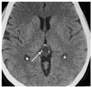
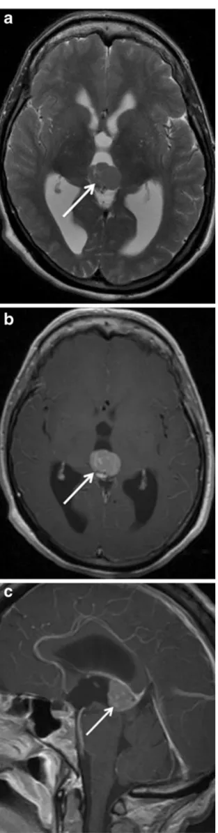
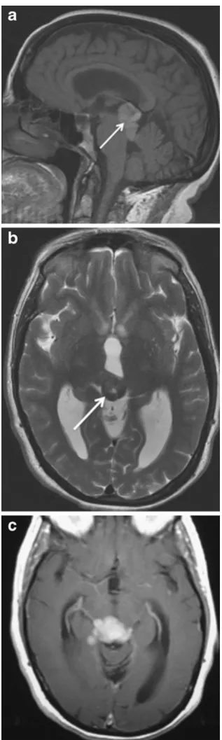
Due to their well-differentiated nature, pineocytomas can be indistinguishable from the normal pineal parenchyma. They are well-circumscribed and unencapsulated tumours that arise and expand from the pineal gland, producing the characteristic “explosion” of normal pineal calcifications towards the periphery (Fig.1) [9]. On magnetic resonance imaging (MRI), pineocytomas usually have low to interme-diate signal on T1-weighted images and intermeinterme-diate to high signal on T2-weighted images (Fig. 2). These lesions typi-cally show prominent contrast enhancement (Fig.2). Occa-sionally cystic or partially cystic pineocytomas are seen, which demonstrate internal or nodular wall enhancement on post-contrast imaging [10, 11]. Pineal apoplexy can occur in rare circumstances.
Pineal parenchyma tumour of intermediate differentiation
Pineal parenchyma tumour of intermediate differentiation (PPTID) accounts for up to 20 % of all pineal parenchymal tumours [8]. They are characterised by diffuse sheets or lobules of cells similar to pineocytomas and pineoblastomas with mild to moderate nuclear atypia and low to moderate mitotic activity. PPTID are of intermediate-grade malignan-cy and are classified as WHO grade II or III neoplasm. The peak prevalence is during adulthood; however, they can occur at all ages and have a female predilection. The 5-year survival is 39–79 %, which is between the outcomes of pineocytoma and pineoblastoma [8].
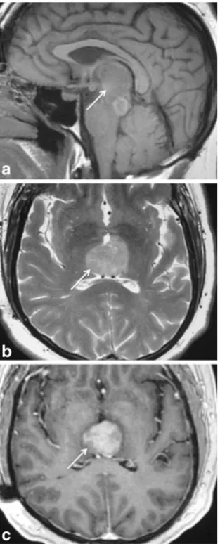
No specific MRI findings distinguish PPTID from pineocytomas or pineoblastomas. PPTID often demonstrate intermediate to high signal on T2-weighted images, may contain cystic areas and typically show contrast enhancement (Fig.3) [9].
Fig. 1 Pineocytoma in a 35-year-old woman. A well-circumscribed low-attenuated lesion arising from the pineal gland producing the characteristic “explosion”of normal pineal calcifications (arrow) to-wards the periphery
Table 1 Pineal lesions
Pineal parenchymal lesions Pineocytoma
Pineal parenchymal tumour of intermediate differentiation Papillary tumour of the pineal region
Pineoblastoma Germ cell tumours
Germinoma Teratoma Choriocarinoma Yolk sac tumour Embryonal carcinoma
Other lesions within or adjacent to the pineal gland Metastasis
Pineal cyst Pineal meningioma Pineal melanoma Embryonal tumours
Primitive neuroectodermal tumours (PNETs) Atypical teratoid/rhabdoid tumours (AT/RTs) Ependymoma
Arachnoid cyst
Papillary tumour of the pineal region
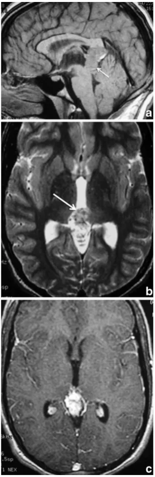
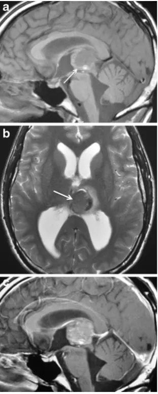
Papillary tumour of the pineal region (PTPR) is a rare neuroepithelial neoplasm that originates from specialised ependymocytes of the sub-commissural organ within the pineal region [12–14]. Recently recognised by the WHO in 2007, they correspond to WHO grade II or III tumours [8]. Children and adults are affected with the reported age Fig. 2 Pineocytoma in a 65-year-old woman with worsening
headaches. A circumscribed slightly-lobulated lesion (arrow) with low to intermediate signal on axial T2-weighted image (a) in-volves the pineal gland. The lesion shows prominent diffuse contrast enhancement on axial (b) and sagittal (c) T1-weighted images (arrow)
range of presentation between 5 and 66 years (mean age is 29 years) with a slight female predominance. The 5-year survival rate is 73 % and local recurrence is common even after complete resection and radiation therapy [12].
PTPRs are often difficult to distinguish from other pineal region tumours, such as choroid plexus papillomas, papillary pineal parenchymal tumours, and papillary ependymomas, and often require immunohistochemical techniques for further diagnosis [13]. PTPRs are well-circumscribed and can dem-onstrate slightly-high to high signal on T1- and T2-weighted images (Fig.4) due to the presence of secretory inclusions containing protein and glycoprotein [13, 15]. Post-contrast images demonstrate moderate enhancement (Fig.4) and cystic regions are commonly present [12]. Neuroimaging of the
brain and spine is often recommended because of frequent local recurrence of tumour and occasional occurrence of leptomeningeal tumour spread [12,16].
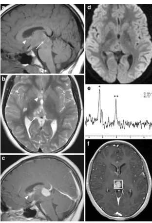
Pineoblastoma
Pineoblastomas are highly malignant WHO Grade IV neo-plasms and account for 40 % of pineal parenchymal tumours [8]. They are considered a primitive type of neuroectodermal tumour. These tumours are comprised of densely packed small cells with irregular nuclei, scant cytoplasm, high mitotic ac-tivity and necrosis. On immunohistological evaluation, they demonstrate increased Ki-67 labelling index, which is an indicator of tumour proliferation and consistent with high-grade tumours [17]. Most pineoblastomas present in the first 2 decades of life with equal predominance in both sexes. The 5-year survival is dismal at 58 % despite chemoradiotherapy [8]. In some patients, bilateral retinoblastomas with associated pineoblastoma may occur, which is referred to as “trilateral retinoblastoma.”
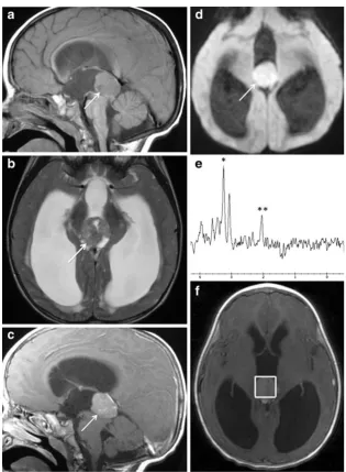
On MRI, pineoblastomas show low to intermediate signal on T1-weighted images, intermediate to high signal on T2-weighted images and demonstrate contrast enhancement (Fig. 5). Given their highly malignant nature, it is not uncommon to see haemorrhage and necrosis within the lesion, as well as infiltration into adjacent structures with cerebral spinal fluid (CSF) seeding and dissemination within the subarachnoid space [9]. Pineoblastomas usually have low minimum apparent diffusion coefficient (minADC) and restricted diffusion on diffusion-weighted imaging (DWI) (Fig. 5). On MR spectroscopy (Fig. 5), there is elevated choline, decreased N-acetylaspartate (NAA), as well as slightly elevated glutamate and taurine peaks (∼3.4 ppm).
Germ cell tumours
Germ cell tumours (GCTs) make up more than half of all pineal lesions and are comprised of residual primordial ectodermal, mesodermal or endodermal tissue. They are classified into germinomas and non-germinomatous GCTs, which include teratomas, embryonal carcinomas, yolk sac tumours, choriocarcinomas and mixed GCTs. The majority of intracranial GCTs are germinomas and teratomas [2].
Germinoma
Germinomas account for 3–5 % of paediatric intracranial tumours, 0.4–1 % of adult intracranial tumours, and 50– 70 % of all pineal neoplasms [9,18,19]. They are malignant tumours composed of large cells that appear undifferentiated and resemble primordial germinal elements. Germinomas Fig. 4 Papillary tumour of the pineal region (PTPR) in a 6-year-old
can be separated into two types: pure germinoma and germinoma with syncytiotrophoblastic cells, which are as-sociated with higher recurrence, decreased survival rate and elevated CSF levels of human chorionic gonadotropin (hCG) [20]. The majority of intracranial germinomas (65 %) occur in the pineal region, while others (25–35 %) are found in the suprasellar region [9]. Most patients (90 %) are less than 20 years old at the time of diagnosis and the male-to-female ratios vary between 5:1 and 22:1. Patients often present with endocrine dysfunction secondary to in-volvement of the sellar region by tumour. Disseminated disease is commonly seen at the time of presentation as well as subependymal spread of the tumour along the third ven-tricle. Germinomas have a good prognosis, with a 5-year survival rate of at least 90 % and are highly responsive to radiation therapy [9,18,21].
On MRI, germinomas can appear as a solid pineal mass with intermediate to slightly-high signal compared with grey
matter on both T1- and T2-weighted images (Fig.6). Cystic components are commonly identified in 20–52 % of cases [9,10,22] and post-contrast enhancement is usually present on MRI (Fig.6). Germinomas demonstrate ADC values that overlap those for pineocytoma, PPTID and PTPR. Dumrongpisutikul et al. [23] demonstrated that when the region of interest includes both solid and cystic portions of germinomas, the ADC values are often higher than those of the more densely cellular pineal cell tumours, particularly pineoblastomas, which is likely due to the abundance of cytoplasm, presence of cystic components, less tumour cel-lularity and lesser nuclear/cytoplasmic ratio. However, when only the solid portions of germinomas are included, normal, decreased or increased ADC values can be demon-strated relative to normal brain tissue [24]. Leptomeningeal and intraventricular tumour deposits typically show contrast enhancement (Fig.6) and occur in 13 % of patients at the time of diagnosis [22].
Teratoma
Teratomas account for approximately 15 % of all intracra-nial GCTs and are the most common non-germinomatous GCTs [9]. They are commonly derived from multi-potential cells from at least two (usually all three) embryological layers involving the ectodermal, endodermal or mesodermal lines. Teratomas can be classified into three types: mature teratoma, immature teratoma and teratoma with malignant transformation [9]. Most patients with teratomas occur in the first 2 decades of life, and there is a male predilection.
MRI findings demonstrate a multi-loculated and lobulat-ed lesion with mixlobulat-ed signal, including areas of high signal intensity on T1-weighted images due to the presence of fat or lipid components, and areas of low signal from calcifica-tion (Fig. 7). T2-weighted images often have a low to
intermediate signal soft tissue component (Fig. 7). Post-contrast images can show enhancement of the soft tissue component (Fig. 7). Teratomas with malignant transfor-mation have a more homogeneous imaging appearance with fewer cysts and calcifications [9].
Other GCTs
Choriocarcinoma, yolk sac tumours and embryonal carcino-ma carcino-make up the other nongerminocarcino-matous GCTs and each account for 10 % of intracranial GCTs [19]. Patients often have elevated serum oncoproteins that may help in making the appropriate diagnosis. However, they are not specific to these tumours. Patients with choriocarcinoma, which are characterised by extra-embryonic differentiation along tro-phoblastic lines, typically have elevated CSF and plasmaβ -Fig. 6 Germinoma in a
hCG [9,25]. Pineal yolk sac tumours made up of primitive-appearing epithelial cells may demonstrate elevated concen-trations ofα-fetoprotein (AFP). Embryonal carcinoma com-posed of large cells that proliferate in cohesive nests and sheets often shows absent CSF and plasma concentrations of AFP andβ-hCG.
No specific imaging features alone are capable of differ-entiating among these various tumours, although most of these lesions demonstrate a higher rate of cystic changes compared with germinomas (Figs.8and9) [26]. In partic-ular, choriocarcinoma is prone to haemorrhage, and can
demonstrate low signal with blooming effects on T2*-weighted sequences and intrinsic high T1 signal on MRI [9].
Metastases
Pineal metastasis is uncommon, occurring in 0.4–3.8 % of patients with solid tumours as seen on autopsy reports and in Fig. 7 Teratoma in a 56-year-old man with worsening confusion and
ataxia. The pineal lesion (arrow) is hyperintense relative to brain with foci of very low signal on sagittal T1-weighted image (a) and mixed low, intermediate and high signal on axial T2-weighted image (b). Internal foci and peripheral rim of low signal on T1-weighted and T2-weighted images
5 % of patients referred to surgical management of pineal tumours [27,28]. Lung cancer is the most frequent cancer to metastasise to the pineal gland; however, other tumours such as breast, kidney, esophagus, stomach, colon, melanoma and myeloma have also been reported [28]. Metastatic disease is thought to be related to hematogenous spread due to the absence of a blood-brain barrier in the pineal gland. In the majority of cases, metastasis to the pineal gland is asymptom-atic. However, a few patients have presented with clinical features, such as headache, encephalopathy, Parinaud’s syn-drome and hydrocephalus [28]. Leptomeningeal seeding is a common finding with pineal metastases, occurring in 67 % of patients, and may be secondary to the proximity of the pineal gland to the CSF pathways in the third ventricle and quadrigeminal cistern [28].
Cells types adjacent to the pineal gland
Pineal cyst
Pineal cysts are found in 1–4 % of MRIs and between 20 % and 40 % of patients in an autopsy series [10, 29]. Histo-logically, they are composed of an inner layer of gliotic
tissue, middle layer of pineal parenchymal tissue and an outer layer of connective tissue. Pineal cysts occur predom-inantly in adults between the ages of 40 and 49 years old and are more prevalent among females compared with men.
On MRI, they may be round or oval, thin-walled, uniloc-ular or multi-loculated collections filled with proteinaceous and haemorrhagic components. A thin rim of calcification is seen in 25 % of cases. They have signal intensity similar to CSF (low signal on T1-weighted images and high signal on T2-weighted images) (Fig.10). However, 50–60 % of pineal
Fig. 9 Pineal embryonal carcinoma. The tumour involving the pineal gland (arrow) has lobulated margins with intermediate to high signal on axial T2-weighted image (a). The tumour shows heterogeneous contrast enhancement on sagittal T1-weighted image (b) related to its solid and cystic portions
cysts are slightly hyperintense to CSF [10]. The signal of these cysts often does not fully suppress on fluid-attenuated inversion-recovery (FLAIR) (Fig. 10). The cyst contents typically show no restricted diffusion. Post-contrast images (Fig.10) demonstrate a thin rim of wall enhancement usu-ally <2-mm thickness that is seen as a result of fragmenta-tion of the pineal parenchyma, which occurs as the cyst enlarges [10,11]. The cyst can show with internal enhance-ment on delayed images.
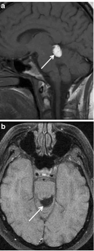
Pineal lipoma
Pineal lipomas are rare lesions, which represent 10 % of intracranial lipomas, account for 0.02 % of intracranial operated lesions and 0.8 % of patients in an autopsy and neuroradiological series [30,31]. They are benign mesen-chymal tumours, which arise from the abnormal differenti-ation of the meninx primitive. Men and women are equally affected.
On MRI, pineal lipomas have homogeneously high sig-nal on T1-weighted images and low sigsig-nal on fat-suppressed T1-weighted and T2-weighted images (Fig. 11) [30, 31]. There is typically no enhancement post contrast.
Pineal meningioma
Pineal meningiomas make up 6–8 % of pineal region tu-mours and 0.3 % of intracranial meningiomas [32,33]. They are classified as a WHO grade I lesion and considered as a meningothelial cell neoplasm that are attached to the surface of the dura mater. They may arise from cells within the tela choroidea, velum interpositum or falcotentorial junction. The average age at diagnosis is 53 years (range of 41– 69 years), and there is a male-to-female ratio of 3:7 [33].
Meningiomas characteristically are highly cellular in na-ture and calcifications are present within the lesion in 15– 20 % of cases. Meningiomas are dural-based lesions, which exhibit a dural tail. They demonstrate low to intermediate signal on T1-weighted images and intermediate to slightly-high signal on T2-weighted images (Fig. 12). On post-contrast images, there is typically avid enhancement of the lesion (Fig.12).
Pineal melanoma
Primary pineal melanoma is exceedingly rare. The majority of melanocytic lesions are found in the central nervous system (CNS) secondary to metastatic disease. Malignant melanin-producing cells predominately arise from cells within the leptomeninges that surround the pineal gland and secondarily invade and replace it. This melanin-containing tumour has a wide age range of presentations (20–77 years), and women are more commonly affected
than men. The outcome in patients is poor, especially when leptomeningeal dissemination occurs [34].
Embryonal tumours
Embryonal tumours, which include primitive neuroectodermal tumours (PNETs) and atypical teratoid/rhabdoid tumours (AT/RTs), are a heterogeneous group of immature-appearing
neoplasms that most often arise in children and can involve the pineal gland.
PNET is a WHO grade IV tumour composed of undifferentiated or poorly differentiated neuroepithelial Fig. 12 Tentorial meningioma in a 45-year-old woman with
papilloedema. A meningioma (arrow) is seen involving the tentorium which extends into the pineal recess. The lesion is isointense to grey matter on axial T2-weighted image (a) and sagittal T1-weighted image (b) and shows prominent contrast enhancement on coronal T1-weight-ed image (c)
cells. These tumours have a variable age range of presenta-tion from 4 weeks to 20 years (mean age is 5.5 years) and a male-to-female ratio of 1.2:1. Most of these tumours are found in the cerebrum, but they can also be found in the spinal cord, pineal or suprasellar region [8].
On MRI, PNETs demonstrate low signal relative to cor-tical grey matter on T1-weighted images and can have high signal on T2-weighted images from cystic or necrotic areas. These tumours can present with areas of restricted diffusion [36]. Haemorrhage may also occur and can appear as areas with low signal on T2-weighted images. These lesions often show heterogeneous enhancement with contrast (Fig.14).
Fig. 14 Primitive neuroectodermal tumour (PNET) involving the pi-neal gland in a 3-year-old boy. Well-circumscribed, tumour arising from the pineal gland (arrow), which has mostly intermediate signal as well as high signal foci on sagittal T1-weighted image (a) and intermediate, low and high signal on axial T2-weighted image (b). The pineal tumour demonstrates heterogeneous contrast enhancement on sagittal T1-weighted image (c). The tumour causes obstructive hydrocephalus by exerting localised mass effect and compression of the quadrigeminal plate, cerebral aqueduct and third ventricle
AT/RTs are highly malignant WHO grade IV embryonal tumours that account for 10 % of CNS tumours in infants and 1–2 % of paediatric brain tumours [35–38]. These tumours are comprised of rhabdoid cells with variable com-bination of primitive neuroectodermal, mesenchymal, and epithelial components. They are associated with inactivation
of hSNF5/INI1 gene on chromosome 22q11.2. AT/RTs often present in infants and young children less than 3 years old and rarely in adults. There is a male predominance ranging from 1.6 to 2:1 [39]. They can occur in the posterior fossa (cerebellum and cerebello-pontine angle) or cerebrum [37, 39]. AT/RTs infrequently arise in the spinal cord.
On MRI, AT/RTs often have heterogeneous intermediate signal on T1-weighted and T2-weighted images as well as areas of low and high signal from haemorrhagic, cystic and/or necrotic zones. Restricted diffusion is also commonly seen involving these tumours. On post-contrast images, there is variable enhancement (Fig. 15). In a quarter of patients with AT/RT, leptomeningeal dissemination is found at the time of presentation [37,39,40].
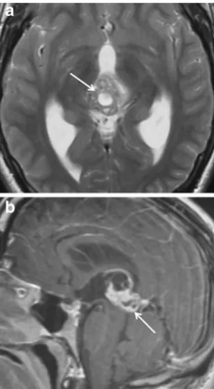
Ependymoma
Ependymomas make up 6–12 % of all intracranial tumours in children and are slow-growing WHO grade II neoplasms derived from ependymal cells [8]. They can be classified into the following subtypes: cellular, papillary, clear cell and tanycytic. They have a bimodal age of distribution com-monly presenting around 1–5 years and in the mid 30s. Males are more often affected than females. Approximately two-thirds of ependymomas are infratentorial, occurring
Fig. 16 Ependymoma in a 54-year-old man causing headaches, nau-sea, vomiting, and hydrocephalus. Sagittal T1-weighted image (a) shows a large lesion involving the pineal gland (arrow), which con-tains areas of low and intermediate signal. Axial T2-weighted image (b) demonstrates cystic areas within the tumour (*) that have high signal and solid portions (arrow) with mixed intermediate and high signal. Solid portions of the tumour (arrow) show contrast enhance-ment on coronal T1-weighted image (c). The tumour causes hydro-cephalus from compression of the midbrain and cerebral aqueduct, pons and cerebellum
predominantly within the fourth ventricle, while one-third occur supratentorially in the fronto-parietal region.
On MRI, ependymomas appear as heterogeneous, usually low to intermediate signal on T1-weighted images and in-termediate to high signal on T2-weighted images with areas of cystic foci, calcifications and/or blood products (Fig.16). Variable enhancement is seen on post-contrast images (Fig. 16). Diffusion weighted imaging demonstrates rela-tively lower cellularity and high ADC. On MR spectrosco-py, elevated choline and lactate peaks with decreased levels of NAA can be seen.
Arachnoid cyst
Arachnoid cysts are the most common congenital intracra-nial cystic abnormality, occurring in 1 % of all intracraintracra-nial masses. They are intra-arachnoid CSF-filled sacs, which do not communicate with the ventricular system. Arachnoid cysts can be found at any age; however, 75 % present in children. Men are more commonly affected than women. The majority of arachnoid cysts are supratentorial. Up to 50 % are seen anterior to the temporal lobes in the middle cranial fossa. Approximately 10 % are found in the suprasellar cistern and posterior fossa [10].
On MRI, arachnoid cysts appear as a sharply marginated extra-axial fluid collection, which is similar in signal with CSF on T1-weighted and T2-weighted images (Fig. 17). There is no post-contrast enhancement. The signal of arach-noid cysts is identical to CSF and suppresses completely on FLAIR (Fig.17) No restricted diffusion is seen with these lesions [10].
Conclusions
Pineal lesions can represent a heterogeneous collection of tumours and congenital/developmental abnormalities. While sometimes often difficult to differentiate, each of these varying pineal lesions may have unique clinical and imaging characteristics. Pineal parenchymal tumours often produce an“explosion”of normal pineal calcifications towards the periphery and may demonstrate areas of cystic changes. The most common type of pineal lesion is GCT, particularly germinoma, which tends to engulf pineal calcifications. Pineal teratomas and pineal lipomas demonstrate fat signal characteristics on MRI. Although pineal metastasis is un-common, patients with pineal lesions who present with a known malignancy should raise suspicion of metastatic involvement. Both pineal cysts and arachnoid cysts show MRI signal characteristics similar to CSF; however, arach-noid cysts demonstrate signal suppression on FLAIR, whereas pineal cysts often do not. Pineal meningiomas are dural-based lesions that enhance with contrast. Pineal
ependymomas can have distinct MR spectroscopy charac-teristics with elevated choline and lactate peaks. Pineoblastomas and embryonal tumours, such as PNETs and AT/RTs, involving the pineal gland, are high-grade neoplasms that can show restricted diffusion. Basic under-standing of these imaging characteristics can assist in narrowing the differential diagnosis of varying pineal lesions and allow for accurate and rational therapeutic planning.
Disclosures The authors do not have any financial relationship with a commercial organisation that may have a direct or indirect interest in the content of this article.
Open AccessThis article is distributed under the terms of the Creative Commons Attribution License which permits any use, distribution, and reproduction in any medium, provided the original author(s) and the source are credited.
References
1. Drummond KJ, Rosenfeld JV (1999) Pineal region tumours in childhood: a 30-year experience. Childs Nerv Syst 15(2-3):119– 126, discussion 127
2. Matsutani M, Sano K, Takakura K et al (1997) Primary intracranial germ cell tumors: a clinical analysis of 153 histologically verified cases. J Neurosurg 86(3):446–455
3. Sumida M, Barkovich A, Newton T (1996) Development of the pineal gland: measurement with MR. AJNR Am J Neuroradiol 17:233–236
4. Zimmerman RA, Bilaniuk LT (1982) Age-related incidence of pineal calcification detected by computed tomography. Radiology 142:659–662
5. Klein P, Rubinstein LJ (1989) Benign symptomatic glial cysts of the pineal gland: a report of seven cases and review of the litera-ture. J Neurol Neurosurg Psychiatry 52:991–995
6. Patel AJ, Fuller GN, Wildrick DM, Sawaya R (2005) Pineal cyst apoplexy: case report and review of the literature. Neurosurgery 57(5):1066
7. Surawicz TS, McCarthy BJ, Kupelian V et al (1999) Descriptive epidemiology of primary brain and CNS tumors: results from the Central Brain Tumor Registry of the United States, 1990-1994. Neuro Oncol 1(1):14–25
8. Louis DN, Ohgaki H, Wiestler OD, Cavenee WK (2007) WHO classification of tumors of the central nervous system. IARC Press, Lyon
9. Smith AB, Rushing EJ, Smirniotopoulos JG (2010) Lesions of the pineal region: radiologic-pathologic correlation. Radiographics 30:2001–2020
10. Osborn AG, Preece MT (2006) Intracranial cysts: radiologic-pathologic correlation and imaging approach. Radiology 239(3):650– 664
11. Fakhran S, Escott EJ (2008) Pineocytoma mimicking a pineal cyst on imaging: true diagnostic dilemma or a case of incomplete imaging? AJNR Am J Neuroradiol 29(1):159–163
12. Fèvre-Montange M, Hasselblatt M, Figarella-Branger D et al (2006) Prognosis and histopathologic features in papillary tumors of the pineal region: a retrospective multicenter study of 31 cases. J Neuropathol Exp Neurol 65(10):1004–1011
14. Jouvet A, Fauchon F, Liberski P et al (2003) Papillary tumor of the pineal region. Am J Surg Pathol 27:505–512
15. Sato TS, Kirby PA, Buatti JM et al (2009) Papillary tumor of the pineal region: report of a rapidly progressive tumor with possible multicentric origin. Pediatr Radiol 39(2):188–190
16. Kim YH, Kim JW, Park CK et al (2010) Papillary tumor of pineal region presenting with leptomeningeal seeding. Neuropathology 30(6):654–660
17. Zhu L, Ren G, Li K et al (2011) Pineal parenchymal tumors: minimum apparent diffusion coefficient in prediction of tumour grading. J Int Med Res 39:1456–1463
18. Villano JL, Propp JM, Porter KR et al (2008) Malignant pineal germ-cell tumors: an analysis of cases from three tumor registries. Neuro Oncol 10:121–130
19. Horowitz MB, Hall WA (1991) Central nervous system germinomas: a review. Arch Neurol 48:652–657
20. Echevarría ME, Fangusaro J, Goldman S (2008) Pediatric central nervous system germ cell tumors: a review. Oncologist 13(6):690–699 21. Shibamoto Y, Sasai K, Oya N et al (2001) Intracranial germinoma: radiation therapy with tumor volume-based dose selection. Radiology 218:452–456
22. Moon W, Chang K, Han M et al (1999) Intracranial germinomas: correlation of imaging findings with tumor response to radiation therapy. Am J Roentgenol 172:713–716
23. Dumrongpisutikul N, Intrapiromkul J, Yousem DM (2012) Distinguishing between germinomas and pineal cell tumors on MR imaging. AJNR Am J Neuroradiol 33:550–555
24. Douglas-Akinwande AC, Ying J, Momin Z, Mourad A, Hattab EM (2009) Diffusion-weighted imaging characteristics of primary cen-tral nervous system germinoma with histopathologic correlation: a retrospective study. Acad Radiol 16:1356–1365
25. Page R, Doshi B, Sharr MM (1986) Primary intracranial chorio-carcinoma. J Neurol Neurosurg Psychiatry 49:93–95
26. Hayashida Y, Hirai T, Korogi Y et al (2004) Pineal cystic germinoma with syncytiotrophoblastic giant cells mimicking MR imaging findings of a pineal cyst. AJNR Am J Neuroradiol 25:1538– 1540
27. King AB, Ford FR (1942) Clinical and anatomical study of neu-rological conditions resulting from metastases in the central
nervous system due to carcinoma of the lung: review of 100 cases. Bull Johns Hopkins Hosp 70:124–156
28. Lassman AB, Bruce JN, Fetell MR (2006) Metastases to the pineal gland. Neurology 67:1303–1304
29. Pu Y, Mahankali S, Hou J et al (2007) High prevalence of pineal cysts in healthy adults demonstrated by high resolution, noncontrast brain MR imaging. AJNR Am J Neuroradiol 28(9):1706–1709
30. Maiuri F, Cirillo S, Simonetti L, De Simone MR, Gangemi M (1988) Intracranial lipomas: diagnostic and therapeutic consider-ations. J Neurosurg Sci 32:161–167
31. Friedman RB, Segal R, Latchaw RE (1986) Computed tomograph-ic and magnettomograph-ic resonance imaging of intracranial lipoma. J Neurosurg 65:407–410
32. Stein BM (1979) Surgical treatment of pineal tumors. Clin Neurosurg 26:490–510
33. Konovalov AN, Spallone A, Pitzkhelauri DI (1996) Meningioma of the pineal region: a surgical series of 10 cases. J Neurosurg 85:586–590
34. Arantes M, Castro AF, Romao H et al (2011) Primary pineal malignant melanoma: case report and literature review. Clin Neurol Neurosurg 113:59–64
35. Biegel JA (2006) Molecular genetics of atypical teratoid/rhabdoid tumor. Neurosurg Focus 20:E11
36. Rickert CH, Paulus W (2001) Epidemiology of central nervous system tumors in childhood and adolescence based on the new WHO classification. Childs Nerv Syst 17:503–511
37. Meyers SP, Khademian ZP, Biegel JA et al (2006) Primary intracra-nial atypical teratoid/rhabdoid tumors of infancy and childhood: MRI features and patient outcomes. AJNR Am J Neuroradiol 27:962–971 38. Wong TT, Ho DM, Chang KP et al (2005) Primary pediatric brain tumors: statistics of Taipei VGH, Taiwan (1975-2004). Cancer 102:2156–2167
39. Hilden JM, Meerbaum S, Burger P et al (2004) Central nervous system atypical teratoid/rhabdoid tumor: results of therapy in chil-dren enrolled in a registry. J Clin Oncol 22:2877–2884