ULTRASOUND
Open Access
R E S E A R C H
Bio
Med
Central
© 2010 Previtali et al; licensee BioMed Central Ltd. This is an Open Access article distributed under the terms of the Creative CommonsAttribution License (http://creativecommons.org/licenses/by/2.0), which permits unrestricted use, distribution, and reproduction in any medium, provided the original work is properly cited.Research
Dobutamine stress echocardiography for assessing
the role of dynamic intraventricular obstruction in
left ventricular ballooning syndrome
Mario Previtali*, Rita Camporotondo, Alessandra Repetto and Stefania Panigada
Abstract
Background: Dynamic intraventricular obstruction has been observed in patients with left ventricular ballooning syndrome (LVBS) and has been hypothesized as a possible mechanism of the syndrome. The aim of this study was to assess the prevalence and significance of dynamic intraventricular obstruction in patients with LVBS.
Methods and Results: Dobutamine stress echocardiography was carried out in 22 patients with LVBS (82% apical), all women, aged 68 ± 9 years. At baseline 1 patient had a > 30 mmHg LV gradient; during stress a LV gradient > 30 mm Hg developed in 6/21 patients (28%) and was caused by systolic anterior motion of the mitral valve in the 3 patients with severe gradient (mean 116 ± 29 mmHg), who developed mitral regurgitation and impaired apical wall motion and by obstruction at mid-ventricular level in the other 3 with a moderate gradient (mean 46 ± 16 mmHg). Compared with patients without obstruction those with obstruction had a greater mean septal thickness (11.6 ± .6 vs 9.8. ± 3, p < .01), a higher prevalence of septal hypertrophy (71% vs 7%, p < .005) and a higher peak wall motion score index (1.62 ± .4 vs 1.08 ± .4, p < .01).
Conclusion: Spontaneous or dobutamine-induced dynamic LV obstruction is documented in 32% of patients with LVBS, is correlated with the presence of septal hypertrophy and may play a role in the development of LVBS in this subset of patients. In those without septal hypertrophy a dynamic obstruction is rarely induced with dobutamine and is unlikely to be a major pathogenetic factor of the syndrome.
Left ventricular ballooning syndrome (LVBS) is a recently described acute cardiac syndrome mimicking acute myo-cardial infarction, characterized by reversible regional apical or midventricular dysfunction whose pathogenetic mechanisms are as yet undefined [1-4]. Sympathetic stimulation and increased catecholamine release second-ary to emotional stress leading to myocardial stunning are likely to play an important role [5,6]. Recently it has been hypothesized that in the presence of a localized proximal-mid septal hypertrophy, stress-related increased sympathetic tone may induce dynamic intra-ventricular obstruction, that in turn increases myocardial wall stress distal to the obstruction and decreases regional subendocardial perfusion leading to the develop-ment of akinesia and dilation in the apical region [7].
Dobutamine administration can induce a dynamic LV obstruction in a significant number of patients undergo-ing the stress testundergo-ing [8-11] and could therefore repro-duce the spontaneous event in patients with LVABS. We therefore carried out dobutamine stress echocardiogra-phy in a group of patients with LVBS to assess the preva-lence and pathogenetic role of dynamic intraventricular obstruction in LVBS.
Methods
Study population
The study population was composed of 22 consecutive patients admitted with an acute coronary syndrome who fulfilled the diagnostic criteria for LVBS proposed by Mayo Clinic group [4] including: 1) Chest pain associated with new ST-segment elevation or T wave inversion in ≥ 2 contiguous leads; 2) Absence of significant (≥ 50%)obstructive coronary artery disease and of an acute plaque rupture at coronary angiography carried out
* Correspondence: marprevi@hotmail.com
1 From Department of Cardiology, IRCCS Fondazione Policlinico San Matteo,
within 48 hours from onset of symptoms; 3) Transient reversible akinesia or dyskinesia and dilatation involving either the midventricular and apical segments with basal LV hyperkinesia (apical ballooning) or the midventricular segments with normal or hyperkinetic basal and apical segments (midventricular variant); 4) No evidence of recent major head trauma, intracranial bleeding, pheo-chromocytoma, myocarditis or hypertrophic cardiomyo-pathy. All patients were included in the Italian Multicenter Registry on Takotsubo syndrome.
Dobutamine stress echocardiography
The test was carried out at a mean of 9 ± 14 days from admission. Beta-blocking therapy was withdrawn 24 hours before the test. At baseline LV wall thickness and dimensions were measured by M-mode echocardiogra-phy in parastemal long axis view according to standard methods; a ≥ 12 mm end-diastolic wall thickness was considered indicative of wall hypertrophy. Dobutamine was administered at an initial dose of 5 mcg/kg/min for 5 minutes, with increase to 10 and 20, 30 and 40 mcg/kg/ min for 5 minutes each under electrocardiographic and echocardiographic monitoring. The peak LV velocity was measured at rest, at the end of each stage and at peak stress by continuous wave Doppler from apical 4-or 5 chamber view. A pressure gradient >30 mmHg with a late peaking was considered significant for a dynamic intra-ventricular obstruction [1]; the site of LV obstruction was identified by pulsed and color Doppler; systolic anterior motion of the anterior mitral leaflet was searched for in multiple views. LV end-diastolic and end-systolic volume using the area-length method and ejection fraction were measured in the apical 4 chamber view at baseline and at peak stress. Digitized basal, low dose, peak dose and recovery images were displayed in a quad screen format for off-line comparison. Regional wall motion was assessed on a 16-segment LV model [12] as previously described and a wall motion score index was calculated at baseline and at peak stress [13]. A new or worsening wall motion abnormality compared to baseline was consid-ered diagnostic of myocardial ischemia. Myocardial via-bility was judged to be present in basally hypokinetic or akinetic segments when normalization or improvement in myocardial thickening and motion was observed in at least 2 contiguous segments after dobutamine. The test was interrupted according to previously described crite-ria [13]. At the end of the test intravenous propranolol (1-5 mg) was administered. All patients gave their informed consent to the test.
Coronary Angiography
Left ventriculography and multiplane coronary angiogra-phy were carried out according to standard methods. Ejection fraction was calculated by the area-length
method. Coronary artery disease was defined as a ≥ 50% reduction in the luminal diameter of a major coronary artery. The TIMI flow grade classification and TIMI frame count were used to assess coronary blood flow in the 3 coronary arteries and calculated as previously described [14]. A TIMI frame count ≤ 27 frames was con-sidered normal. The study complied with the Declaration of Helsinki on the research on humans; the study proto-col was approved by the local ethical committee. All patients gave written informed consent.
Statistical analysis
Data are presented as mean ± SD. Continuous variables were compared using a paired or unpaired Student's t test; categorical variables were compared by Fischer exact test.
Results
Clinical findings
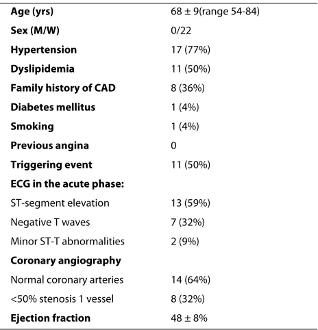
The main clinical findings of the patients studied are shown in table 1. All patients were women with a mean age of 68 ± 9 years (range 54-84). No patient had a history of angina. Presenting symptoms were chest pain in 21 patients, dyspnea in 2 and dizziness or syncope in 4. Cor-onary angiography showed normal corCor-onary arteries in 14 patients (64%) and <50% stenosis of 1 vessel in 8 (36%); TIMI frame count in the left anterior descending, left cir-cumflex and right coronary arteries was abnormally pro-longed in 32, 32 and 36% of patients respectively. Mean LV ejection fraction was 48 ± 8%; 10/22 patients (45%)
Table 1: Clinical characteristics of the patients studied
Age (yrs) 68 ± 9(range 54-84)
Sex (M/W) 0/22
Hypertension 17 (77%)
Dyslipidemia 11 (50%)
Family history of CAD 8 (36%)
Diabetes mellitus 1 (4%)
Smoking 1 (4%)
Previous angina 0
Triggering event 11 (50%)
ECG in the acute phase:
ST-segment elevation 13 (59%)
Negative T waves 7 (32%)
Minor ST-T abnormalities 2 (9%)
Coronary angiography
Normal coronary arteries 14 (64%) <50% stenosis 1 vessel 8 (32%)
Ejection fraction 48 ± 8%
had an ejection fraction <50%. LV angiography showed a typical apical ballooning pattem in 18 patients (82%) and a midventricular ballooning with sparing of the apical segments in 4 (18%). In the acute phase a dynamic intra-ventricular gradient was documented in 3 patients (14%).
Dobutamine stress echocardiography
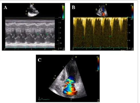
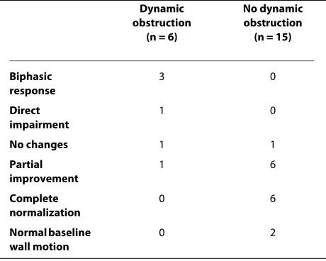
In basal conditions 5 patients showed a complete recov-ery of regional function while apical wall motion abnor-malities were still present in 17. A localized proximal-mid septal hypertrophy was found in 6/22 patients (27%); no patient had a concentric LV hypertrophy. One patient showed a dynamic intraventricular gradient of 150 mmHg associated with systolic anterior motion of the mitral valve and mitral regurgitation and did not undergo DSE. In the remaining 21 patients heart rate increased from 67 ± 10 at baseline to 106 ± 18 beats/min at peak stress (p < .001) and systolic blood pressure increased from l30 ± 30 to 133 ± 27 mmHg (NS). Due to reduced diastolic filling time LV end-diastolic volume decreased from 99 ± 27 at baseline to 74 ± 30 ml at peak stress (p < .001) and end-systolic volume decreased from 48 ± 15 to 39 ± 23 ml (p < .002); mean walI motion score index improved from 1.4 ± .3 to 1.2 ± .4 (p < .01). A dynamic intraventricular gradient > 30 mmHg (mean 81 ± 44 mmHg, range 150-35 mmHg) with a late peaking devel-oped in 6/21 patients (28%); the obstruction was localized in the outflow tract and caused by systolic anterior motion of the anterior mitral leaflet in 3 patients (mean gradient 116 ± 29 mmHg) (Figure 1) and at papillary mus-cle level in the other 3 (mean gradient 46 ± 16 mmHg) (Figure 2). The changes in regional wall motion in rela-tion to dobutamine-induced obstrucrela-tion are shown in Table 2. Of the 2 patients with a mild to moderate obstruction (gradient < 40 mmHg), both at papillary muscle level, 1 had normal baseline regional function and showed no change with dobutamine and 1 showed a par-tial recovery of regional wall motion abnormalities. Of the 4 patients with a severe obstruction (gradient > 40 mm Hg) 3 showed a biphasic response (improvement at low dobutamine doses followed by impaired apical wall motion associated with the development of obstruction) and 1 a direct deterioration of function in the apical region. In 3/4 of these patients the obstruction was local-ized in the outflow tract and associated with severe mitral regurgitation secondary to systolic anterior motion of the mitral valve (Additional files 1 and 2). No correlation was found between stenosis of left anterior descending coro-nary artery and stress echo positivity. On the other hand, of the 15 patients without obstruction, 6 showed a com-plete recovery and 6 a partial recovery of baseline regional wall motion abnormalities, 1 had no significant change and 2 had a normal regional function already at baseline. During the test > l mm ST-segment depression
occurred in 1/6 (17%) patients with and in 0/15 without obstruction and positive T waves in the precordial leads developed in 3/6 patients with and in 4/15 without obstruction. No patients complained of chest pain or dys-pnea during the test. In all cases dynamic obstruction and regional wall motion abnormalities rapidly disappeared after intravenous propranol. No major complication occurred during the test. Both patients with and those without contractile reserve during the test showed a com-plete recovery of regional function during follow-up.
Comparison or patients with and without obstruction
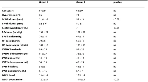
Table 3 compares the clinical, angiographic and stress echocardiographic findings of the 7 patients with sponta-neous or dobutamine induced obstruction with those of the 15 without obstruction. The main clinical characteris-tics, baseline and peak stress heart rate, blood pressure, LV volumes and ejection fraction were not significantly different between the 2 groups. Compared with patients without obstruction those with obstruction had a signifi-cantly greater mean septal thickness (11.6 ± .6 vs 9.8 ± .3, p < .01) and a higher prevalence of localized septal hyper-trophy (71% vs 7%, p < .005). Despite similar baseline wall motion score index, patients with a dynamic gradient had a significantly higher wall motion score index at peak stress (1.62 ± 4 vs 1.08 ± .4, p < 0.01) indicating an impair-ment in regional wall motion associated with the obstruc-tion. In relation to the site of LV ballooning, a dynamic obstruction was documented in 6/18 patients with apical and in 1/4 with midventricular ballooning (33% vs 25%, NS).
Discussion
Prevalence and significance of dynamic LV obstruction in LVBS
Figure 1 Severe dobutamine induced-obstruction. Severe dobutamine-induced dynamic LV obstruction (peak velocity 5 m/sec corresponding to a gradient of 100 mm Hg, panel B) caused by systolic anterior motion of the mitral valve (panel A) and associated with severe mitral regurgitation (panel C) in a patient with apical ballooning.
microvascular dysfunction is a primary or secondary phenomenon. Coronary epicardial spasm has been docu-mented in a significant proportion of Japanese patients [1,2], but is rare in Caucasian patients [3-6,19]. During the acute phase a transient dynamic intraventricular gra-dient was documented in 18% of patients in the largest population of LVBS so far reported [1]; in other smaller studies the prevalence of dynamic obstruction in the acute phase ranged from 12.5% [16] to 23% [5]. These findings and the association of dobutamine-induced dynamic intraventricular gradient with worsening of api-cal wall motion recently reported in 2 patients with LVBS and septal hypertrophy [7] suggested a possible pathoge-netic role for dynamic intraventricular obstruction in LVBS. The prevalence of dobutamine-induced dynamic obstruction in our patients with LVBS is similar to that reported by previous studies in patients undergoing dob-utamine stress echocardiography for evaluation of coro-nary artery disease [8-11,20-22], that showed a prevalence ranging from 13% in the study by Heinle et al [10] to 53% by Wagner et al [9]. Therefore, the develop-ment of a mild to moderate intraventricular gradient dur-ing dobutamine stress is frequent and may have no major clinical significance in the majority of patients with LVBS as in the general population. The significant association found in our patients between dynamic intraventricular gradient and septal hypertrophy is in keeping with the studies by Khanal et al [21] and Wagner et al [9], who showed that dobutamine-induced LV cavity obliteration was associated with female sex and LV hypertrophy. Thus, the dynamic intraventricular obstruction docu-mented in our patients does not seem to be a specific fea-ture of LVBS, but rather depends upon the presence of a localized septal hypertrophy that is frequently found in
elderly women. On the other hand, in a minority of patients with LVBS a severe dynamic intraventricular obstruction can play a role in the development of LVBS by increased myocardial wall stress and decreased suben-docardial perfusion in the apical region leading to myo-cardial ischemia on a hemodynamic basis; this hypothesis is supported by the fact that all 4 patients with severe dobutamine-induced obstruction showed a significant impairment in apical wall motion. The hypothesis of a coronary spasm during dobutamine stress or at the time of beta-blocking infusion as the mechanism of the wall motion abnormalities observed in these patients is unlikely because none of these patients developed ST-segment elevation, which is the hallmark of coronary artery spasm and the wall motion changes occurred before administration of beta-blockers and were abol-ished by them. On the basis of our data, it is not possible to assess whether dynamic LV obstruction plays a pri-mary pathogenetic role, leading to the development of an apical ballooning as suggested by Merli et al [7], or is sec-ondary to the distortion of the LV geometry caused by apical akinesia and basal hyperkinesia and acts as a per-petuating mechanism of the ballooning. Another impor-tant finding of the study is the association of a major dynamic obstruction with the development of severe mitral regurgitation due to systolic anterior motion of the anterior mitral leaflet in 15% of patients with LVBS. Severe mitral regurgitation increases pulmonary capillary pressure and decreases LV forward stroke volume and may therefore be an important yet unrecognized mecha-nism contributing to acute LV failure and shock reported in 20% to 40% of patients with LVBS [1,4,17]. Acute mitral regurgitation due either to systolic anterior motion of the anterior mitral leaflet or to displacement of the papillary muscle with impaired leaflet coaptation has been recently reported in 20% of patients with LVBS and has been associated with a worse prognosis [23]. On the other hand, our study demonstrates the absence of a spontaneous or dobutamine-induced dynamic LV gradi-ent in >65% of patigradi-ents with LVBS; this finding suggests that in the majority of patients with LVBS the develop-ment of the syndrome is not related to a dynamic intra-ventricular obstruction elicited by increased sympathetic tone.
Clinical implications
Dobutamine stress echocardiography may be a useful tool for assessing the presence and significance of LV dynamic obstruction in patients with LVBS and for clini-cal decision making. However, the safety of the test in this setting has not been as yet assessed and further studies on a larger number of patients are warranted. Beta-block-ing drugs decrease LV contractility and can prevent the development of dynamic intraventricular gradient and
Table 2: Changes in apical wall motion in relation to dobutamine-induced obstruction in the population studied.
Dynamic obstruction
(n = 6)
No dynamic obstruction
(n = 15)
Biphasic response
3 0
Direct impairment
1 0
No changes 1 1
Partial improvement
1 6
Complete normalization
0 6
Normal baseline wall motion
should therefore be the treatment of choice for patients with dobutamine-induced dynamic LV obstruction. The subgroup of patients who develop a severe outflow obstruction associated with major mitral regurgitation are at a higher risk of acute LV failure in the case of recur-rences and should benefit from chronic beta-blocking therapy at maximal tolerated doses. and from intravenous beta-blockade if episodes of apical ballooning with dynamic obstruction recur.
Additional material
Competing interests
The authors declare that they have no competing interests.
Authors' contributions
MP planned the study, performed and interpreted the echocardiographic stress tests and wrote the manuscript; RC collected the clinical data, performed and interpreted the echocardiographic stress tests and reviewed the manu-script; AR performed the angiographic studies and reviewed the manumanu-script; SP collected the clinical data, performed the echocardiographic stress tests
and reviewed the manuscript. All the authors have red and approved the final manuscript.
Author Details
From Department of Cardiology, IRCCS Fondazione Policlinico San Matteo, University of Pavia School of Medicine, Pavia, Italy
References
1. Tsuchihashi K, Ueshima K, Uchida T, Ohmura N, Kimura K, Owa M, Yoshiyama M, Miyazaki S, Haze K, Hogawa H, Honda T, Hase M, Kai R, Morii L: Transient Left Ventricular Apical Ballooning without coronary artery stenosis: a novel heart syndrome mimicking acute myocardial infarction. Angina pectoris-myocardial infarction investigations in Japan. J Am Coll Cardiol 2001, 38:11-18.
2. Kurisu S, Sato H, Kawagoe T, Ishihara M, Shimatani Y, Nishioka K, Kono Y, Umemura T, Nakamura S: Tako-tsubo-like left ventricular dysfunction with ST-segment elevation: a novel cardiac syndrome mimicking acute myocardial infarction. Am Heart J 2002, 143:448-455.
3. Desmet WJR, Adriaenssens BFM, Dens JAY: Apical ballooning of the left ventricule: first series in white patients. Heart 2003, 89:1027-1031. 4. Bybee K, Kara T, Pradas A, Lerman A, Barsness G, Wright S, Rihal C:
Systematic review: Transient Left Ventricular Apical Ballooning: a syndrome that mimics ST-segment elevation myocardial infarction. Ann Intern Med 2004, 141:858-865.
5. Sharkey SW, Lesser JR, Zenovich AG, Maron MS, Lindberg J, Longe TF, Maron BJ: Acute and reversible cardiomyopathy provoked by stress in women from the United States. Circulation 2005, 111:472-479. 6. Wittstein IS, Thiemann DR, Lima JAC, Baughman KL, Schulman SP,
Gerstenblith G, Wu KC, Rade JJ, Bivalacqua T Jr, Champion HC:
Neurohumoral features of myocardial stunning due to sudden emotional stress. N Eng J Med 2005, 352:539-548.
Additional file 1 Example of dobutamine-induced dynamic obstruc-tion in one of the patient studied. Apical 4-chamber view in basal condi-tions showing no evidence of dynamic obstruction and mitral regurgitation and normal apical wall motion.
Additional file 2 Apical 4-chamber view during dobutamine stress showing severe mitral regurgitation due to mitral SAM and impaired apical wall motion.
Received: 22 January 2010 Accepted: 9 April 2010 Published: 9 April 2010
This article is available from: http://www.cardiovascularultrasound.com/content/8/1/11 © 2010 Previtali et al; licensee BioMed Central Ltd.
This is an Open Access article distributed under the terms of the Creative Commons Attribution License (http://creativecommons.org/licenses/by/2.0), which permits unrestricted use, distribution, and reproduction in any medium, provided the original work is properly cited. Cardiovascular Ultrasound 2010, 8:11
Table 3: Comparison of patients with (group 1) and without (group 2) dynamic intraventricular obstruction.
Group 1 Group 2 p-value
Age (years) 67 ± 9 68 ± 9 ns
Hypertension (%) 85 73 ns
IVS thickness (mm) 11.6 ± .6 9.8 ± .3 <0.01
PW thickness (mm) 9.8 ± .6 8.7 ± .1 ns
Septal hypertrophy (%) 71 7 <0.01
BPs basal (mmHg) 131 ± 29 129 ± 27 ns
BPd basal (mmHg) 74 ± 10 69 ± 14 ns
HR basal (b/min) 70 ± 8 66 ± 12 ns
HR dobutamine (b/min) 101 ± 18 108 ± 18 ns
LVEDV basal (ml) 99 ± 29 99 ± 28 ns
LVEDV dobutamine (ml) 81 ± 29 72 ± 32 ns
LVESV basal (ml) 44 ± 19 48 ± 14 ns
LVESV dobutamine (ml) 34 ± 23 40 ± 24 ns
LVEF basal (%) 54 ± 5 48 ± 14 ns
LVEF dobutamine (%) 61 ± 16 67 ± 7 ns
WMSI basal 1.44 ± .4 1.29 ± .4 ns
WMSI dobutamine 1.62 ± .4 1.08 ± .4 <0.01
The patient with dynamic obstruction in basal conditions is included in group 1.
7. Merli E, Sutcliffe S, Gori M, Sutherland GR: Tako-Tsubo cardiomyopathy: new insights into the possible underlying pathophysiology. Eur J Echocardiography 2006, 7:53-61.
8. Pellikka P, Oh J, Bailey K, Nichols B, Monahan K, Tajik A: Dynamic intraventricular obstruction during dobutamine stress
echocardiography. A new observation. Circulation 1992, 86:1429-1432. 9. Wagner S, Mohr-Kahaly S, Nixdorff U, Kuntz S, Menzel T, Kolsch B, Meinert
R, Meyer J: Intraventricular obstruction in dobutamine stress echocardiography: determinants of their development and clinical sequelae. Z Kardiol 1997, 86:327-335.
10. Heinle SK, Tice FD, Kisslo J: Hypotension during dobutamine stress echocardiography: is it related to dynamic intraventricular obstruction? Am Heart J 1995, 130:314-317.
11. Dawn B, Paliwal VS, Raza ST, Mastali K, Longaker RA, Stoddard MF: Left ventricular outflow tract obstruction provoked during dobutamine stress echocardiography predicts future chest pain, syncope, and near syncope. Am Heart J 2005, 179:908-916.
12. Schiller NB, Shah PM, Crawford M, DeMaria A, Devereux R, Feigenbaum H, Gutgesell H, Reichek N, Sahn D, Schnittiger I: Recommendations for quantification of the left ventricle by two-dimensional
ehocardiography. American Society of Echocardiography Committee on Standards, Subcommittee on quantification of two-dimensional echocardiogram. J Am Soc Echocardiogr 1989, 2:358-367.
13. Previtali M, Lanzarini L, Fetiveau R, Poli A, Ferrario M, Falcone C, Mussini A:
Comparison of dobutamine stress echocardiography, dipyridamole stress echocardiography and exercise stress testing for diagnosis of coronary artery disease. Am J Cardiol 1993, 72:865-870.
14. Gibson CM, Shoming A: Coronary and myocardial angiography. Angiographic assessment of both epicardial and myocardial perfusion. Circulation 2004, 109:3096-3105.
15. Madhavan M, Borlaug BA, Lerman A, Rihal CS, Prasead A: Stress hormone and circulating biomarker profile of apical ballooning syndrome (Takotsubo cardiomyopathy). Insights into the clinical significance of B-natriuretic peptide and troponin levels. Heart 2009, 95:1436-1441. 16. Bybee KT, Prasad A, Barsness GW, Lerman A, Jaffe AS, Murphy JG, Scott
Wright R, Rihal CS: Clinical characteristics and thrombolysis in myocardial infaction frame count in women with transient left ventricular apical ballooning syndrome. Am J Cardiol 2004, 94:343-346. 17. Rigo F, Sicari R, Citro R, Ossena G, Buja P, Picano E: Diffuse, marked
impairment in coronary microcirculation in stress cardiomyopathy: a Doppler transthoracic echo study. Ann Med 2009, 2:1-9.
18. Meimoun P, Malaquin D, Sayah S, Benali T, Luycx-Bore A, Levy F, Zemir H, Tribouilloy C: The coronary flow reserve is transiently impaired in tako-tsubo cardiomyopathy: a prospective study using serial Doppler transthoracic echocardiography. J Am Soc Echocardiogr 2008, 1:72-77. 19. Gianni M, Dentali F, Grandi AM, Sumner G, Hiralal R, Lonn E: Apical
Ballooning Syndrome or Tako-Tsubo cardiomyopathy: a systematic review. Eur Heart J 2006, 27:1523-1529.
20. Hashimoto Y, Reid CL, Gardin JM: Left ventricular cavitary geometry and dynamic intracavitary left ventricular obstruction during dobutamine stress echocardiography. Am J Card Imaging 1996, 10:163-169. 21. Khanal S, Daggubati R, Gaalla A, Shah PM, Pai RG: Left ventricular cavity
obliteration during dobutamine stress echocardiography is associated whit female sex and left ventricular size and function. J Am Soc Echocardiogr 1998, 11:957-960.
22. Luria D, Klutstein W, Rosenmann D, Shaheen J, Sergey S, Tzivoni D:
Prevalence and significance of left ventricular outflow gradient during dobutamine echocardiography. Eur Heart J 1999, 2:386-392.
23. Parodi G, Del Pace S, Salvadori C, Carrabba N, Olivotto I, Gensini GF: Left Ventricular Apical Ballooning Syndrome as a novel cause of acute mitral regurgitation. J Am Coll Cardiol 2007, 50:647-651.
doi: 10.1186/1476-7120-8-11