R E V I E W
Modulating the biochemical and biophysical
culture environment to enhance osteogenic
differentiation and maturation of human
pluripotent stem cell-derived mesenchymal
progenitors
Giuseppe Maria de Peppo and Darja Marolt
*Abstract
Advances in the fields of stem cell biology, biomaterials, and tissue engineering over the last decades have brought the possibility of constructing tissue substitutes with a broad range of applications in regenerative medicine, disease modeling, and drug discovery. Different types of human stem cells have been used, each presenting a unique set of
advantages and limitations with regard to the desired research goals. Whereas adult stem cells are at the frontier of research for tissue and organ regeneration, pluripotent stem cells represent a more challenging cell source for clinical translation. However, with their unlimited growth and wide differentiation potential, pluripotent stem cells represent an unprecedented resource for the construction of advanced human tissue models for biological studies and drug discovery. At the heart of these applications lies the challenge to reproducibly expand, differentiate, and organize stem cells into mature, stable tissue
structures. In this review, we focus on the derivation of mesenchymal tissue progenitors from human
pluripotent stem cells and the control of their osteogenic differentiation and maturation by modulation of the biophysical culture environment. Similarly to enhancing bone development, the described principles can be applied to the
construction of other mesenchymal tissues for basic and applicative studies.
* Correspondence:[email protected]
The New York Stem Cell Foundation, 1995 Broadway, New York, NY 10032, USA
Introduction
Engineering of viable human tissue substitutes has been pursued as a promising alternative to the transplantation of tissue grafts and alloplastic materials [1]. In the case of bone, one of the most commonly transplanted tissues, there is a variety of bone substitute materials available for surgical treatments [2,3]. However, in complex bone re-constructions, most of these display limitations and often fail to provide a desired clinical outcome [4]. In a tissue engineering (TE) approach, osteogenic cells are combined with biomaterial scaffolds and signaling molecules– and, in some cases, subjected to dynamic in vitro culture in bioreactors – for the construction of three-dimensional bone substitutes [5,6]. Adult human mesenchymal stem cells (hMSCs) have largely been explored for bone TE and show encouraging results in preclinical models of bone healing [7] and in several clinical case report series [5].
However, hMSCs can exhibit drawbacks, such as limited availability, inadequate regenerative potential (such as con-tributing to the regeneration of vasculature in the healing bone), and a decrease in functionality associated with in vitro expansion and increasing donor age [8-11]. Pluripo-tent stem cells (PSCs), which possess an unlimited growth potential and ability to differentiate toward all specialized cell types in the body, can provide an alternative cell source [12,13]. To minimize the risks of immune responses and teratoma formation, autologous human induced PSCs (hiPSCs) are derived by using nuclear reprogramming tech-nologies [14,15] and are induced to lineage-specific progen-itors with restricted differentiation potential [16] prior to the construction of tissue substitutes. It is crucial to provide an appropriate culture environment with precisely con-trolled biochemical and biophysical signals to guide the
different stages of PSC differentiation toward specialized cells and allow the development of functional tissue substi-tutes [5,17].
Several groups have recently demonstrated that pro-genitors of the mesenchymal lineages (MPs) can be de-rived from both human embryonic stem cells (hESCs) and hiPSCs [8,16,18-23] and can be further differentiated toward the osteogenic lineage both in vitro and in vivo [8,18,21,24-26]. We discuss the principal strategies for the derivation of MPs, their characteristics in relation to adult hMSCs, and recent advances in constructing bone substitutes from MPs, based on the TE principles devel-oped with hMSCs. In particular, we highlight the effects of biophysical signals on the derivation of MPs as well as their differentiation toward the osteogenic lineage and maturation into bone-like tissue.
Background: tissue-engineered bone substitutes
The intrinsic capacity of bone to self-repair and regenerate is limited to small fractures, and therapeutic solutions are needed to restore tissue integrity and functionality in larger bone deficiencies, resulting from congenital and traumatic defects, degenerative disorders, and surgical resection after neoplastic transformation and chronic infection [2]. The number of bone-grafting procedures reached 2.2 million worldwide in 2006 and is expected to increase because of the increasing number of conditions associated with aging [2].
Current treatments include the transplantation of autolo-gous and allogeneic bone grafts or implantation of biocom-patible materials with osteoconductive and osteoinductive properties [27]. However, owing to limitations (including availability, mechanical properties, slow integration, and implant failure [4]), engineering of viable bone substitutes has been pursued as a promising alternative strategy. Fol-lowing a ‘biomimetic principle’ (reproducing the key ele-ments that induce and guide native bone development), environments are designed to induce osteogenic cell devel-opment into bone tissue. Scaffolds provide a structural and logistic template for tissue development and direct cell-cell and cell-matrix interactions and provide biochemical and biophysical signaling. The dynamic culture systems– bio-reactors–promote cell survival, proliferation, and differen-tiation in three-dimensional scaffolds by facilitating the transport of nutrients and soluble signals, maintaining the physiological milieu, and providing biophysical condition-ing to the developcondition-ing tissue [28]. The goals are to engineer three-dimensional substitutes that could provide the neces-sary function upon transplantation into the bone defect and to enhance the process of healing [5].
Beyond preparation of grafts for transplantation, engineered tissues could also serve as advanced research models [29]. With the possibility to control cell-cell and cell-matrix interactions and provide control over spatial and temporal gradients of biochemical/biophysical signals,
three-dimensional-engineered tissues represent experimen-tal models that are more adequate than monolayer cul-tures, as the former reproduce crucial aspects of the native tissue environment. Compared with experimental animals, engineered tissues lack the systemic control of cell growth and development and the complexity of native tissue set-ting but offer advanced control over experimental parame-ters and the specifics of human (patho)physiology [30].
Cell sources for bone engineering
Stem cells support bone formation during fetal develop-ment and bone regeneration throughout the lifetime [31] and therefore are suitable for engineering bone sub-stitutes. hMSCs residing in the bone marrow drive bone repair and regeneration throughout the lifetime and have been extensively studied for the construction of TE sub-stitutes [7]. Stem cells of the mesenchymal lineage have also been derived from other adult tissues and used to study osteogenesis and bone development in vitro as well as repair skeletal defects in vivo [32,33]. Import-antly, hMSCs derived from adult tissues can be used in an autologous fashion for personalized clinical applica-tions and do not form tumors upon transplantation in vivo [34]. Nevertheless, from a TE perspective, hMSCs manifest important limitations, including limited avail-ability and a high degree of heterogeneity [35,36], limited proliferative ability [8], and a decrease in regenerative properties associated with extended expansion [8,37] and increasing age [10,38]. The last of these is restricting their use for the treatment of bone defects in older pa-tients, who represent a major segment of patients in need of bone replacement therapies [2].
hESCs display excellent regenerative potential and are generating great excitement in relation to their potential use for the treatment of several pathological conditions and engineering of biological tissues. An increasing body of literature demonstrates the successful differentiation of hESCs toward the osteogenic lineage using different ex-perimental strategies both in vitro and in vivo [26,39-41]. Despite this, the ability to form teratomas, elaborate cul-ture conditions [12], and unclear immunogenic properties [42,43] are hindering their potential for clinical translation.
and processed to remove unwanted cellular contaminants (that is, other cell lineages or undifferentiated PSCs). MPs are highly proliferative [8] and, owing to their restricted differentiation potential, do not form teratomas after transplantation [16,18,21,23,44] and this is essential for clinical applications. Autologous MPs can be employed to engineer bone substitutes for personalized applications. Although different protocols and strategies have been adopted, the derivation of MPs from PSCs relies essen-tially on the promotion of the early phases of embryonic mesodermal development and on subsequent expansion and selection of cells exhibiting characteristics of the mes-enchymal lineage [17,18,21].
Derivation of mesenchymal progenitors from pluripotent stem cells
A variety of protocols have been reported for the derivation of the mesenchymal lineage from hESC and hiPSC lines. In earlier studies, undifferentiated hESC colonies were lifted and cultured on non-adherent dishes as suspended embryoid bodies to induce differentiation. After 4 to 5 days of culture, cells capable of osteogenesis were present in mixed popula-tions of progenitors dissociated from the embryoid bodies [39,45]. Karp and colleagues [41] demonstrated that osteo-genic differentiation can be induced in two-dimensional cul-ture, by plating the hESC colony fragments directly on tissue culture plastic in medium containing serum and osteogenic factors, typically used for the osteogenic induction of hMSCs. Osteogenic populations were also induced in embryoid bod-ies co-cultured with primary bone cells in the absence of additional osteogenic factors [46], suggesting that primary cells release morphogens that can affect osteogenic specifica-tion of hESCs. Together, these studies demonstrated the po-tential to derive the osteogenic lineage from hESCs in vitro; however, further characterization of osteogenic progenitors was not attempted.
An alternative approach has been the induction of hMSC-like progenitors by co-culturing hESCs with bone marrow stromal cells [19], by cultivation of cells mechanic-ally isolated from differentiated areas surrounding hESC colonies [20], or by incubation in media conditioned with primary chondrocytes [22] or supplemented with growth factors [21]. MPs were isolated either in one step by immunoselection [19,21] or in several steps by passaging the induced hESCs until a morphologically uniform popula-tion of adherent progenitors developed [22]. More recently, Kuznetsov and colleagues [47] tested a number of MP in-duction protocols and evaluated the frequency and reliabil-ity of bone-forming potential in vivo. In several cases, transplanted populations formed teratomas, suggesting the presence of cells with broad differentiation potential or un-stable phenotype or both. This study also showed that seeding of MPs on osteoconductive biomaterial particles at high densities favors osteogenic development [47].
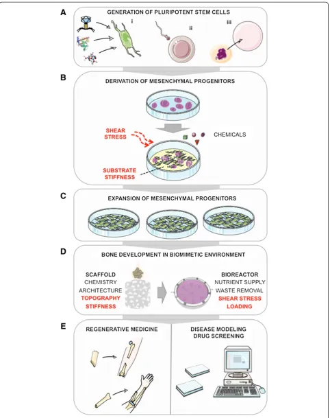
Efforts to develop simple, reproducible protocols for MP derivation have also been made (that is, by exposing feeder-free hESCs to low-serum, growth factor-supplemented medium) [48-50]. Taking into account these and previous reports, our strategy to derive MPs has involved a similar stepwise induction, in which PSC cultures were first switched to serum-supplemented medium, followed by subculture of adherent progenitors for three to five passages to obtain a uniform population [18,51] (Figure 1). The described approaches are gener-ally in agreement with strategies to derive other meso-dermal lineages by stepwise induction of molecular signaling involved in specific stages of early embryonic development [52,53]. The time frame of MP ation (3 to 4 weeks) and the yields of MPs in differenti-ating cultures (not more than 40%) [21,49,50] are comparable to those reported for other mesodermal lin-eages [52,53].
Less is understood about the influence of biophysical factors on early differentiation of PSCs into MP popula-tions. Culture dimensionality (that is, two-dimensional monolayer culture versus three-dimensional embryoid body culture) seems not to be critical for MP induction. Recently, Zoldan and colleagues [54] demonstrated that the variation in scaffold stiffness can affect differenti-ation of hESCs to specific germ layers. Their finding that polymer scaffolds with high elastic moduli (1.5 to 6 MPa) promoted mesoderm development, intermediate elastic moduli (0.1 to 1 MPa) promoted primitive streak and endoderm development, and low elastic moduli (less than 0.1 MPa) promoted neuroectoderm develop-ment [54] is in agreedevelop-ment with other studies [55]. Thus, substrates of adequate stiffness could be used in PSC induction protocols to enhance MP derivation. Efforts to scale-up the culture and differentiation of mouse ESCs and hESCs in stirred suspension bioreactors also indicated that the hydrodynamic forces (and associated shear stresses) in different mixing regimes affect meso-dermal differentiation and could potentially be tested for derivation of MPs [56,57] (Figure 1). With the dem-onstrated ability to derive MPs from several PSC lines, the current studies are aimed at evaluating the reprodu-cibility of MP induction protocols and properties of MPs from a large number of lines as well as achieving mesenchymal/osteogenic lineage specification in com-pletely defined culture conditions.
Properties of pluripotent stem cell-derived mesenchymal progenitors in relation to adult human mesenchymal stem cells from the bone marrow
After subculture, the majority of cells assume an elong-ate fibroblastic morphology and exhibit stable prolifera-tion [8,18]. A direct comparison between hESC-derived MPs and adult hMSCs from the bone marrow showed a sustained high proliferation of hESC-derived MPs over 30 passages in comparison with a stark decline in growth potential after 15 to 20 passages in hMSCs [8]. Similarly, a fast, stable proliferation was observed in MPs from hESC and hiPSC lines in recent studies [18,49,51].
Evaluation of surface antigens is typically conducted to test the mesenchymal phenotype [8,18,19,21,50,51]. Studies report high expression of a comprehensive set of markers that are considered to define adult hMSCs, including CD29, CD44, CD54, CD73, CD90, CD105, STRO-1, CD106, and CD166. MPs are negative for pluripotent, hematopoietic, endothelial, neuroectodermal, epithelial, and muscle cell markers [8,18,19,21]. In a subpopulation of MPs, expression of stage-specific embryonic antigen-4 can be detected, similarly to subpopulations of hMSCs [51,58]. Our recent study showed that MPs of different PSC lines derived with the same protocol exhibited some variation in surface marker expression profile, which correlated to the differentiation capacity. However, the overall expression profile was very similar to that of hMSCs derived from the bone marrow [51].
Interestingly, MPs also seem to express low levels of immunological markers. de Peppo and colleagues [8] found that both hESC-derived MPs and hMSCs were negative for CD80 and CD86 and that MPs exhibited a lower expression of HLA-ABC than hMSCs and were negative for HLA-DR. In our recent study, similar ex-pression patterns were observed in hESC-derived MPs and hiPSC-derived MPs [51], suggesting the potential of allogeneic MPs for use in cell therapies. Currently,
in vitro and in vivo studies are assessing the MPs for their immunosuppressive and anti-inflammatory proper-ties [49,59].
Global transcriptome comparisons also suggest a high degree of similarity between the MPs and adult hMSCs from the bone marrow. Lian and colleagues [21] com-pared MP lines with their parent hESC lines and with hMSC lines from the bone marrow and adipose tissue. The authors found that the gene expression profiles of hESC-derived MPs were more closely related to those of adult tissue hMSCs than to their parent hESCs. Cell
lines also clustered according to their tissue of origin, and adult cells clustered away from embryonic MPs, suggesting subtle differences between the cells at differ-ent developmdiffer-ental stages [21]. These findings were con-firmed by recent studies [8,51].
The potential of MPs to differentiate toward osteo-genic, chondroosteo-genic, and adipogenic lineages is com-monly verified by using in vitro culture models [16,18,19,23,24,48]. In most studies, the derived MPs exhibited three-lineage differentiation potential; thus far, however, quantitative comparison between the func-tional capacity of different MP lines has been limited. In our recent studies, we observed quantitative differences in differentiation potentials of MPs from different hESC and hiPSC lines, similar to the extent observed between hMSC lines. Interestingly, the MP line exhibiting low or negligible differentiation also exhibited a lower expres-sion of mesenchymal surface markers and clustered away in global transcriptome comparisons [18,51]. These findings suggest that MP lines displaying poor differenti-ation could be identified prior to TE studies. It is unclear whether the differences arise from different genetic backgrounds, source tissues used for cellular reprogram-ming to hiPSCs, or the reprogramreprogram-ming method itself, and a larger number of MP lines needs to be evaluated to address these questions.
Another question is the maintenance of differentiation potential during extended proliferation in vitro. de Peppo and colleagues [60] noted that hESC-derived MPs exhibit a stronger osteogenic differentiation potential compared with hMSCs after 20 passages. However, the authors also noted a decrease compared with earlier pas-sages, suggesting that the culture environment for cell expansion needs to be optimized to maintain the differ-entiation potential (similar to that observed with hMSCs) [37].
Effects of biophysical environment on osteogenic differentiation and bone tissue formation: translating from human mesenchymal stem cells to pluripotent stem cell-derived mesenchymal progenitors
Bone formationin vitrofrom adult hMSCs can be mod-ulated by the factors known to regulate native tissue de-velopment [28]. Considering the similarities between adult hMSCs and PSC-derived MP populations, we and
(See figure on previous page.)
others postulated that TE models supporting bone for-mation from hMSCs could generally be applied to in-duce differentiation and maturation of MPs into functional bone tissue (Figure 1). Furthermore, the com-bination of biochemical and biophysical stimulatory sig-nals in a three-dimensional setting could potentially enhance the development of MPs into mature osteo-blasts. The finding that MPs display slight differences to hMSCs in gene and protein expression (see previous sec-tion) warrants an exploration of specific culture condi-tions supporting stable osteogenic commitment and maturation of MPs into bone tissue.
The mechanical properties and surface topography of the scaffold [61,62] can present biophysical signals that influence hMSC growth and osteogenic responses, as can the scaffold geometry and surface chemistry [63-65]. It was found that scaffolds with rough inner surfaces (that is, grooves, grids, and disordered nanopits) made of osteoconductive materials (that is, bone protein, hy-droxyapatite, and synthetic polymers) facilitate cell at-tachment and osteogenesis [66]. Scaffold mechanical properties similar to those of native bone (that is, stiffer matrices mimicking collagenous bone) stimulate osteo-genesis, support maintenance of the construct shape during in vitro growth, and enable load-bearing. In addition, appropriate scaffold porosity and sufficiently large, interconnected pores are needed to facilitate cell infiltration, matrix deposition, and ingrowth of vascula-ture after implantation [63].
Scaffolds of select properties have been tested for their effects on growth, differentiation, and maturation of MPsin vitro. An early study by Tian and colleagues [45] confirmed that three-dimensional porous poly(lactic-co-glycolic) osteoconductive scaffolds, with pore sizes of ap-proximately 1 mm and a compressive strength and Young’s modulus of 7.8 ± 3.1 MPa and 77.2 ± 10.8 MPa, respectively, support growth and enhance osteogenesis of hESC-derived progenitors, as indicated by the in-creased alkaline phosphatase activity and osteocalcin se-cretion. Considering the findings with hMSCs, our group has been using decellularized bovine trabecular bone scaffolds for TE with hESC- and hiPSC-derived MPs. We found that MPs attach, proliferate, and deposit dense bone matrix in these scaffolds [18,51]. In a separ-ate study, we evalusepar-ated the influence of bone scaffold architecture on bone formation by hESC-derived MPs [67]. We sorted the scaffolds into three density groups (low: 0.281 ± 0.018 mg/mm3; medium: 0.434 ± 0.015 mg/ mm3; and high: 0.618 ± 0.027 mg/mm3) and found that the compressive elastic moduli and internal surface areas were the highest in scaffolds of high density (55 ± 3.3 MPa and 7.7 ± 2.6 cm2, respectively) but that the po-rosities and pore sizes were the highest in scaffolds of low density (88.3% ± 1.0% and 376 ± 21μm, respectively).
The medium-density scaffold group supported formation of constructs with the highest densities of cells and new bone matrix, as indicated by increased deposition of osteopontin, osteocalcin, and bone sialoprotein. Presum-ably this was due to the best balance between the trans-port of nutrients and metabolites, space for cell infiltration, surface for cell attachment, and the mechan-ical strength of the scaffolds [67]. Hu and colleagues [68] compared the effect of nanofibrous architecture of polylactic acid matrices to flat films and found that nanofibrous architecture enhances the differentiation of hESC-derived MPs in osteogenic medium. Subsequently, culture of MPs on nanofibrous three-dimensional polylactic scaffolds on an orbital shaker at 75 revolutions per minute resulted in increased gene expression of bone sialoprotein and osteocalcin and the formation of highly mineralized tissue, presumably as a result of syn-ergistic effects of biochemical and biophysical signaling [68]. The influence of scaffold stiffness on osteogenic de-velopment was reported for a mouse embryonic MP cell line [69]. This study indicated that stiffer core shell poly (ether sulfone)-poly(ε-caprolactone) scaffolds (with a mechanical modulus of approximately 30 MPa) pro-moted osteogenesis, as demonstrated by approximately twofold increases in Runx2, alkaline phosphatase, and osteocalcin gene expression. Softer pure poly(ε -caprolactone) scaffolds (approximately 7 MPa) promoted chondrogenesis, as demonstrated by significantly higher Sox9, aggrecan, and collagen type 2 gene expression and increased glycosaminoglycan deposition [69]. Import-antly, the identical microstructure and surface chemis-tries of the scaffolds allowed the evaluation of specific effects of scaffold stiffness on cell differentiation.
complete healing of bone defects, and a small incidence of teratoma formation (2 out of 42 animals) after 8 weeks. Also, Hwang and colleagues [70] recently dem-onstrated that a scaffold-mediated microenvironment can regulate the mechanism of bone formation by hESC-derived MPs in vivo. Hydroxyapatite/polymer composite scaffolds, exhibiting nano-scale surface topog-raphy and exposed hydroxyapatite particles, and control polymer scaffolds were seeded with hESC-derived MPs, pre-cultured in osteogenic medium, and implanted sub-cutaneously in immunodeficient mice. In composite scaffolds, MPs formed bone by intramembranous ossifi-cation, whereas in polymer scaffolds, cells differentiated by endochondral ossification, as evidenced by the forma-tion of cartilaginous tissue followed by calcificaforma-tion and increased blood vessel invasion [70].
In addition to scaffolds, bioreactors can be designed to recapitulate one or more of the developmentally relevant biophysical signals in a time-controlled manner to pro-mote in vitro bone formation [28]. Ideally, a bioreactor system should coordinate biological, physiological, and mechanical stimuli and apply them in a spatially and temporally controlled manner to provide lineage-specific stimulation. In MSCs, increased mass transport and fluid shear (ranging between 0.1 and 0.3 dynes/cm2) by medium perfusion [71] and mechanical loading (short bouts of 5% strain) [72] were shown to improve osteo-genesis and enhance formation of homogenous bone constructs.
Considering these reports, we developed a perfusion bioreactor for bone TE and tested how different perfu-sion conditions affect bone formation by hMSCs seeded on decellularized bone scaffolds [73]. We found that in-creasing the perfusing medium flow velocity (from 80 to 1800 μm/second) significantly affected cell morphology, cell-cell interactions, matrix production and compos-ition, and the expression of osteogenic genes and that intermediate flow velocities (400 to 800 μm/second) yielded the best osteogenic outcome [73]. This bone scaffold-perfusion bioreactor model was then used to engineer bone substitutes from hESC-derived MPs [18] and more recently from hiPSC-derived MPs [51]. We have found that perfusion was critical for the develop-ment of large, compact bone substitutes and that bone protein and mineral content was significantly higher compared with static cultures. Engineered bone tissue was quantitatively comparable between hESC-derived MPs and the control hMSC cultures [18]. Similarly, a study by de Peppo and colleagues [24] showed that cul-ture of MPs on natural coral scaffolds in packed bed/col-umn bioreactors with a flow perfusion rate of 10 mL/ minute and an estimated average shear stress of 0.001 Pa significantly increased cell numbers and bone tissue for-mation in comparison with static culture. However, MPs
formed a thicker, denser tissue than hMSCs, reflecting the higher proliferation and biosynthetic activity of the former [24].
Together, these studies underline that adequate selec-tion of culture on three-dimensional scaffolds in bioreac-tors is fundamental to guide the maturation of PSC-derived MPs into macroscopic (over 1 cm large) func-tional bone substitutes. Further studies are needed to in-vestigate the specific effects of biophysical signals on MPs and to evaluate the stability and functionality of engineered tissuesin vivo.
Maturation and stability of mesenchymal progenitor-derived bone tissue
With a growing number of studies demonstrating the po-tential of PSC-derived MPs for bone TE, it is critical to understand the MP maturation and development after transplantation in vivo. Several studies showed a lack of teratoma formation by transplanted hESC-derived MPs [16,18,21,23,44], presumably as a result of downregulation of genes involved in pluripotency, stemness, and cell pro-liferation and increased expression of lineage-specific genes [8]. It is noteworthy that, in these studies, MP popu-lations were monitored in vivo for short periods (1 to 4 months) and that MPs derived by using other protocols should also be tested to assess their developmental potential.
Tissues engineered from MPs also need to be evalu-ated for their stability and developmental potential prior to, or in conjunction with, testing functionality in bone defects [23]. Presumably, the extended culture time in conditions promoting differentiation should further de-crease the pluripotent phenotype and induce cell com-mitment and maturation into specialized phenotype. We have evaluated hESC-engineered bone after 8 weeks of subcutaneous transplantation in immunodeficient mice and found a stable bone phenotype with signs of further tissue maturation (evidenced by increased mineralized tissue content), vascular invasion, and initiation of re-modeling [18].
hESC-and hiPSC-derived MPs follow similar differentiation pathways under the investigated culture conditions. Interestingly, genes commonly downregulated in hESC-and hiPSC-derived MPs encoded for a set of interacting proteins with roles in cell cycle, DNA replication, spin-dle assembly, mitotic division, and carcinogenesis, suggesting a strong repression of proliferation associated with culture in bioreactors. In contrast, genes upregulated during bioreactor culture encoded for pro-teins involved in extracellular matrix synthesis and re-modeling, osteoblast differentiation, and bone formation, suggesting a strong commitment toward the osteogenic lineage. Together, these findings could indicate the oc-currence of a proliferation/differentiation switch associ-ated with the progression of osteogenesis and tissue maturation during bioreactor culture. It remains to be determined whether common molecular changes could be identified by using a larger number of hESC- and hiPSC-derived MPs to understand their role in guiding MP maturation into phenotypically stable bone substi-tutes. In line with the gene microarray data, transplant-ation experiments confirmed a stable phenotype of engineered bone after 12 weeks of subcutaneous im-plantation into immunodeficient mice [51], similar to our previous work [18].
High-throughput gene expression profiling and stan-dardized in vivo transplantation assays using a larger number of MP lines could potentially allow the identifi-cation of common molecular mechanisms guiding MP maturation into phenotypically stable bone substitutes and identification of markers predicting their functional potentialin vivo.
Future directions and challenges
As discussed in the previous section, culture in scaffold-bioreactor systems allows reproducible, large-scale pro-duction of bone tissue substitutes with the potential to translate into clinical settings [74]. The proper combin-ation of osteogenic cells, scaffolding materials, and cul-ture conditions is paramount for engineering macul-ture bone substitutes for replacement therapies of the skeletal system. However, there are a number of remaining chal-lenges, including reproducibility of osteogenic induction protocols from different PSC lines; the influences of gen-etic background, source tissue, and methods of repro-gramming on regenerative potential; and development of defined differentiation protocols. Another concern is that the current TE approaches involving‘custom-made’ bioreactors, which differ in maintenance and running re-quirements [24,71,73], limit broad implementation of specific strategies, compared with the universality of well-plate culture designs for both experimentation and analytics.
Some of the open questions could be addressed by the development of high-throughput TE strategies on a common platform (for instance, by designing stem cell culture environments in micro-bioreactor formats) [30]. These miniaturized versions of three-dimensional bio-reactor systems allow a step toward accurate, multifac-torial control of cultured cells and tissues, allowing high-throughput studies with increased numbers of ex-perimental conditions and replicates while reducing the amounts of cell and culture materials used. For instance, different culture conditions affecting PSC and MP differ-entiation could be studied, as could the effects of various chemicals and biologics on cell survival, growth, and dif-ferentiated phenotype [75]. The integration of automated handling, on-line analytical reouts, and imaging ad-vances the usefulness of high-throughput platforms [30]. In one example, Figallo and colleagues [76] developed a simple device composed of an array of culture wells to enable systematic and precise variation of mass transport and hydrodynamic shear and used it to study the differ-entiation of hESCs into vascular and cardiac cells by using standard imaging systems. In a recent study, Gobaa and colleagues [75] developed a micro-platform that simultaneously probes the role of biochemical and biophysical niche factors in stem cell fate. Their device consisted of a high-throughput hydrogel microwell sys-tem, where the hydrogel stiffness could be controlled, and the hydrogels could be functionalized with proteins using robotic technology. The fate of single cells, ex-posed to variations in cell density, substrate mechanics, and protein incorporation, could be tested in high throughput (more than 2,000 experiments on a single glass slide) [75].
demonstrate how PSCs could be used in conjunction with TE strategies to construct advanced tissue models, holding the potential to greatly improve the process of drug discovery by testing the substances/biologics dir-ectly on the cell types affected by a particular condition.
Conclusions
A number of studies have demonstrated the potential of using PSCs for the derivation of MPs. Via TE strategies, biophysical signals can be integrated with biochemical factors to enhance and control PSC differentiation and maturation into three-dimensional bone tissue. It is cru-cial to assess the role of individual stimuli on cell devel-opment, such as biomaterial scaffold properties and biophysical conditioning in bioreactors, to develop func-tional bone substitutes of clinical relevance. Current studies with PSC-derived MPs suggest that culture in scaffold-bioreactor systems, optimized by using adult hMSCs, allows the development of stable substitutes exhibiting functional properties typical of mature bone tissue, with unprecedented potential for future recon-structive therapies. In addition, using TE approaches for PSC culture offers a possibility to develop advanced cul-ture models for use in basic biological studies, disease modeling, and drug discovery.
Abbreviations
BMP-2:Bone morphogenetic protein 2; hESC: Human embryonic stem cell; hiPSC: Human induced pluripotent stem cell; hMSC: Human mesenchymal stem cell; MP: Mesenchymal progenitor; PSC: Pluripotent stem cell; TE: Tissue engineering.
Competing interests
The authors declare that they have no competing interests.
Acknowledgments
This work was supported by the New York Stem Cell Foundation’s Helmsley Investigator award (to DM), the Leona M. and Harry B. Helmsley Charitable Trust, Robin Chemers Neustein, Goldman Sachs Gives (with the
recommendations of Alan and Deborah Cohen), New York State Stem Cell Science (Shared Facility Grant C024179), and the New York Stem Cell Foundation.
Published: 4 September 2013
References
1. Langer R, Vacanti JP:Tissue engineering.Science1993,260:920–926. 2. Hing KA:Bone repair in the twenty-first century: biology, chemistry or
engineering?Philos Transact A Math Phys Eng Sci2004,362:2821–2850. 3. Laurencin C, Khan Y, El-Amin SF:Bone graft substitutes.Expert Rev Med
Devices2006,3:49–57.
4. Neovius E, Engstrand T:Craniofacial reconstruction with bone and biomaterials: review over the last 11 years.J Plast Reconstr Aesthet Surg
2010,63:1615–1623.
5. Marolt D, Knezevic M, Novakovic GV:Bone tissue engineering with human stem cells.Stem Cell Res Ther2010,1:10.
6. Meijer GJ, de Bruijn JD, Koole R, van Blitterswijk CA:Cell-based bone tissue engineering.PLoS Med2007,4:e9.
7. Frohlich M, Grayson WL, Wan LQ, Marolt D, Drobnic M, Vunjak-Novakovic G: Tissue engineered bone grafts: biological requirements, tissue culture and clinical relevance.Curr Stem Cell Res Ther2008,3:254–264. 8. de Peppo GM, Svensson S, Lenneras M, Synnergren J, Stenberg J, Strehl R,
Hyllner J, Thomsen P, Karlsson C:Human embryonic mesodermal progenitors highly resemble human mesenchymal stem cells and display high potential for tissue engineering applications.Tissue Eng Part A2010,16:2161–2182.
9. Wagner W, Horn P, Castoldi M, Diehlmann A, Bork S, Saffrich R, Benes V, Blake J, Pfister S, Eckstein V, Ho AD:Replicative senescence of
mesenchymal stem cells: a continuous and organized process.PLoS One
2008,3:e2213.
10. Zhou S, Greenberger JS, Epperly MW, Goff JP, Adler C, Leboff MS, Glowacki J:Age-related intrinsic changes in human bone-marrow-derived mesenchymal stem cells and their differentiation to osteoblasts.Aging Cell2008,7:335–343.
11. Bertram H, Mayer H, Schliephake H:Effect of donor characteristics, technique of harvesting andin vitroprocessing on culturing of human marrow stroma cells for tissue engineered growth of bone.Clin Oral Implants Res2005,16:524–531.
12. Thomson JA, Itskovitz-Eldor J, Shapiro SS, Waknitz MA, Swiergiel JJ, Marshall VS, Jones JM:Embryonic stem cell lines derived from human blastocysts.
Science1998,282:1145–1147.
13. Takahashi K, Tanabe K, Ohnuki M, Narita M, Ichisaka T, Tomoda K, Yamanaka S:Induction of pluripotent stem cells from adult human fibroblasts by defined factors.Cell2007,131:861–872.
14. Warren L, Manos PD, Ahfeldt T, Loh YH, Li H, Lau F, Ebina W, Mandal PK, Smith ZD, Meissner A, Daley GQ, Brack AS, Collins JJ, Cowan C, Schlaeger TM, Rossi DJ:Highly efficient reprogramming to pluripotency and directed differentiation of human cells with synthetic modified mRNA.
Cell Stem Cell2010,7:618–630.
15. Zhou H, Wu S, Joo JY, Zhu S, Han DW, Lin T, Trauger S, Bien G, Yao S, Zhu Y, Siuzdak G, Schöler HR, Duan L, Ding S:Generation of induced pluripotent stem cells using recombinant proteins.Cell Stem Cell2009, 4:381–384.
16. Karlsson C, Emanuelsson K, Wessberg F, Kajic K, Axell MZ, Eriksson PS, Lindahl A, Hyllner J, Strehl R:Human embryonic stem cell-derived mesenchymal progenitors - potential in regenerative medicine.Stem Cell Res2009,3:39–50.
17. Murry CE, Keller G:Differentiation of embryonic stem cells to clinically relevant populations: lessons from embryonic development.Cell2008, 132:661–680.
18. Marolt D, Campos IM, Bhumiratana S, Koren A, Petridis P, Zhang G, Spitalnik PF, Grayson WL, Vunjak-Novakovic G:Engineering bone tissue from human embryonic stem cells.Proc Natl Acad Sci U S A2012,109:8705– 8709.
19. Barberi T, Willis LM, Socci ND, Studer L:Derivation of multipotent mesenchymal precursors from human embryonic stem cells.PLoS Med
2005,2:e161.
20. Olivier EN, Rybicki AC, Bouhassira EE:Differentiation of human embryonic stem cells into bipotent mesenchymal stem cells.Stem Cells2006, 24:1914–1922.
21. Lian Q, Lye E, Suan Yeo K, Khia Way Tan E, Salto-Tellez M, Liu TM, Palanisamy N, El Oakley RM, Lee EH, Lim B, Lim SK:Derivation of clinically compliant MSCs from CD105+, CD24- differentiated human ESCs.Stem Cells2007,25:425–436.
22. Hwang NS, Varghese S, Lee HJ, Zhang Z, Ye Z, Bae J, Cheng L, Elisseeff J:In
vivocommitment and functional tissue regeneration using human embryonic stem cell-derived mesenchymal cells.Proc Natl Acad Sci U S A
2008,105:20641–20646.
23. Villa-Diaz LG, Brown SE, Liu Y, Ross AM, Lahann J, Parent JM, Krebsbach PH: Derivation of mesenchymal stem cells from human induced pluripotent stem cells cultured on synthetic substrates.Stem Cells2012,30:1174– 1181.
Note:This article is part of a thematic series onPhysical influences on stem cellsedited by Gordana Vunjak-Novakovic. Other articles in the series can be found online at http://stemcellres.com/series/
24. de Peppo GM, Sladkova M, Sjovall P, Palmquist A, Oudina K, Hyllner J, Thomsen P, Petite H, Karlsson C:Human embryonic stem cell-derived mesodermal progenitors display substantially increased tissue formation compared to human mesenchymal stem cells under dynamic culture conditions in a packed bed/column bioreactor.Tissue Eng Part A2013, 19:175–187.
25. Kim S, Kim SS, Lee SH, Eun Ahn S, Gwak SJ, Song JH, Kim BS, Chung HM:In
vivobone formation from human embryonic stem cell-derived osteogenic cells in poly(d,l-lactic-co-glycolic acid)/hydroxyapatite composite scaffolds.Biomaterials2008,29:1043–1053.
26. Levi B, Hyun JS, Montoro DT, Lo DD, Chan CK, Hu S, Sun N, Lee M, Grova M, Connolly AJ, Wu JC, Gurtner GC, Weissman IL, Wan DC, Longaker MT:In
vivodirected differentiation of pluripotent stem cells for skeletal regeneration.Proc Natl Acad Sci U S A2012,109:20379–20384.
27. Finkemeier CG:Bone-grafting and bone-graft substitutes.J Bone Joint Surg Am2002,84-A:454–464.
28. Vunjak-Novakovic G, Meinel L, Altman G, Kaplan D:Bioreactor cultivation of osteochondral grafts.Orthod Craniofac Res2005,8:209–218.
29. Rouwkema J, Gibbs S, Lutolf MP, Martin I, Vunjak-Novakovic G, Malda J:In
vitroplatforms for tissue engineering: implications for basic research and clinical translation.J Tissue Eng Regen Med2011,5:e164–e167.
30. Tandon N, Marolt D, Cimetta E, Vunjak-Novakovic G:Bioreactor engineering of stem cell environments.Biotechnol Adv2013:.pii: S0734-9750(13)00066-9.
31. Bruder SP, Fink DJ, Caplan AI:Mesenchymal stem cells in bone development, bone repair, and skeletal regeneration therapy.J Cell Biochem1994,56:283–294.
32. Gastaldi G, Asti A, Scaffino MF, Visai L, Saino E, Cometa AM, Benazzo F: Human adipose-derived stem cells (hASCs) proliferate and differentiate in osteoblast-like cells on trabecular titanium scaffolds.J Biomed Mater Res A2010,94:790–799.
33. Roberts SJ, Geris L, Kerckhofs G, Desmet E, Schrooten J, Luyten FP:The combined bone forming capacity of human periosteal derived cells and calcium phosphates.Biomaterials2011,32:4393–4405.
34. Tarte K, Gaillard J, Lataillade JJ, Fouillard L, Becker M, Mossafa H, Tchirkov A, Rouard H, Henry C, Splingard M, Dulong J, Monnier D, Gourmelon P, Gorin NC, Sensebé L:Société Française de Greffe de Moelle et Thérapie Cellulaire: Clinical-grade production of human mesenchymal stromal cells: occurrence of aneuploidy without transformation.Blood2010, 115:1549–1553.
35. Ho AD, Wagner W, Franke W:Heterogeneity of mesenchymal stromal cell preparations.Cytotherapy2008,10:320–330.
36. Wagner W, Ho AD:Mesenchymal stem cell preparations - comparing apples and oranges.Stem Cell Rev2007,3:239–248.
37. Mauney JR, Kirker-Head C, Abrahamson L, Gronowicz G, Volloch V, Kaplan DL:Matrix-mediated retention ofin vitroosteogenic differentiation potential andin vivobone-forming capacity by human adult bone marrow-derived mesenchymal stem cells duringex vivoexpansion.J Biomed Mater Res A2006,79:464–475.
38. Mareschi K, Ferrero I, Rustichelli D, Aschero S, Gammaitoni L, Aglietta M, Madon E, Fagioli F:Expansion of mesenchymal stem cells isolated from pediatric and adult donor bone marrow.J Cell Biochem2006,97:744–754. 39. Bielby RC, Boccaccini AR, Polak JM, Buttery LD:In vitrodifferentiation and
in vivomineralization of osteogenic cells derived from human embryonic stem cells.Tissue Eng2004,10:1518–1525.
40. Inanc B, Elcin AE, Elcin YM:Effect of osteogenic induction on thein vitro differentiation of human embryonic stem cells cocultured with periodontal ligament fibroblasts.Artif Organs2007,31:792–800. 41. Karp JM, Ferreira LS, Khademhosseini A, Kwon AH, Yeh J, Langer RS:
Cultivation of human embryonic stem cells without the embryoid body step enhances osteogenesisin vitro.Stem Cells2006,24:835–843. 42. Li L, Baroja ML, Majumdar A, Chadwick K, Rouleau A, Gallacher L, Ferber I,
Lebkowski J, Martin T, Madrenas J, Bhatia M:Human embryonic stem cells possess immune-privileged properties.Stem Cells2004,22:448–456. 43. Swijnenburg RJ, Schrepfer S, Govaert JA, Cao F, Ransohoff K, Sheikh AY,
Haddad M, Connolly AJ, Davis MM, Robbins RC, Wu JC:
Immunosuppressive therapy mitigates immunological rejection of human embryonic stem cell xenografts.Proc Natl Acad Sci U S A2008, 105:12991–12996.
44. Mahmood A, Harkness L, Schroder HD, Abdallah BM, Kassem M:Enhanced differentiation of human embryonic stem cells to mesenchymal
progenitors by inhibition of TGF-beta/activin/nodal signaling using SB-431542.J Bone Miner Res2010,25:1216–1233.
45. Tian XF, Heng BC, Ge Z, Lu K, Rufaihah AJ, Fan VT, Yeo JF, Cao T: Comparison of osteogenesis of human embryonic stem cells within 2D and 3D culture systems.Scand J Clin Lab Invest2008,68:58–67. 46. Ahn SE, Kim S, Park KH, Moon SH, Lee HJ, Kim GJ, Lee YJ, Cha KY, Chung
HM:Primary bone-derived cells induce osteogenic differentiation without exogenous factors in human embryonic stem cells.Biochem Biophys Res Commun2006,340:403–408.
47. Kuznetsov SA, Cherman N, Robey PG:In vivobone formation by progeny of human embryonic stem cells.Stem Cells Dev2011,20:269–287. 48. Boyd NL, Robbins KR, Dhara SK, West FD, Stice SL:Human embryonic stem
cell-derived mesoderm-like epithelium transitions to mesenchymal progenitor cells.Tissue Eng Part A2009,15:1897–1907.
49. Sánchez L, Gutierrez-Aranda I, Ligero G, Rubio R, Muñoz-López M, García-Pérez JL, Ramos V, Real PJ, Bueno C, Rodríguez R, Delgado M, Menendez P: Enrichment of human ESC-derived multipotent mesenchymal stem cells with immunosuppressive and anti-inflammatory properties capable to protect against experimental inflammatory bowel disease.Stem Cells
2011,29:251–262.
50. Tran NT, Trinh QM, Lee GM, Han YM:Efficient differentiation of human pluripotent stem cells into mesenchymal stem cells by modulating intracellular signaling pathways in a feeder/serum-free system.Stem Cells Dev2012,21:1165–1175.
51. de Peppo GM, Marcos-Campos I, Kahler D, Alsalman D, Shang L, Vunjak-Novakovic G, Marolt D:Engineering bone tissue substitutes from human induced pluripotent stem cells.Proc Natl Acad Sci U S A2013,110:8680– 8685.
52. James D, Nam HS, Seandel M, Nolan D, Janovitz T, Tomishima M, Studer L, Lee G, Lyden D, Benezra R, Zaninovic N, Rosenwaks Z, Rabbany SY, Rafii S: Expansion and maintenance of human embryonic stem cell-derived endothelial cells by TGFbeta inhibition is Id1 dependent.Nat Biotechnol
2010,28:161–166.
53. Yang L, Soonpaa MH, Adler ED, Roepke TK, Kattman SJ, Kennedy M, Henckaerts E, Bonham K, Abbott GW, Linden RM, Field LJ, Keller GM: Human cardiovascular progenitor cells develop from a KDR + embryonic-stem-cell-derived population.Nature2008,453:524–528.
54. Zoldan J, Karagiannis ED, Lee CY, Anderson DG, Langer R, Levenberg S:The influence of scaffold elasticity on germ layer specification of human embryonic stem cells.Biomaterials2011,32:9612–9621.
55. Keung AJ, Asuri P, Kumar S, Schaffer DV:Soft microenvironments promote the early neurogenic differentiation but not self-renewal of human pluripotent stem cells.Integr Biol (Camb)2012,4:1049–1058.
56. Niebruegge S, Bauwens CL, Peerani R, Thavandiran N, Masse S, Sevaptisidis E, Nanthakumar K, Woodhouse K, Husain M, Kumacheva E, Zandstra PW: Generation of human embryonic stem cell-derived mesoderm and cardiac cells using size-specified aggregates in an oxygen-controlled bioreactor.Biotechnol Bioeng2009,102:493–507.
57. Sargent CY, Berguig GY, Kinney MA, Hiatt LA, Carpenedo RL, Berson RE, McDevitt TC:Hydrodynamic modulation of embryonic stem cell differentiation by rotary orbital suspension culture.Biotechnol Bioeng
2010,105:611–626.
58. Riekstina U, Cakstina I, Parfejevs V, Hoogduijn M, Jankovskis G, Muiznieks I, Muceniece R, Ancans J:Embryonic stem cell marker expression pattern in human mesenchymal stem cells derived from bone marrow, adipose tissue, heart and dermis.Stem Cell Rev2009,5:378–386.
59. Giuliani M, Oudrhiri N, Noman ZM, Vernochet A, Chouaib S, Azzarone B, Durrbach A, Bennaceur-Griscelli A:Human mesenchymal stem cells derived from induced pluripotent stem cells down-regulate NK-cell cytolytic machinery.Blood2011,118:3254–3262.
60. de Peppo GM, Sjovall P, Lenneras M, Strehl R, Hyllner J, Thomsen P, Karlsson C:Osteogenic potential of human mesenchymal stem cells and human embryonic stem cell-derived mesodermal progenitors: a tissue engineering perspective.Tissue Eng Part A2010,16:3413–3426.
61. Dalby MJ, Gadegaard N, Tare R, Andar A, Riehle MO, Herzyk P, Wilkinson CD, Oreffo RO:The control of human mesenchymal cell differentiation using nanoscale symmetry and disorder.Nat Mater2007,6:997–1003. 62. Shih YR, Tseng KF, Lai HY, Lin CH, Lee OK:Matrix stiffness regulation of
integrin-mediated mechanotransduction during osteogenic
63. Karageorgiou V, Kaplan D:Porosity of 3D biomaterial scaffolds and osteogenesis.Biomaterials2005,26:5474–5491.
64. Comisar WA, Kazmers NH, Mooney DJ, Linderman JJ:Engineering RGD nanopatterned hydrogels to control preosteoblast behavior: a combined computational and experimental approach.Biomaterials2007,28:4409– 4417.
65. Kasemo B, Lausmaa J:Material-tissue interfaces: the role of surface properties and processes.Environ Health Perspect1994,102:41–45. 66. Nava MM, Raimondi MT, Pietrabissa R:Controlling self-renewal and
differentiation of stem cells via mechanical cues.J Biomed Biotechnol
2012,2012:797410.
67. Marcos-Campos I, Marolt D, Petridis P, Bhumiratana S, Schmidt D, Vunjak-Novakovic G:Bone scaffold architecture modulates the development of mineralized bone matrix by human embryonic stem cells.Biomaterials
2012,33:8329–8342.
68. Hu J, Smith LA, Feng K, Liu X, Sun H, Ma PX:Response of human embryonic stem cell-derived mesenchymal stem cells to osteogenic factors and architectures of materials duringin vitroosteogenesis.Tissue Eng Part A2010,16:3507–3514.
69. Nam J, Johnson J, Lannutti JJ, Agarwal S:Modulation of embryonic mesenchymal progenitor cell differentiation via control over pure mechanical modulus in electrospun nanofibers.Acta Biomater2011, 7:1516–1524.
70. Hwang NS, Varghese S, Lee JH, Zhang Z, Elisseeff J:Biomaterials directed
in vivoosteogenic differentiation of mesenchymal cells derived from human embryonic stem cells.Tissue Eng Part A2013,19:1723–1732. 71. Sikavitsas VI, Bancroft GN, Holtorf HL, Jansen JA, Mikos AG:Mineralized
matrix deposition by marrow stromal osteoblasts in 3D perfusion culture increases with increasing fluid shear forces.Proc Natl Acad Sci U S A2003, 100:14683–14688.
72. Sittichokechaiwut A, Edwards JH, Scutt AM, Reilly GC:Short bouts of mechanical loading are as effective as dexamethasone at inducing matrix production by human bone marrow mesenchymal stem cell.Eur Cell Mater2010,20:45–57.
73. Grayson WL, Marolt D, Bhumiratana S, Frohlich M, Guo XE, Vunjak-Novakovic G:Optimizing the medium perfusion rate in bone tissue engineering bioreactors.Biotechnol Bioeng2011,108:1159–1170.
74. Salter E, Goh B, Hung B, Hutton D, Ghone N, Grayson WL:Bone tissue engineering bioreactors: a role in the clinic?Tissue Eng Part B Rev2012, 18:62–75.
75. Gobaa S, Hoehnel S, Roccio M, Negro A, Kobel S, Lutolf MP:Artificial niche microarrays for probing single stem cell fate in high throughput.Nat Methods2011,8:949–955.
76. Figallo E, Cannizzaro C, Gerecht S, Burdick JA, Langer R, Elvassore N, Vunjak-Novakovic G:Micro-bioreactor array for controlling cellular
microenvironments.Lab Chip2007,7:710–719.
77. Quarto N, Leonard B, Li S, Marchand M, Anderson E, Behr B, Francke U, Reijo-Pera R, Chiao E, Longaker MT:Skeletogenic phenotype of human Marfan embryonic stem cells faithfully phenocopied by patient-specific induced-pluripotent stem cells.Proc Natl Acad Sci U S A2012,109:215– 220.
78. Deyle DR, Khan IF, Ren G, Wang PR, Kho J, Schwarze U, Russell DW:Normal collagen and bone production by gene-targeted human osteogenesis imperfecta iPSCs.Mol Ther2012,20:204–213.
doi:10.1186/scrt317