BioMedCentral
Cardiovascular Ultrasound
Open Access
Review
Myocardial contractility in the echo lab: molecular, cellular and
pathophysiological basis
Tonino Bombardini*
Address: Department of Echocardiography, Institute of Clinical Physiology, National Council of Research, Pisa, Italy
Email: Tonino Bombardini* - [email protected] * Corresponding author
Abstract
In the standard accepted concept, contractility is the intrinsic ability of heart muscle to generate force and to shorten, independently of changes in the preload or afterload with fixed heart rates. At molecular level the crux of the contractile process lies in the changing concentrations of Ca2+ ions in the myocardial cytosol. Ca2+ ions enter through the calcium channel that opens in response to the wave of depolarization that travels along the sarcolemma. These Ca2+ ions "trigger" the release of more calcium from the sarcoplasmic reticulum (SR) and thereby initiate a contraction-relaxation cycle.
In the past, several attempts were made to transfer the pure physiological concept of contractility, expressed in the isolated myocardial fiber by the maximal velocity of contraction of unloaded muscle fiber (Vmax), to the in vivo beating heart. Suga and Sagawa achieved this aim by measuring pressure/volume loops in the intact heart: during a positive inotropic intervention, the pressure volume loop reflects a smaller end-systolic volume and a higher end-systolic pressure, so that the slope of the pressure volume relationship moves upward and to the left. The pressure volume relationship is the most reliable index for assessing myocardial contractility in the intact circulation and is almost insensitive to changes in preload and after load. This is widely used in animal studies and occasionally clinically. The limit of the pressure volume relationship is that it fails to take into account the frequency-dependent regulation of contractility: the frequency-dependent control of transmembrane Ca2+ entry via voltage-gated Ca2+ channels provides cardiac cells with a highly sophisticated short-term system for the regulation of intracellular Ca2+ homeostasis. An increased stimulation rate increases the force of contraction: the explanation is repetitive Ca2+ entry with each depolarization and, hence, an accumulation of cytosolic calcium. As the heart fails, there is a change in the gene expression from the normal adult pattern to that of fetal life with an inversion of the normal positive slope of the force-frequency relation: systolic calcium release and diastolic calcium reuptake process is lowered at the basal state and, instead of accelerating for increasing heart rates, slows down. Since the force-frequency relation uncovers initial alteration of contractility, as an intermediate step between normal and abnormal contractility at rest, a practical index to measure it is mandatory.
Measuring end-systolic elastance for increasing heart rates is impractical: increasing heart rates with atrial pacing has to be adjunct to the left ventricular conductance catheter, to the left ventricular pressure catheter, to the vena cava balloon, and to afterload changes. Furthermore, a noninvasive index is needed. Noninvasive measurement of the pressure/volume ratio for increasing heart rates during stress in the echo lab could be the practical answer to this new clinical demand in the current years of a dramatic increase in the number of heart failure patients.
Published: 08 September 2005
Cardiovascular Ultrasound 2005, 3:27 doi:10.1186/1476-7120-3-27
Received: 27 July 2005 Accepted: 08 September 2005
This article is available from: http://www.cardiovascularultrasound.com/content/3/1/27
© 2005 Bombardini; licensee BioMed Central Ltd.
Calcium channels: evolutional aspects
When ~3,500,000,000 years ago prokaryotes appeared, the selection of an intracellular messenger preceded the appearance of ionic channels of enveloping lipid membranes.
Calcium had conformational and stechiometric advan-tages to be chosen as an intracellular messenger (=good messenger and modulator of intracellular processes), due to its high coordination number and irregular coordina-tion geometry. If calcium was the first intracellular mes-senger, ionic channels for calcium had to appear for the cells to maintain constant intracellular calcium concentra-tions [1]. Ionic channels, other than function of intracel-lular ionic concentration surveyors, became to have the first function of reactive capability to outer stimulus, changing abruptly their functions. The primitive Ca2+
channels were activated by mechanical stimuli, present but slow and low efficient in reactions. But faster reactions are essential for survival.
When ~1,500,000,000 years ago the hydrosphere became aerobic and the primitive unicellular organisms devel-oped mechanisms of energy production, eukaryotes developed active transport and voltage-gated channels as a result of selection for faster signaling [1,2].
When in this new evolutional cell a mechanical stimulus involving a portion of the membrane hits the cell, a depo-larization produced by the mechanic-sensitive Ca2+
chan-nels subsequently extends the effect to the entire cell by voltage gated Ca2+ channels.
Toward the Na channel: high-speed signaling required by multicellularity
With the appearance of multicellularity, even faster sign-aling had to appear for life competition. As a possible solution to this new requirement, increase in density of Ca2+ channels would increase the speed of the
depolariz-ing wave, but would have compromised the role of Ca2+
as modulator of intracellular function.
The appearance of Na+ channels, capable of carrying
greater ionic fluxes without interfering with intracellular processes, would be evolutionarily favorable. And, in fact, this model was the one chosen by evolution in nerve cells: earlier selection of Na+ channels to sustain potential
changes [1]. In fact, also for a maximum concentration on the cell membrane of channels, Ca2+ channels mediated
maximum velocity is equal to 0.10 m/sec. For Na+
chan-nels mediated max. velocity is 3 m/sec.
Muscle fibers: a particular evolutionary aspect
In primitive muscle fibers all the activating Ca2+ ions for
contraction came from outside the cell.
High density of Ca2+ channels would increase the speed of
the depolarizing wave and at the same time would speed up the activation of the contractile machinery.
But with the evolutionary appearance of intracellular cal-cium stores (sarcoplasmic reticulum) in supplying Ca2+
ions for contraction, muscle fibers acquired the capability of stronger contraction with less energy consumption. As for force of contraction, also signaling speed was a prob-lem in muscle cells: but in these cells less pronounced advantages are provided by an exclusively Na+
-channels-based membrane potential changes. The simultaneous presence of fast Na+ channels for conduction function and
slow Ca2+ channels for beginning the contraction
mecha-nism through inward calcium flow was maintained [3].
At the present evolutionary state, once the impulse has formed in the sinus node, it spreads very rapidly through-out the atrium to reach the atrioventricular (AV) node and ventricles. In atrial tissue, the pattern of the action poten-tial is dominated by a fast sodium channel. The action potential duration of atrial tissue is short when compared to that of ventricles, and the inward flow of calcium ions is less, with a lower force of contraction developed in the atria and lower activity of L-type calcium channels. Con-duction of the wave of depolarization is rapid through conduction tissues where the action potential goes through fast sodium channel activity, whereas conduction is lower through the ventricular myocardium, where there is chiefly calcium channel activity with a slower rate of depolarization.
High frequency-induced upregulation of human cardiac calcium currents: the final evolution
Ultimately developed, the frequency-dependent control of transmembrane Ca2+ entry via voltage-gated Ca2+
chan-nels provides mammalian cardiac cells with a highly sophisticated short-term system for regulation of intracel-lular Ca2+ homeostasis.
Up-regulation of Ca2+ entry through Ca2+ channels by
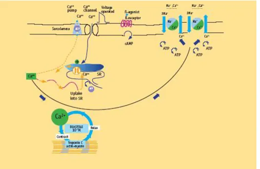
high rates of beating (HFIUR of ICa) is involved in the fre-quency-dependent regulation of contractility: this process is crucial in adaptation to exercise and stress [4,5]. This regulation is rapid, (the steady state is reached rapidly within few seconds for each heart rate level), intrinsic to the myocardium cell, with no necessity to be driven from neuronal or hormonal controls (Fig. 1).
Contractility
Molecular aspects: calcium ion fluxes in cardiac contraction-relaxation cycle
Cardiovascular Ultrasound 2005, 3:27 http://www.cardiovascularultrasound.com/content/3/1/27
Crucial features are [6] entry of Ca2+ ions through the
volt-age-sensitive L-type Ca2+ channels, acting as a trigger for
the release of Ca2+ ions from the sarcoplasmic reticulum
(SR).
Relatively small amounts of calcium ions actually enter and leave the cell during each cardiac cycle, whereas much
larger amounts move in and out of the sarcoplasmic retic-ulum. Calcium-induced calcium release explains most of the current available data. This process elevates by about tenfold the concentration of calcium ions in the cytosol. The result is the increasing interaction of calcium ions with troponin C to trigger the contractile proteins [6] (Fig. 2).
High frequency-induced upregulation of human cardiac calcium currents in isolated cardiomyocytes
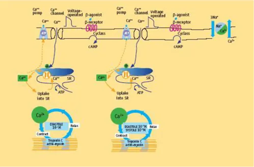
Figure 1
Left ventricular contraction
Left ventricular pressure starts to builds up when the arrival of calcium ions at the contractile proteins starts to trigger actin-myosine interaction. The thin actin filament interacts with the myosin head when Ca2+ ions arrive at
troponin C (TnC). As more and more myofibers enter the contracted state, pressure development in the left ventricle proceeds. The interaction of actin and myosine increases, and cross-bridge cycling augments. As long as enough cal-cium ions are bound to troponin C, many repetitive cycles of this nature occur. The enhanced force development in response to a greater calcium ion concentration is due to recruitment of additional cross bridges.
When calcium ions depart from their binding sites on tro-ponin C, cross-bridge cycling cannot occur and the diasto-lic phase of the cardiac cycle sets in.
Left ventricular relaxation
At the end of systole, calcium stops interacting with tro-ponin C and calcium ions are taken up into the SR by the activity of the SERCA (sarcoplasmatic reticulum Ca2+
ATPase) pump that constitutes nearly 90% of the protein component of the SR. Calcium taken up into the SR by the calcium uptake pump is stored within the SR before fur-ther release. To balance the small quantity of calcium ions entering the heart cell with each depolarization, a similar
Molecular basis of contractility in normal heart
Figure 2
Cardiovascular Ultrasound 2005, 3:27 http://www.cardiovascularultrasound.com/content/3/1/27
Pressure-volume loops in the cath lab
Figure 3
quantity must leave the cell. First, calcium can be exchanged for sodium ions entering by the Na+/Ca2+
exchange and, second, an ATP-consuming sarcolemmal calcium pump can transfer calcium into this extra cellular space against a concentration gradient.
As the cytosolic calcium ion concentration starts to decline because of the uptake of calcium into the SR under the influence of activated phospholamban, more and more myofibers enter the state of relaxation (Fig. 2).
Preload and after load
The preload is the load present before contraction has started, at the end of diastole. When the preload increases, the left ventricle distends during diastole, and the stroke volume rises according to Starling's law [7]. The proposed explanation for the Starling effect, whereby a greater end-diastolic fiber length develops a greater force, is explained
by an interaction between sarcomere length and calcium ions (length sensitization of the sarcomere): 1) increase in end-diastolic fiber length at any given free Ca2+
concentra-tion would increase force by a small amount on the basis of the change in filament overlap; 2) when the fiber is stretched and the sarcomere length increases, for any given number of Ca2+ ions binding to TnC, there is greater
force development. Length sensitization of the sarcomere explains how the sarcomere can "upgrade itself" to a higher force-length curve [6].
The afterload is the systolic load on the left ventricle after it has started to contract.
Increased afterload means that an increased intraventricu-lar pressure has to be generated first to open the aortic valve and then during the ejection phase [8].
In the nonfailing heart, the left ventricle can overcome any physiological acute increase in load [6].
Contractility: how can it be defined?
"Contractility is the inherent capacity of the myocardium to contract independently of changes in the preload or afterload. Whatever the problems of measuring it, con-tractility remains an essential corner concept to separate the effects of a primary change in loading conditions from an intrinsic change in the force of contraction [6]". It is a basic property of cardiac muscle and is strictly linked to the activation quantity of actin myosin transverse bridges in the myocardial fibers, and to the velocity of cross-bridge activation at the systole onset [6,9]. Cytosolic cal-cium level is the determinant of:
- The myocardial fiber number involved in the contraction process.
- The maximal velocity of myocardial fibers shortening.
Increased contractility, is reflected in higher myocardial fiber shortening velocity, with a more highly developed tension peak and a steeper pressure rise, when preload, afterload, and heart rate are constant: in the cytosol cal-cium release is more and faster from SR with a higher cytosol calcium concentration in systole: more troponin is activated from higher levels of calcium with more acitn-myosin cross-bridges in the time unit, and ultimately myocardial fiber contraction is more and faster.
Decreased contractility is reflected in lower myocardial fiber shortening velocity, with a lower tension peak and a blunted pressure rise, when preload, after load, and heart rate are constant: in the cytosol calcium release is less and slower from SR with a lower cytosol calcium concentra-tion in systole: less troponin is activated from lower levels
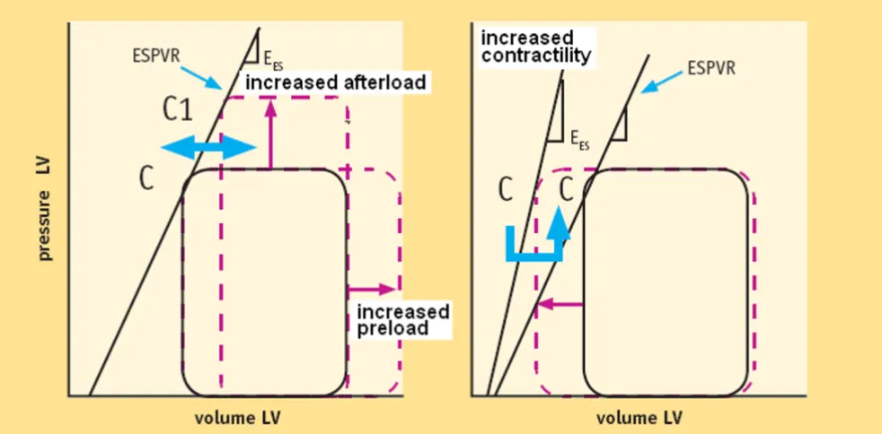
The end-systolic pressure-volume relationship (ESPVR)
Figure 4
Cardiovascular Ultrasound 2005, 3:27 http://www.cardiovascularultrasound.com/content/3/1/27
of calcium with less actin-myosin cross-bridges in time unit, and ultimately myocardial fiber contraction is less and slower.
The isolated myocardial fiber: idealized contractility in the physio lab
Contractility expressed in the isolated myocardial fiber is the maximal velocity of contraction of unloaded muscle fiber (Vmax). This value is defined as the maximal velocity of contraction, when there is no load on the isolated mus-cle. This strictly preload and after load independent index, fulfills the theoretical requirements for contractility quan-tification and greatly contributes to this research field [9,10]. Nevertheless, this model is not usable in in-vivo conditions.
The in vivo, beating heart: how to measure contractility
In the past attempts were made to transfer the purely physiological concept of contractility expressed in the
iso-lated myocardial fiber by the maximal velocity of contrac-tion of unloaded muscle fiber (Vmax), to the in vivo beating heart. Suga and Sagawa achieved this aim by measuring pressure/volume loops in the intact heart: dur-ing a positive inotropic intervention, the pressure volume loop reflects a smaller end systolic volume and a higher end-systolic pressure, so that the slope of the pressure vol-ume relationship (Ees) moves upward and to the left [11,12]. Ees is the most reliable index for assessing (stand-ard) myocardial contractility at rest in the intact circula-tion and is almost insensitive to changes in preload, and after load.
This is widely used in animal studies and occasionally clinically [6].
A now-time conductance catheter is used for human stud-ies [13]. The method is highly correct, but invasive, com-plex, and technically demanding. (Fig. 3)
Load changes at constant contractility (left) and contractility changes at constant load (right)
Figure 5
Focusing on cytosol calcium concentrations along the pressure-volume loop, (Fig 3) in diastole (D-A tract) cytosolic calcium is reuptake from cytoplasm and stored in the SR [6]. At the A end-diastolic point, the end-diasto-lic volume (or maximal myocardial fiber length) predicts contractile-proteins calcium-sensitivity of the upcoming systole according to the Starling's law [7]. The velocity of the pressure development in the isovolumic systole (A-B tract), and the ejection force in the isotonic systole (B-C tract) are both strictly linked to the contractile state. When LV systolic emptying ends (C point), the aortic valve closes, and isovolumic diastolic relaxation starts. (C-D tract).
More highly developed systemic pressure simultaneously with lower end-systolic volume is typical of higher con-tractility. Counter-directional changes identify compro-mised contractility.
If end-systolic volume is measured for different end-systo-lic pressure values, sequential end systoend-systo-lic pressure/vol-ume values can be recorded (C, C1, C2, C... points). The upper left corners (C, C1, C2, and C... points) of the loops define the LV end-systolic pressure-volume relation (ESPVR). (Fig. 4) The ESPVR predicts in a heart with con-stant contractility the systolic volume when end-systolic pressure changes, and ultimately predicts the left ventricle ability to empty for different afterload values [14].
Contractility is quantified by the angular coefficient (or slope) of the ESPVR relation: the Ees (end systolic elastance) (Fig. 5).
Contractility and heart rate
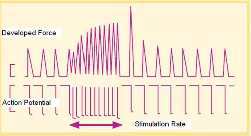
The heart contractility dependence on increasing heart rates has been established in most mammalians.
Force-frequency relation or Bowditch treppe
Figure 6
Cardiovascular Ultrasound 2005, 3:27 http://www.cardiovascularultrasound.com/content/3/1/27
The inherent ability of ventricular myocardium to increase its strength of contraction independently of neu-rohormonal control, in response to an increase in contrac-tion frequency is known as frequency treppe [4] (Fig. 6). In humans this myocardial property causes the contractile force to rise, as contraction frequency is increased from 60 to about 180 bpm and to then decline with further increase in frequency (the force-frequency relation "FFR") [6].
Molecular basis
Heart rate is a leading determinant of cytosol calcium con-centration, and strictly linked to the contractility levels. In the healthy heart, a frequency increase up to 180 beats per
minute provides systolic faster calcium SR release (increased contractility or developed force) and diastolic faster SR calcium reuptake (positive lusitropic effect).
Up-regulation of Ca2+ entry through Ca2+ channels by
high rates of beating (HFIUR of ICa) is involved in the fre-quency-dependent regulation of contractility: this process is crucial in adaptation to exercise and stress [5]. This reg-ulation is rapid, (the steady state is reached rapidly within few seconds for each heart rate level), intrinsic to the myo-cardium cell, with no need to be driven from neuronal or hormonal controls (Fig. 1, Fig. 6).
Plots of average steady-state isometric twitch tension versus stimulation frequency in non-failing and failing myocardium
Figure 7
Cellular and myocardial fiber level
This property has been definitively established in the human heart in experimental settings using cardiomyo-pathic myocardial strips.
Measurements of twitch tension in isolated left-ventricu-lar strips from explanted cardiomyopathic hearts com-pared with non-failing hearts show reduction in peak rates of generation and relaxation of twitch tension and a decrease in slope of tension rate vs. contraction frequency [15,16] (Fig. 7).
The FFR of these failing groups both exhibit a negative treppe at contraction frequencies above about 100 bpm.
Presence of a negative treppe in the working range of heart rates may constitute an additional liability beyond mere depression of the wall tension since this may contribute to an accelerated progression of heart failure. In patients in end stage failure the peak of the FFR occurs at such a low frequency that there is a negative treppe over the entire in vivo range of heart rates. The contraction frequency at which the FFR begins its descending limb ("optimum
Molecular basis of contractility in failing heart
Figure 8
Cardiovascular Ultrasound 2005, 3:27 http://www.cardiovascularultrasound.com/content/3/1/27
Molecular pathopysiology, action potentials and calcium transients in isolated myocytes of normal (A) vs. failing (B) hearts
Figure 9
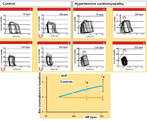
Force-frequency relationship in the cath lab
Figure 10
Cardiovascular Ultrasound 2005, 3:27 http://www.cardiovascularultrasound.com/content/3/1/27
stimulation frequency") declines progressively in the order: atrial septal defect, coronary artery disease, diabetic myopathy, mitral regurgitation, dilated cardiomyopathy. This suggests that a correlation between severity of myo-cardial disease and optimum contraction frequency may exist [15,16].
In more severe heart failure the peak of the FFR is shifted sufficiently to lower frequencies so that it has a negative slope over the entire range of in vivo heart rates (i.e., 80– 150 bpm). The weakening of contractile strength as heart rate rises suggest the possibility that in vivo, a sudden increase in heart rate could predispose the ventricle to being stretched by venous return.
While the FFR is well known in the physiological lab [5,17,18], with extensive studies in isolated strips of fail-ing myocardium [15,16], in animal models of heart failure [19,20], till now its knowledge and use in the clin-ical setting is extremely limited [21-26].
Fetal gene program: back from the future
"As the ventricle fails, there is a change in the ventricular gene expression pattern from the normal adult pattern to that normally observed only during fetal life. There is a down regulation of the calcium uptake pump (SERCA2) and of the fast-contracting myosin heavy chain. The fetal program may be activated from cytosolic calcium over-load, by adding phosphate groups to enzymes that nor-mally inhibit the fetal program [6]" (Fig. 8).
Changes in the calcium cycle are fundamental to the impaired contractile performance of the failing heart. The SR calcium stores are severely depleted because of the combined effects of depressed calcium uptake into the SR resulting from decreased SERCA activity, both down-regu-lated and inhibited. Thus, the calcium ions entering with depolarization are unable to trigger the release of enough calcium to generate a normal calcium transient (Fig. 9). There is a close relationship between the depression of SERCA in human heart failure and the depressed force-fre-quency relationship. Paradoxically, the diastolic calcium level is higher than normal. Starting from this higher level, as the heart rate increases, the calcium ions enter more
Table 1: Force-frequency relationship from the experimental lab to clinical applications
Author Feldman Bhargava Hasenfusss Liu Inagaki Schuler Dehmer Lavie
Journal J Clin Invest Am J Cardiol Eur Heart J Circ Circ Am j Cardiol Am J Cardiol Chest
Year 1988 1988 1994 1993 1999 1982 1981 1989
Method cath lab cath lab cath lab cath lab cath lab nuc nuc nuc
FORCE SP/ESV dP/dt dP/dt Ees dP/dt SP/ESV SP/ESV SP/ESV
TREPPE Yes Yes Yes Yes Yes Base- peak Base-peak Base-peak
HR increase PM PM PM PM PM
EX ISO
EX EX EX
PTS# Disease DC 7 DC 5 DC 9 HYP 10 HYP 17 AR 14 AR 17 MR 11
FFR Upsloping 3 - - - 7 7 11 7
Flat-Biph - 2 - 10 10 7 2 2
Neg 4 3 9 - - - 4 2
Control # 6 3 8 8 10 9 15
-FFR Upsloping 6 3 8 8 10 9 15
-Flat-Biph - - -
-Neg - - -
-PM = atrial pacing; EX = exercise; ISO = isoproterenol ; DC = dilated cardiomyopathy; HYP = hypertensive cardiomyopathy; CHD = coronary artery disease; AR = aortic regurgitation; MR = mitral regurgitation; FFR = force-frequency relation
Several attempts have been made to transfer the force-frequency relationship from the experimental lab to clinical applications. Such attempts have been based on invasive evaluation in cath lab (Feldman 1988, Bhargava 1988, Hasenfuss 1994, Liu 1993, Inagaki 1999), or noninvasive evaluation with radionuclide scintigraphy (Schuler 1982, Dehmer 1981, Lavie 1989). The extensively adopted maximum rate of pressure rise (max dP/dt) for force measurement is largely preload and afterload dependent. Since End-systolic elastance (Ees), is almost insensitive to changes in preload and afterload, Ees should be measured at each heart rate step increase, as done by Liu and coworkers.
But measuring Ees for increasing heart rates is impractical: increasing heart rates obtained with atrial pacing has to be adjunct to the LV
conductance catheter, the LV pressure catheter, the vena cava balloon, and the afterload changes. Proof of this is that only Liu adopted this method in humans.
rapidly through the calcium channels than can be extruded through the Na+/Ca2+ exchange, so that the
diastolic levels rise, as does the diastolic tension.
Muscle strips prepared from patients with severe heart failure behave very differently from normal muscle, in that there is hardly any response to an increased stimula-tion frequency. Whereas in strips from normal hearts, optimal force development is reached at rates of about 150 to 180 beats/min, in patients with cardiomyopathy
an increased heart rate produces a decreased twitch ten-sion (Fig. 7). In addition, the diastolic tenten-sion rises mark-edly with the stimulation frequency, compatible with a rate-induced cytosolic calcium overload causing diastolic dysfunction [6].
Present limits of end systolic elastance (ESPVR slope) for contractility measurement
This standard, historically accepted, rest-assessed contrac-tility, is limited because it is an invasive index, but
The Suga (SP/ESV) index instead of end-systolic elastance for FFR measurement
Figure 11
The Suga (SP/ESV) index instead of end-systolic elastance for FFR measurement. Since End-systolic elastance (Ees), expressing the slope of the in-vivo, end-systolic ventricular pressure vs. chamber volume relation, is the most "foolproof' window into in vivo myocardial contractility, Ees should be measured at each heart rate step increase. A simpler approach was utilized by Feld-man and co-workers by measuring SP/ESV ratio at baseline, and for pacing induced heart rate increase to 25 and 50 bpm beyond basal heart rate. Feldman showed that 7 patients with dilated cardiomyopathy (DCM) demonstrated little or no signifi-cant enhancement in SP/ESV ratio during atrial pacing tachycardia. The lack of improvement in cardiomyopathy patients has been contrasted to patients with normal ventricular function (Control) who demonstrated significant increase in SP/ESV ratio. SP/ESV ratio is simpler than Ees measurement, and equally provides knowledge of up-sloping vs flat-biphasic force-frequency relationship. (Modified from: Feldman MD, Alderman JD, Aroesty JM, Royal HD, Ferguson JJ, Owen RM, et al. Depression of systolic and diastolic myocardial reserve during atrial pacing tachycardia in patients with dilated cardiomyopathy. J Clin Invest
Cardiovascular Ultrasound 2005, 3:27 http://www.cardiovascularultrasound.com/content/3/1/27
especially because it fails to take into account frequency-dependent regulation of contractility: ultimately devel-oped in the evolutional scale, as a typical feature of more advanced mammalian species, absent in fetal life and in adults with heart failure-induced regression of the contractile mechanism, the frequency-dependent control of transmembrane Ca2+ entry via voltage-gated Ca2+
chan-nels provides mammalian cardiac cells with a highly sophisticated short-term system for regulation of intracel-lular Ca2+ homeostasis.
The impossibility of separating the cellular mechanism of contractility changes from those of load or heart rate is now clear. "Thus, there is a clear overlap between contrac-tility, which should be independent of load or heart rate, and the effects of load and heart rate on the cellular mech-anism. Hence, the traditional separation of inotropic state from load or heart rate effects as two independent
regula-tors of cardiac muscle performance is no longer simple now that the underlying cellular mechanisms have been uncovered [6]." This topic is not important only as a spec-ulative concept, but especially clinically: in fact as the heart fails, there is a change in the ventricular gene expres-sion pattern from the normal adult pattern to that nor-mally observed only during fetal life, as a memory of primordial contraction patterns, with an inversion of the normal positive slope of the relation: the systolic calcium release and diastolic calcium reuptake process is lowered at the basal state and, instead of accelerating for increasing heart rates, it slows down. Since the assessment of FFR shows initial alteration of contractility, as an intermediate step between normal and abnormal contractility at rest, a practical index to measure it is mandatory.
Since end-systolic elastance (Ees), expressing the slope of the in-vivo, end-systolic ventricular pressure vs chamber
Stress echo lab: contractility me too?
Figure 12
volume relation, is the most "foolproof' window into in vivo myocardial contractility, Ees should be measured at each heart rate step increase, as made by Liu and cowork-ers [21] (Fig. 10).
But measuring Ees for increasing heart rates is impractical: increasing heart rates obtained with temporarily pacing has to be adjunct to the LV conductance catheter, the LV
FFR, from myocardial strips to the echo lab
Figure 13
Cardiovascular Ultrasound 2005, 3:27 http://www.cardiovascularultrasound.com/content/3/1/27
Force-frequency curve with stress echo in a subject with latent LV dysfunction without dilation
Figure 15
Force-frequency curve with stress echo in a normal subject
Figure 14
Cardiovascular Ultrasound 2005, 3:27 http://www.cardiovascularultrasound.com/content/3/1/27
Force-frequency curve with stress echo in a subject with dilated cardiomyopathy and depressed baseline left ventricular func-tion (EF% = 30%)
Figure 16
pressure catheter, the vena cava balloon, and to afterload changes. Proof of this is that only Liu [21] adopted this method in humans. (Table 1).
If assessment of Ees is difficult under clinical conditions at fixed heart rates, assessment of Ees for increasing heart rates is much more difficult.
The Suga index (SP/ESV ratio) for increasing heart rates: the link toward the stress echo lab FFR measurement in a practical clinical method
A simpler approach was utilized by Feldman and co-work-ers [26] in DCM pts vs. normal hearts, by measuring the SUGA index (SP/ESV ratio, instead of Ees) at baseline, and for pacing induced heart rate increase to 25 and 50 bpm beyond basal heart rate. SP/ESV ratio measurement is sim-pler than Ees measurement, and equally provides knowledge of an up-sloping, flat, or biphasic Bowditch treppe (Fig. 11).
Force-frequency relationship in the stress echo
lab: a practical, noninvasive, modern approach
to contractility
Non-invasive methods [27-30] have been proposed to assess the rest-peak stress change in inotropic state, based upon the assumption that positive inotropic interventions are mirrored by smaller end-systolic volumes and higher end-systolic pressures (Table 1). During bicycle stress echocardiography, dobutamine or pacing stress, continuous 2D echo monitoring is performed by protocol and blood pressure, ECG and left ventricular volumes are obtained at each step, providing the basic information required to build a force-frequency relationship over a wide range of frequencies (Fig. 12). A totally noninvasive estimation of force-frequency relation during stress in the echo lab is theoretically appealing for the identification of limited contractile reserve and latent global left ventricu-lar dysfunction.
This method is similar to the previously proposed ones but is totally noninvasive, with echocardiography used to assess LV volumes during exercise and cuff blood pressure to estimate peak systolic pressure as an index of end-systo-lic pressure [31-33].
Bowditch treppe and stress echo. Methodology
During (exercise, DOB or pacing) stress echocardiography continuous 2D echo monitoring is performed by protocol and blood pressure, ECG and left ventricular volumes are obtained at each step, providing the basic information required to build a force-frequency relation over a wide range of frequencies [34].
This approach is based on serial assessment of these vari-ables at different exercise steps so that the force-frequency
pattern (up sloping, flat, and biphasic) can be assessed (Fig 13).
Baseline and stress echocardiography
The patient undergoes transthoracic echocardiography at baseline and at each 10 beat frequency increase during stress. This is performed using conventional two-dimen-sional echocardiography and tissue harmonic imaging, and digitized on-line into a quad screen, cineloop format. Images are also recorded on half-inch S-VHS videotape. Left ventricular end-diastolic and end-systolic volumes are measured from apical four and two chamber view, by an experienced observer using the biplane discs-method [35,36] (Fig. 12). Only representative cycles are measured and the average of three measurements is taken. The endo-cardial border is traced, excluding the papillary muscles. The frame captured at the R wave of the ECG is considered to be the end diastolic frame, and the frame with the smallest left ventricular cavity the end systolic frame.
Blood pressure analysis
One investigator records all blood pressures at rest and during exercise during the study. The blood pressure recording is made using a manometer sphygmomanome-ter and the diaphragm of a standard stethoscope (Fig. 12).
End-systolic pressure-volume determination
To build the force-frequency relationship, the force is determined at each step as the ratio of the systolic pressure (cuff sphygmomanometer)/end-systolic volume index (biplane Simpson rule/body surface area). The force frequency relation is built off line (Fig. 13). The slope of the relationship is calculated as the ratio between SP/ESV (Systolic Pressure/End-Systolic Volume) index increase (from baseline to peak exercise)/heart rate increase (from baseline to peak exercise). The force-frequency relationship is defined up-sloping when peak exercise SP/ ESV index is higher than baseline and intermediate stress values (Fig. 14); biphasic, with an initial up-sloping followed by a later down-sloping trend, when peak exer-cise systolic pressure/end-systolic volume index is lower than intermediate stress values [6,25] (Fig. 15); flat or neg-ative, when peak exercise systolic pressure/end-systolic volume index is equal to or lower than baseline stress val-ues (Fig. 16). The critical heart rate (or optimum stimula-tion frequency) is defined as the heart rate at which systolic pressure/end-systolic volume index reaches the maximum value during progressive increase in heart rate; in biphasic pattern, the critical heart rate is the heart rate beyond which systolic pressure/end-systolic volume index has declined by 5%; in a negative pattern the critical heart rate is the starting heart rate [25].
Cardiovascular Ultrasound 2005, 3:27 http://www.cardiovascularultrasound.com/content/3/1/27
phenomenon; "in situ, the optimal heart rate is not only the rate that would give maximal mechanical performance of an isolated muscle twitch, but also is determined by the need for diastolic filling [6]" (Fig. 13)
Conclusion
This proposed approach allows the assessment of a theo-retically robust and sophisticated index of left ventricular contractility with an absolute minimum extra-burden of data acquisition and analysis, since all the basic parame-ters (heart rate, blood pressure and left ventricular volumes) are routinely acquired during exercise stress echo testing [34]. The extra measurements consist of serial evaluation of ventricular volumes and linear interpola-tion of the force-frequency relainterpola-tionship. This approach is simple, not time-consuming, and highly feasible [31-33]. This index of global contractility is theoretically appealing for the identification of limited contractile reserve and latent global left ventricular dysfunction.
These are all prerequisites for a larger scale testing in the clinical subsets in which the contractility information can be more important – such as patients with latent ventricu-lar dysfunction [37] or advanced chronic heart failure [33].
Noninvasive measurement of pressure/volume relation (the Suga index) [11,26,31] for increasing heart rates dur-ing stress in the echo lab could be the practical answer to this new clinical demand in recent years of a dramatic increase in the number of heart failure patients.
Acknowledgements
TB is funded by a PhD program on Cardiovascular Pathophysiology of the Scuola Superiore S. Anna, Pisa
References
1. Franciolini F, Petris A: Evolution of ionic Channels of biological membranes. Mol Biol Evol 1989, 6:503-513.
2. Alpert NR, Blanchard EM, Mulieri LA, Nagai R, Zarain-Herberg A, Periasamy M: Genetic and non-genetic control of myocardial calcium. Basic Res Cardiol 1989, 84:55-56.
3. Reuter H, Stevens CF, Tsien RW, Yellen G: Properties of single channels in cardiac cell culture. Nature 1982, 279:501-504. 4. Bowditch HP: Über die Eigenthüm-lichkeiten der Reizbarkeit,
welche die Muskelfasern des Herzens zeigen. Ber Sächs Akad Wiss 1871, 23:652-689.
5. Piot C, Lemaire S, Albat B, Seguin J, Nargeot J, Richard S: High fre-quency-induced upregulation of human cardiac calcium currents. Circulation 1996, 93:120-128.
6. Opie LH: Mechanisms of cardiac contraction and relaxation. In Heart DiseaseVolume Chap 19. 7th edition. Edited by: Braunwald E, Zipes DP, Libby P, Bonow RO. WB Saunders Company; 2005:457-489. page 480
7. Starling EH: Linacre lecture on the law of the heart (1915). London, Longmans; 1918.
8. Sonnenblick EH, Downing SE: Afterload as a primary determi-nant of ventricular performance. Am J Physiol 1962, 204:604-610.
9. Braunwald E: On the difference between the heart's output and its contractile state (Editorial). Circulation 1971, 43:171. 10. Ross J Jr: Cardiac function and myocardial contractility: a
perspective. J Am Coll Cardiol 1983, 1:52-62.
11. Suga H, Sagawa K, Shoukas AA: Load independence of the instan-taneous pressure/volume ratio of the canine left ventricle and effects of epinephrine and heart rate on the ratio. Circu-lation Research 1973, 32:314-322.
12. Suga H, Sagawa K: Instantaneous pressure/volume relation-ships and their ratio in the excised, supported canine left ventricle. Circulation Research 1974, 35:117-126.
13. Kass DA, Midei M, Graves W, Brinker JA, Maughan WL: Use of a conductance (volume) catheter and transient inferior vena cava occlusion for rapid determination of pressure-volume relationship in man. Cathet Cardiovasc Diagn 1988, 15:192-202. 14. Grossman W, Braunwald E, Mann T, McLaurin LP, Green LH:
Con-tractile state of the left ventricle in man as evaluated from end-systolic pressure-volume relations. Circulation 1977, 56:845-852.
15. Mulieri LA, Hasenfuss G, Leavitt B, Allen PD, Alpert NR: Altered myocardial force-frequency relation in human heart failure.
Circulation 1992, 85:1743-1750.
16. Mulieri LA, Leavitt BJ, Martin BJ, Haeberle JR, Alpert NR: Myocardial force-frequency defect in mitral regurgitation heart failure is reversed by forskolin. Circulation 1993, 88:2700-2704.
17. Layland J, Kentish JC: Positive force- and [Ca2+] i-frequency relationships in rat ventricular trabeculae at physiological frequencies. Am J Physiol Heart Circ Physiol 1999, 276:H9-H18. 18. Lewartowski B, Pytkowski B: Cellular mechanism of the
rela-tionship between myocardial force and frequency of contractions. Prog Biophys Mol Biol 1987, 50:97-120.
19. O'Rourke B, Kass DA, Tomaselli GF, Kaab S, Tunin R, Marban E: Mechanisms of altered excitation-contraction coupling in canine tachycardia-induced heart failure I. Circ Res 1999, 84:562-570.
20. Ross J, Miura T, Kambayashi M, Eising GP, Ryu KH: Adrenergic con-trol of the force-frequency relation. Circulation 1995, 92:2327-2332.
21. Liu CP, Ting CT, Lawrence W, Maughan WL, Chang MS, Kass DA: Diminished contractile response to increased heart rate in intact human left ventricular hypertrophy: systolic versus diastolic determinants. Circulation 1993, 88:1893-1906. 22. Hasenfuss G, Holubarsch C, Hermann HP, Astheimer K, Pieske B, Just
H: Influence of the force-frequency relationship on haemody-namics and left ventricular function in patients with non-fail-ing hearts and in patients with dilated cardiomyopathy. Eur Heart J 1994, 15:164-170.
23. Bhargava V, Shabetai R, Mathiasen RA, Dalton N, Hunter JJ, Ross J Jr: Loss of adrenergic control of the force-frequency relation in heart failure secondary to idiopathic or ischemic cardiomyopathy. Am J Cardiol 1998, 81:1130-1137.
24. Izawa H, Yokota M, Takeichi Y, Inagaki M, Nagata K, Iwase M, Sobue T: Adrenergic control of the force-frequency and relaxation-frequency relations in patients with hypertrophic cardiomyopathy. Circulation 1997, 96:2959-2968.
25. Inagaki M, Yokota M, Izawa H, Ishiki R, Nagata K, Iwase M, Yamada Y, Koide M, Sobue T: Impaired force-frequency relations in patients with hypertensive left ventricular hypertrophy. Cir-culation 1999, 14:1822-1830.
26. Feldman MD, Alderman JD, Aroesty JM, Royal HD, Ferguson JJ, Owen RM, Grossman W, McKay RG: Depression of systolic and diasto-lic myocardial reserve during atrial pacing tachycardia in patients with dilated cardiomyopathy. J Clin Invest 1988, 11:1661-1669.
27. Schuller G, Olshausen K, Schwarz F, Mehmel H, Hofmann M, Her-mann HJ, Lange D, Kubler W: Noninvasive assessment of myo-cardial contractility in asymptomatic patients with severe aortic regurgitation and normal left ventricular ejection fraction at rest. Am J Cardiol 1982, 50:45-52.
28. Lavie CJ, Lam JB, Gibbons RJ: Effects of exercise on left ventricu-lar volume and output changes in severe mitral regurgita-tion. A radionuclide angiographic study. Chest 1989, 96(5):1086-1091.
29. Dehmer GJ, Firth BG, Hillis LD, Corbett JR, Lewis SE, Parkey RW, Willerson JT: Alterations in left ventricular volumes and ejec-tion fracejec-tion at rest and during exercise in patients with aor-tic regurgitation. Am J Cardiol 1981, 48:17-27.
two-dimen-Publish with BioMed Central and every scientist can read your work free of charge "BioMed Central will be the most significant development for disseminating the results of biomedical researc h in our lifetime."
Sir Paul Nurse, Cancer Research UK
Your research papers will be:
available free of charge to the entire biomedical community
peer reviewed and published immediately upon acceptance
cited in PubMed and archived on PubMed Central
yours — you keep the copyright
Submit your manuscript here:
http://www.biomedcentral.com/info/publishing_adv.asp
BioMedcentral sional echocardiographic indicator of left ventricular
function. J Am Coll Cardiol 1984, 4:509-516.
31. Bombardini T, Correia MJ, Cicerone C, Agricola E, Ripoli A, Picano E: Force-frequency Relationship in the Echocardiography Lab-oratory: A Noninvasive Assessment of Bowditch Treppe? J Am Soc Echocardiogr 2003, 16:646-655.
32. Bombardini T, Agrusta M, Natsvlishvili N, Solimene F, Pap R, Coltorti F, Varga A, Mottola G, Picano E: Noninvasive assessment of left ventricular contractility by pacemaker stress echocardiography. Eur J Heart Failure 2005, 2:173-181.
33. Grosu A, Bombardini T, Senni M, Duino V, Gori M, Picano E: End-systolic Pressure/Volume relations during dobutamine stress echo: a prognostically useful noninvasive index of left ventricular contractility. Eur Heart J 2005 in press.
34. Picano E: Stress echocardiography. From pathophysiological toy to diagnostic tool. Circulation 1992, 85:1604-1612.
35. Armstrong WF, Pellikka PA, Ryan T, Crouse L, Zoghbi WA: Stress echocardiography: recommendations for performance and interpretations of stress echocardiography. Stress Echocar-diography Task Force of the Nomenclature and Standards Committee of the American Society of Echocardiography. J Am Soc Echocardiogr 1998, 11:97-104.
36. Cerqueira MD, Weissman NJ, Dilsizian V, Jacobs AK, Kaul S, Laskey WK, Pennell DJ, Rumberger JA, Ryan T, Verani MS, American Heart Association Writing Group on Myocardial Segmentation and Registration for Cardiac Imaging: Standardized myocardial seg-mentation and nomenclature for tomographic imaging of the heart: a statement for healthcare professionals from the Cardiac Imaging Committee of the Council on Clinical Car-diology of the American Heart Association. Circulation 2002, 105:539-542.