D
OROTAZ
YŚKO1, J
ACEKG
AJEK2, W
IOLETAK
UCHARSKA3, J
AKUBW
ILCZYŃSKI2,
M
ARTAN
EGRUSZ−K
AWECKA2, W
ALENTYNAM
AZUREK2Changes in QT Interval During Tilt−Induced
Vasovagal Syncope
Ocena okresu repolaryzacji komór serca u chorych
z omdleniami wazowagalnymi w czasie testu pochyleniowego
1Teaching Department for Emergency Medical Service, Silesian Piasts University of Medicine in Wrocław,
Poland
2Department of Cardiology, Silesian Piasts University of Medicine in Wrocław, Poland
3Department of Pediatric Cardiology and Allergology, Silesian Piasts University of Medicine in Wrocław,
Poland
Adv Clin Exp Med 2006, 15, 4, 613–618 ISSN 1230−025X
ORIGINAL PAPERS
© Copyright by Silesian Piasts University of Medicine in Wrocław
Abstract
Background.Orthostatic stress causes changes in the activity of the components of the autonomic nervous system. In patients with neurally mediated syncope (NMS), the balance between the sympathetic and parasympathetic parts of this system is disturbed, which can be the cause of bradycardia and/or asystole and significant fall in blood pres− sure. The QT interval, reflecting the repolarization period of the ventricles, can be altered by the neurocardiogenic reaction because its duration depends mainly on the heart rate and autonomic nervous system activity.
Objectives. Assessing the influence of head−up tilt testing on the duration of the QT interval in patients with NMS. Material and Methods.Fifty−seven patients (group I), aged 46.5 ± 17.9 years (39 women and 18 men) with NMS induced by the tilt test conducted according to the two−phases Raviele protocol, and 33 patients (group II), aged 41.3 ± 16.3 years (18 women and 15 men) diagnosed for unexplained syncope without neurocardiogenic reaction, were evaluated. Durations of the RR, QT, and QTc intervals were assessed after the syncope in group I during the sinus or junctional rhythm, and in group II immediately after the test, five minutes after the test, and during sleep between 4:00 and 4:15 AM.
Results.The duration of the RR interval after the tilt test was significantly longer in group I than in group II (p< 0.001) and the QT interval five minutes after the test was significantly longer in group I than in group II (p< 0.05). In group I the RR interval after syncope was significantly longer than at five minutes after the test (p< 0.01) and during sleep (p< 0.05). The QT intervals after syncope and five minutes after the test did not differ and were sig− nificantly shorter than during sleep (p< 0.05). In group II, the RR intervals immediately after the tilt test and five minutes after the test did not differ and were significantly shorter compared with the values during sleep (p< 0.001). QT after the test and five minutes after the test did not differ and were significantly shorter than during sleep (p< 0.001).
Conclusions.Slowing of the heart rate during neurocardiogenic reaction is not accompanied by adequate QT inter− val prolongation immediately after stopping the tilt test. The results point to the concomitant activation of sympa− thetic and parasympathetic components of the autonomic nervous system in the human heart during neurally medi− ated syncope (Adv Clin Exp Med 2006, 15, 4, 613–618).
Key words:QT interval, vasovagal syncope, tilt test.
Streszczenie
Wprowadzenie. Zmiana aktywności składowych układu autonomicznego wpływa na czas trwania okresu repola− ryzacji komór serca mierzonego jako odstęp QT. Jest to zależne nie tylko od zmian częstotliwości serca, ale także od bezpośredniego wpływu mediatorów układu autonomicznego.
Cel pracy. Ocena okresu repolaryzacji komór u chorych z omdleniami wazowagalnymi.
The relationship between the duration of the QT interval and heart rate is an indispensable property of the ventricular repolarization process. Changes in the activity of the autonomic system and circulating catecholamines may be reflected by the relationship between the RR and OT inter− vals [1]. The effect of parasympathetic system activity on the QT interval duration has not yet been fully established, as the observed QT interval prolongation with decreased heart rhythm may be an indirect consequence of changes in the heart rate and not the direct effect of the activity of the parasympathetic system on the heart muscle [2]. A prolonged QT interval with unchanged heart rate indicates an increase in vagus nerve tone or inhibited activity of the parasympathetic system, which was observed at night compared with the resting period during the day when the patient is alert [3]. On vasovagal syncope, the vagus nerve is activated, which is manifested by bradycardia or even inhibition of heart activity; moreover, strong activation of the sympathetic system is manifested by an increased plasma catecholamine level, espe− cially of adrenalin [4].
Evaluation of changes in the QT interval immediately after vasovagal fainting gives the unique possibility of assessing the effect of the sympathetic and parasympathetic system activa− tion on the QT interval and the relationship between the QT and RR intervals [5]. This is a model which can be used to investigate the effect of autonomic system mediators directly on the heart muscle.
The aim of the study was to evaluate the effect of neurocardiogenic reaction on the activation of both components of the autonomic system on the basis of an analysis of the relationship between the duration of the QT and RR intervals immediately after vasovagal syncope in patients diagnosed for syncope.
Material and Methods
The investigation was performed in 57 patients aged 46.5 ± 17.9 years (39 women and 18 men) who had vasovagal syncope induced by the tilt test according to the Italian (Raviele) protocol (group 1). In 15 patients, syncope occurred in the passive phase of the tilt test, while in 42 patients it was pro− voked by sublingual administration of 400 µg nitroglycerin. According to the VASIS classifica− tion, 23 patients developed type 1 (mixed), 31 patients type 2 (cardio−depressive), and 3 subjects type 3 (vasodepressive) neurocardiogenic reaction. The control group consisted of 33 subjects aged 41.3 ± 16.3 years (18 women, 15 men) diag− nosed for presyncope or fainting who did not develop syncope (group 2). The reasons for the reported complaints were established on the basis of history and further studies. Analysis of the clin− ical picture, accessory investigations, and further observation revealed that 8 subjects who reported faintness did not develop the typical vasovagal reaction; the reaction was found possible on the basis of history findings in 11 patients. The possi− bility of psychogenic fainting was taken into con− sideration in 5 patients. Epileptic seizures were found on further observation in 3 patients; the number of such episodes decreased after institu− tion of anti−epileptic treatment. Three patients were diagnosed with situational syncope, and in a further three patients loss of consciousness was found to be caused by hypoglycemia, migraine, and vertebral syndrome.
The tilt test was performed in all of the inves− tigated subjects in order to diagnose the episodes of syncope. The durations of the RR, QT, and QTc intervals were assessed in the patients after vaso− vagal syncope after two preceding sinus or junc− tional rhythms in the case of the RR interval or during sinus bradycardia or junctional rhythm. In
ło (grupa II). Czas trwania odstępu RR, QT i QTc oznaczano u chorych po omdleniu wazowagalnym w czasie bra− dykardii zatokowej lub zastępczego rytmu węzłowego. U osób bez omdlenia w czasie testu pochyleniowego bada− ne wskaźniki oznaczano po opuszczeniu stołu pionizacyjnego do poziomu. Ponownej oceny badanych wskaźni− ków dokonywano 5 minut po zakończeniu badania oraz w czasie snu, między godz. 4:00 a 4:15.
Wyniki.Czas trwania odstępu RR po zakończeniu testu i czas trwania odstępu QT po 5 minutach od zakończenia badania były istotnie dłuższe u badanych chorych w porównaniu do grupy kontrolnej (odpowiednio p < 0,001 i p < < 0,05). W grupie chorych z omdleniem wazowagalnym odstęp RR po omdleniu był istotnie statystycznie dłuższy niż 5 minut po badaniu (p < 0,01) oraz w czasie snu (p < 0,05). Odstęp QT w okresie po omdleniu oraz 5 minut później nie różnił się istotnie i był istotnie krótszy niż w czasie snu (p < 0,05). W grupie osób bez omdlenia odstę− py RR bezpośrednio po teście pochyleniowym oraz 5 minut po badaniu nie różniły się istotnie między sobą, ale były krótsze od odstępu RR w czasie snu (p < 0,001). Odstęp QT po teście pochyleniowym oraz 5 minut później nie różnił się istotnie i był krótszy niż odstęp QT w godzinach nocnych (p < 0,001).
Wnioski. W czasie omdlenia wazowagalnego nie ma wydłużenia czasu trwania odstępu QT adekwatnego do zwol− nienia częstości serca. Wyniki badania wskazują na jednoczesną aktywację obu składowych układu autonomiczne− go w czasie omdlenia wazowagalnego (Adv Clin Exp Med 2006, 15, 4, 613–618).
patients without syncope during the tilt test, the investigated parameters were assessed after bring− ing the tilt table to the horizontal position. Assessment of the investigated parameters was repeated five minutes after termination of the investigation and during sleep from randomly selected evolutions of the sinus rhythm between 4:00 and 4:15 a.m.
Statistical Analysis
The values of the investigated parameters in corresponding time ranges were compared by means of the Student’s t test for unpaired variables following evaluation of the normality of the distri− bution and the equality of variation of the investi− gated variables. The values of the investigated parameters in the groups were compared by means of parametric variation analysis following evalua− tion of the normality of their distribution. The dif− ferences were then assessed by the NIR test.
Results
The investigated parameters are presented in Table 1 taking into account statistical deviations in the form of arithmetic means and their standard deviations.
The investigated parameters changed signifi− cantly in the course of the examination in a similar degree in the patient and control groups. The dura− tion of the RR interval after termination of the test and the duration of the QT interval five minutes after the test were significantly longer in the patients than in the controls (p < 0.001 and p < < 0.05, respectively).
In the patients with vasovagal syncope, the RR interval after fainting was statistically significant− ly longer than at five minutes after the test (p < 0.01) and during sleep (p< 0.05). The QT intervals immediately after fainting and five minutes after fainting did not differ and were statistically signif− icantly shorter than during sleep (p < 0.05). In patients who did not faint, the RR intervals imme− diately after the tilt test and five minutes after the test did not differ significantly and were statisti− cally significantly shorter that the RR intervals during sleep (p< 0.001). Likewise, the QT inter− vals immediately after the tilt test and five minutes later did not differ, but they were significantly shorter than the QT interval at night (p< 0.001).
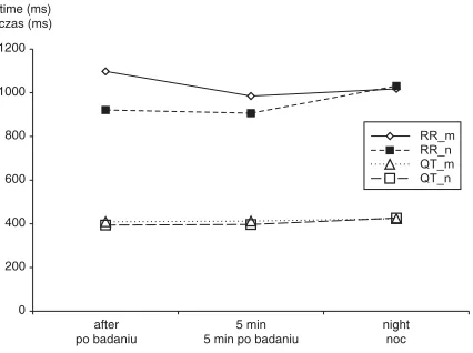
A graphic presentation of the relationship is shown in Figure 1.
It is difficult to interpret the values and changes in the correlated QT interval as the heart rates in the patients of cardiodepressive type dur− ing vasovagal syncope were below the values which could be assessed by Bazett’s formula [6]. Hence the values presented in Table 1 were calcu− lated but not discussed.
Discussion
The QT interval in the electrocardiogram reflects the repolarization of the heart ventricles, and its duration in individual layers of the heart muscle is responsible for the initiation, peak, and termination of the T wave [7]. In physiological conditions, changes in the QT interval duration are mainly brought about by changes in the heart rate, which may be attributable to the effect of the parasympathetic system on the activity of pace−
Table 1.RR, QT, and QTc interval durations immediately after the tilt test, five minutes after the test, and during sleep in vasovagal patients and the control group
Tabela 1. Czas trwania odstępu RR, QT i QTc po zakończeniu testu, po 5 minutach od zakończenia badania i w czasie snu u pacjentów, u których wystąpiło omdlenie i w grupie kontrolnej
Interval Studied group Control group p
(Odstęp) (Grupa badana) (Grupa kontrolna)
RR 1097.7 ± 241.1 921.2 ± 169.5 < 0.001
QT after 409.3 ± 36.8 394.6 ± 39.1 ns.
QTc 0.395 ± 0.037 0.413 ± 0.037 < 0.05
RR 984.9 ± 240.3 906.6 ± 143.1 ns.
QT 5 min. after 412.6 ± 38.5 397.1 ± 25.9 < 0.05
QTc 0.421 ± 0.029 0.418 ± 0.024 ns.
RR 1018.1 ± 162.9 1031.0 ± 182.3 ns.
QT night 423.5 ± 33.2 426.2 ± 35.1 ns.
maker cells of the sinoatrial node [1]. The effect of physical effort on the duration of the QT interval is a complex process which involves not only changes in the heart rate by the inhibition of parasympathetic activity, but also a direct effect of increased sympathetic activity on the repolariza− tion of the ventricular muscle. Although this response results from a number of processes, including presynaptic inhibition of the vagal activ− ity, the effect of circulating and locally secreted catecholamines, and decreased presence of parasympathetic fibers within the heart ventricles, it largely resembles the response observed after administration of atropin [1]. The effect of fluctu− ations in sympathetic−parasympathetic activity on the duration of the action potential of cardiomy− ocytes in various layers of the heart muscle, which determines the duration of repolarization and thus the QT interval of the surface electrocardiogram, is not yet fully understood. The various experi− mental models and a variety of betamimetics (with various effects on the parasympathetic activity) in the performed trials preclude any clinical conclu− sions [1, 7]. Despite a significant effect of the heart rate, it seems that the duration of the QT interval is a relatively constant parameter, not only in the physiological range determined by Bazett’s for−
mula [6], but also in relation to markedly lower frequencies, including paroxysmal ones [2].
Although the vasovagal reflex seems to be a physiological phenomenon, vasovagal syncope is believed to be caused by a disturbed sympathet− ic−parasympathetic balance in response to ortho− static position. The most commonly accepted and well−documented concept of the induction of the neurocardiogenic reaction which leads to syncope assumes excessive sympathetic activation which, by the stimulation of cardiac mechanoreceptors and hypotensive cardiopulmonary region, leads to the activation of vagus nerve centers with inhibi− tion of the tonic sympathetic activity of the hypo− thalamus [8]. The reaction produces significant hypotonia and bradycardia, controlled by the vagus nerve, including episodes of asystole. The clinically described sequel manifests as paroxys− mal presycope or complete loss of consciousness of reflex character [9].
The neurocardiogenic reaction proceeds in several phases which differ in the activity of dif− ferent components of the autonomic nervous sys− tem. The reaction is preceded by potent sympa− thetic activation occurring on different pathways manifested by increased levels of both noradrena− line and adrenaline [10]. Increased levels of the latter are believed by some authors to play the key role in inducing reflex reaction [4, 11], although this has not been confirmed by others [12]. The onset of the neurocardiogenic reaction is charac− terized by an inhibition of sympathetic activity with a relative dominance of parasympathetic activity. The next phase involves the response of the autonomic system to hypotonia and bradycar− dia and/or asystole, which probably results in sym− pathetic stimulation.
The above−described phases of the neurocar− diogenic reaction are reflected in changes in the duration of the QT interval on the electrocardio− grams of patients with vasovagal syncope in the tilt test. Sympathetic activation is associated with a shortening of the ventricular refraction period and the QT interval on the patients’ electrocardio− grams. The relative shortening of the QT interval observed in our trial in the phase of heart rate slowing during vasovagal syncope seems to con− firm the simultaneous activation of both compo− nents of the autonomic system or, what is more probable, parasympathetic activation manifested by bradycardia/asystole and the effect of adrena− line secreted in the presyncopal phase or evoked by the RR pause. Adrenergic stimulation, mainly through the effect on the components of outward potassium currents (Ik), results in shortening of the
ventricular repolarization time with shortening of the QT interval on the electrocardiogram [13]. The
0 200 400 600 800 1000 1200 after po badaniu 5 min 5 min po badaniu
night noc RR_m RR_n QT_m QT_n time (ms) czas (ms)
Fig. 1.RR and QT interval duration in the studied time periods in vasovagal patients and the control group
Ryc. 1.Czas trwania odstępów RR I QT w badanych przedziałach czasu u pacjentów, u których wystąpiło omdlenie wazowagalne I w grupie kontrolnej
RR_m – RR interval in vasovagal patients. RR_n – RR interval in the control group. QT_m – QT interval in vasovagal patients. QT_n – QT interval in the control group.
RR_m – odstęp RR u pacjentów, u których wystąpiło omdlenie wazowagalne.
RR_n – odstęp RR u pacjentów z grupy kontrolnej. QT_m – odstęp QT u pacjentów, u których wystąpiło omdlenie wazowagalne.
answer to the question about the separation of the effect of the autonomic system components at the level of the heart ventricles should be sought in the differentiated autonomic innervation of the atria and ventricles of the heart, with the majority of sympathetic nerves in the latter area. The effect of circulating adrenaline, secreted from the adrenal medulla, cannot be overlooked.
Described observation is consistent both with the study on a similar group of patients suffering from vasovagal syncope reported by Jaeger et al. [5] and with the already quoted study by Castellanos et al. in a group of patients with parox− ysmal atrio−ventricular block [2]. On this basis one can assume that the effect of the autonomic sys− tem, though significant, still merely acts secondar− ily on the electrophysiological properties of the cardiomyocytes, including the effect on the dura− tion of the action potential, and modulates changes in the QT interval. In presented patients, the inter− pretation of the lack of a significant prolongation of the QT interval despite prolonged RR interval may give insight into the sympathetic−parasympa− thetic balance at the level of the ventricular muscle during vasovagal syncope. The findings by Kowallik et al. in healthy men during sleep sug− gest a similar distribution of sympathetic and parasympathetic effects on the sinoatrial node and ventricular muscle [3].
The described phenomenon, apart from cogni− tive value, may reflect the complexity of the path− omechanism of the neurocardiogenic reaction and give insight into the autonomic regulation of the heart muscle itself. The potential arrhythmogenic effect of bradycardia and/or asystole associated with vasovagal syncope is abolished by the relative shortening of the QT interval. However, this obser− vation concerns only patients without any structur− al heart disease, as only these patients were inves− tigated in presented study. Similar reservations concern the earlier mentioned report by Magnano et al. [1]. The presence of severe atherosclerotic lesions in coronary arteries, a history of myocardial infarction, or a significant hypertrophy of the car− diac muscle may significantly modify the response of the QT interval to the described changes in sym− pathetic−parasympathetic activity. Patients with prolonged QT interval syndrome, in whom the shortening of the QT interval in conditions of raised catecholamine levels is disturbed, constitute a similar group. These are patients in whom episodes of vasovagal syncope are at least as com− mon as in the general population, and their loss of consciousness may be a significant prognostic fac− tor. In patients with syncope and severe anginal episodes and/or confirmed atherosclerotic lesions in the coronary arteries, the decision for a tilt test may be taken after carefully considering the indi− vidual risks and expected diagnostic advantages.
References
[1] Magnano AR, Holleran S, Ramakrishnan R, Reiffel JA, Bloomfield DM:Autonomic nervous system influ− ences on QT interval in normal subjects. J Am Coll Cardiol 2002, 39, 1820–1826.
[2] Castellanos A, Moleiro F, Lopera G, Huikuri H, Interian A Jr, Myerburg RJ: Dynamics of the uncorrected QT interval during vagal−induced lengthening of RR intervals. Am J Cardiol 2000, 86, 1390–1392.
[3] Kowallik P, Braun C, Meesmann M: Independent autonomic modulation of sinus node and ventricular myocardium in healthy young men during sleep. J Cardiovasc Electrophysiol 2002, 11, 1063–1070.
[4] Sra JS, Murthy V, Natale A., Jazayeri MR, Dhala A, Deshpande S, Sheth M, Akhtar M: Circulatory and cat− echolamine changes during head−up tilt testing in neurocardiogenic (vasovagal) syncope. Am J Cardiol 1994, 73, 33–37.
[5] Jaeger FJ Jr, Pinski SL, Trohman RG, Fouad−Tarazi FM:Paradoxical failure of QT prolongation during car− dioinhibitory neurocardiogenic syncope. Am J Cardiol 1997, 79, 100–102.
[6] Bazett HC: An analysis of the time relationship of electrocardiograms. Heart 1920, 7, 353–370.
[7] Yan G−X, Antzelevitch C:Cellular basis for the normal T wave and the electrocardiographic manifestations of the long−QT syndrome. Circulation 1998, 98, 1928–1936.
[8] Leonelli FM, Wang K, Evans JM, Patwardhan AR, Ziegler MG, Natale A, Kim CS, Rajikovich K, Knapp CF: False positive head−up tilt: hemodynamic and neurohumoral profile. J Am Coll Cardiol 2000, 35, 188–193. [9] Sutton R, Petersen M, Brignole M:Proposed classification for tilt induced vasovagal syncope. Eur J Cardiac
Pacing Electrophysiol 1992, 3, 180–183.
[10] Fitzpatrick A, Wiliams T, Ahmed R: Echocardiographic and endocrine changes during vasovagal syncope induced by prolonged head−up tilt. Eur J Cardiac Pacing Electrophysiol 1992, 2, 121–128.
[11] Kikushima S, Kobayashi Y, Nakagawa H, Katagiri T:Triggering mechanism for neurally mediated syncope induced by head−up tilt test, role of catecholamines and response to propranolol. J Am Coll Cardioll 1999, 33, 350–357.
[12] Yamanouchi Y, Shehadeh AA, Fouad Tarazi FM:Usefulness of plasma catecholamines during head−up tilt as a measure of sympathetic activation in vasovagal patients. PACE 1998, 21, 1539–1545.
Address for correspondence:
Jacek Gajek
Katedra i Klinika Kardiologii AM ul. Pasteura 4
50−367 Wrocław tel.: +48 71 784 26 11 fax: +48 71 327 09 61 e−mail: [email protected]
Conflict of interest: None declared
Received: 2.06.2005 Revised: 21.04.2006 Accepted: 21.04.2006
Praca wpłynęła do Redakcji: 2.06.2005 r. Po recenzji: 21.04.2006 r.