D
ARIUSZK
UŚMIERZ, M
AŁGORZATAL
ATOCHA, A
LEKSANDRAZ
IELIŃSKA,
D
OMINIKAN
AWROCKA−M
USIAŁ, E
LEKTRAS
LIUPKAS−D
YRDAThe Expression of the Melanogenesis Pathway
Genes
TYR
,
TYRP−1
, and
TYRP−2
and the Synthesis of Melanin in SH−4 Melanoma Cells
After Photodynamic Therapy with Photolon*
Ekspresja genów szlaku melanogenezy
TYR, TYRP−1
i
TYRP−2
oraz synteza melaniny w komórkach SH−4 melanoma
poddanych terapii fotodynamicznej z Fotolonem
Department of Cell Biology, School of Pharmacy, Medical University of Silesia, Poland Adv Clin Exp Med 2009, 18, 5, 449–459
ISSN 1230−025X
ORIGINAL PAPERS
© Copyright by Wroclaw Medical University
Abstract
Background.For many years, melanoma has been considered to be radio−resistant and insensitive to light. This opinion was due, among other things, to the presence of melanins, widely known for their photo− and radio−pro− tective properties, in melanoma cells. Since establishing the substantial heterogeneity of melanomas and their vary− ing resistance to radio− and phototherapy, there have been intensive studies on the molecular processes occurring in these neoplasms during therapy.
Material and Methods.This study assessed the influence of photodynamic therapy (PDT) with the photo−sensi− tizer Photolon (a chlorin e6−PVP complex) on the survival and proliferation of SH−4 human melanoma cells (ATCC), the expressions of the melanogenesis pathway genes TYR, TYRP−1, and TYRP−2, and the synthesis of melanin. The cells were incubated for 1 h with photo−sensitizer and then the cultures were irradiated with doses of 10–50 J/cm2at a laser power output of 500 mW.
Results.The results indicated that PDT with suitable parameters may be an effective tool for restricting the growth of and eliminating SH−4 melanoma cells and that Photolon is an effective photo−sensitizer which may be success− fully used for the PDT of pigmented cells. Changes in the expressions of TYRP, TYRP−1, andTYRP−2were demon− strated, the albuminous products of which actively participate in melanogenesis.
Conclusions.Despite the positive effects of PDT with Photolon related to restricted proliferation and elimination of melanoma cells in culture, the increase in melanin volume in the cells that survived the therapy may become an obstacle to the implementation of fractionated therapy (Adv Clin Exp Med 2009, 18, 5, 449–459).
Key words:photodynamic therapy (PDT), melanoma, melanin, Photolon.
Streszczenie
Wprowadzenie. Czerniak przez wiele lat był uważany za nowotwór promieniooporny i niewrażliwy na światło. Pogląd ten wynikał m.in. z obecności melanin w komórkach czerniaka, znanych szeroko ze swych właściwości fo− to− i radioprotekcyjnych. Od czasu wykazania dużej heterogenności czerniaków i bardzo zróżnicowanej oporności tego nowotworu na radio− i fototerapię są prowadzone intensywne badania procesów molekularnych zachodzących podczas terapii.
Materiał i metody.W pracy oceniano wpływ terapii fotodynamicznej z Fotolonem (kompleks chlorin e6−PVP) ja− ko fotouczulaczem na przeżywalność i proliferację komórek ludzkiego czerniaka SH−4 (ATCC), ekspresję genów szlaku melanogenezy: TYR, TYRP−1iTYRP−2oraz syntezę barwnika melaninowego. Komórki inkubowano z fo−
The substantial problem posed these days by morbid neoplastic conditions has led to an inten− sive search for efficient methods of early diagnos− tics and the devotion of much time and resources on the preparation of effective therapeutic proce− dures. Work is underway to optimize proven ways of identifying and eliminating neoplastic cells in the organism and to find new methods of combat− ing them. In this respect, methods of photodynam− ic diagnostics and therapy (PDD and PDT) have evoked ever−increasing interest. An indispensable element of photodynamic therapy is a substance which sensitizes cells to light, in addition to oxy− gen and the light itself. The result of the coopera− tion of these three constituents is the generation of singlet−state oxygen, which initiates the cascade of free−radical reactions that subsequently leads to extensive oxygenation processes affecting the cell’s constituents and its ultimate death (apoptot− ic or necrotic death) [1]. The efficiency of therapy depends on the properties and concentration of the photo−sensitizer, the irradiation dose, the degree of tissue oxygenation, as well as the sensitivity of the cells and their location in the organism. By manip− ulating the pigment concentration, the time between its application and exposure of the lesion to irradiation, as well as the exposure conditions (type, dose, and exposure time of the light source), it is possible to modify the result. The conditions of therapy leading to the desired result will thus depend mainly on the properties of the cells and their sensitivity. These are connected with the presence of enzymatic and non−enzymatic antioxi− dants and the presence of and the ability to syn− thesize thermal shock proteins, melanin biopoly− mers, mediators of inflammatory processes, and substances that activate immune system cells. In the case of melanin−synthesizing cells, the pig− ment may constitute an efficient barrier to light penetration into the cells and it may also play a role of free−radical scavenger.
For years, melanoma cells’ ability to synthe− size melanins has been considered one of the rea− sons why the neoplasm is poorly susceptible to radio− and phototherapy [2]. As demonstrated many times, cells containing melanin have unique
biological, chemical, and physical properties. Among other things, melanins have high reactivi− ty to various stimuli, such as ion, free radical, and oxygen levels. All melanins, albeit to varying degrees, possess light−absorbing capacity, para− magnetic, donor−acceptor, and ion exchange prop− erties, sorptive properties related to cyclic organic attachment, as well as properties of transducers of electron and photon energy. The synthesis of melanins, i.e. melanogenesis, is connected with the presence of suitable enzymes, in particular tyrosinase. Under physiological conditions, this process is strongly stimulated by light. The occur− rence of suitable hormones or stress−inducing fac− tors is a question of quite some importance here.
Since the demonstration of the substantial het− erogeneity of melanonas and their high variability in resistance to radio− and phototherapy, intensive research has been carried out to investigate the molecular processes taking place during therapy [3]. In the late 1980s, studies began to emerge which demonstrated substantial improvement and perma− nent recovery of patients with melanoma who had undergone radiotherapy. For over 10 years, in vitro
research has been conducted related to various human and animal melanoma cell lines aimed at proving the efficacy of PDT as a method of thera− py in melanoma. These studies investigated cell survival rates, proliferative capacity, invasiveness, as well as molecular effects of the therapy when applying various photo−sensitizers and exposure/ /irradiation conditions. Research has been con− ducted on, for example, Mel25, SK−MEL−2, SKMel−28, SKMEL 188, SKMel−23, BM, SH−4, G−361, Cloudman S91, S91/amel, S91/mel, S91/I3, S91/6, Bomirski hamster melanoma, Me45, B16, B78H1, B16F10, A375, M2R, and C−32 cells [4–10]. Subject to assessment have been photo− sensitizers and their derivatives already known and used in clinical practice in treating other con− ditions, such as Photofrin, ALA, Porphyrin IX, Vertoporphyrin, and others. Research has also been carried out on newly synthesized compounds [11–12]. The most interest has been aroused by photo−sensitizers whose absorption peak is shifted towards the infrared range, which allows the light
touczulaczem 1 godz., a następnie naświetlano hodowlę dawkami 10–50 J/cm2, przy mocy wyjściowej lasera 500 mW.
Wyniki. W wyniku przeprowadzonych badań stwierdzono, że terapia fotodynamiczna z zastosowaniem odpowied− nich parametrów może być skutecznym narzędziem ograniczenia wzrostu i eliminacji komórek melanoma SH−4 oraz że kompleks chlorin e6−PVP (Fotolon) jest skutecznym fotouczulaczem, który może być z powodzeniem za− stosowany do terapii fotodynamicznej komórek upigmentowanych.
Wnioski. Mimo pozytywnych rezultatów PDT−Fotolon – związanych z ograniczeniem proliferacji i eliminacją ko− mórek czerniaka z hodowli, wzrost ilości melaniny w komórkach, które przeżyły terapię, może stać się jednak przeszkodą podczas terapii frakcjonowanej (Adv Clin Exp Med 2009, 18, 5, 449–459).
to penetrate the tissue deeply and also avoids light absorption by melanin. Analyses of interactions between the particles of specific photo−sensitizers and melanin are also performed under laboratory conditions.
The second−generation photo−sensitizer used in the study presented here, Photolon®(Fotolon®;
Belmedpreparaty, Belarus), is a complex of chlo− rin e6 (13−carboxy−17−[2−carboxethyl]15−carboxy− methyl−17,18−trans−dihydro−3vinyl−8ethyl− −2,7,12,18−tetramethyl porphyrin) and polyvinyl− pyrrolidine (PVP) in a proportion of 1:1, with a molar mass of 12,000 [13–16] (Fig. 1). It is char− acterized by better stability, better solubility in water, and thus also better biological availability than chlorin e6 itself [13]. The studies conducted by Parkhots et al. [17] with the use of Photolon demonstrated that the presence of DOPA−melanin in the research system (up to a concentration of 0.1 mg/ml) did not influence the properties of the photo−sensitizer and neither reacts with it nor pro− duces any complexes. However, even relatively low concentrations of melanin in the sample (up to 0.02 mg/ml), although they do not influence the rate of singlet−oxygen production in the environ− ment, may still significantly decrease the efficacy of that reactive oxygen species.
The aim of the study presented here was to assess the influence of photodynamic therapy using Photolon on the expression of the melano− genesis pathway genes TYR, TYRP−1, and TYRP−2
and the synthesis of melanin pigment itself in SH−4 melanoma cell cultures with regard to the me− thod’s efficiency (reduction of survival and possi− ble reduction of proliferation of the cells).
Material and Methods
Cell Cultures
The was an in vitrostudy of cultures of SH−4 human skin melanoma cells (ATCC). The medium applied was Dulbecco’s Modified Eagle’s (Sigma)
with addition of 4 mM L−glutamine (Sigma), 4.5 g/l of glucose, 1.5 g/l of sodium bicarbonate, antibiotics (penicillin 1000 U/ml, streptomycin 10 mg/ml), and 10% fetal bovine serum (FBS; PAA Laboratories).
PDT
Photolon, a complex of chlorin e6 and PVP (Fotolon; Belmedpreparaty, Belarus), was applied as the photo−sensitizing substance at a concentra− tion of 0.01 mg/ml of medium. The cells were incubated with the photo−sensitizer for 1 h and subsequently irradiated with a PDT−662 laser (Kriotechnika, Poland) emitting radiation at a wavelength of 662 nm with an output in the range of 1–500 mW. The radiation doses applied were in the range of 10–50 J/cm2, with laser j
power of 500 mW.
Survival and Proliferation
The cells were cultured on 96−well plates (5000 cells/well), incubated for 24 h, and then the medium was replaced with fresh medium. When assessing survival rate, the medium did not con− tain growth factors (serum), but when assessing proliferation, the medium contained serum. After 24 h the medium was replaced with medium con− taining photo−sensitizer (0.01 mg of Photolon/ml of medium) and the experiment was carried out. Part of the culture was incubated for 24 h and the remaining parts for 48 or 72 h. Staining with try− pan blue (Sigma) was performed to establish the number of dead cells and the WST−1 test (Roche Diagnostics GmbH) for the survival of cells in cul− tures. For the visual assessment of the cells in the cultures an Olympus IX50 inverted phase−contrast microscope equipped with a Sony SSC−DC58 AP camera and an Olympus DP10 digital camera was used. The MultiScan 11.06 computer image analy− sis system (Olympus) was used for qualitative and quantitative assessment of the preparations. Absorbance was measured with a Perkin Elmer Wallac Victor2 1402 and UVM340 micro−plate reader (Biogenet).
Assessment of Transcription
Activity of Genes Participating
in Melanogenesis
Changes were determined at the gene tran− script level of the genes participating in the melanogenesis pathway, i.e. tyrosinase (TYR) [18], oxidase of DHICA (TYRP−1), also called tyrosi− nase−dependent protein 1 or gp75 melanoma anti−
Fig. 1.Complex of chlorin e6–PVP (Photolon) [16]
gen [19], and the gene encoding dopa−chrome tau− tomerase (TYRP−2), also known as tyrosinase− dependent protein 2 [20]. mRNA was extracted 6 h after completing the irradiation of the cells. Gene expression at the transcript level was assessed by the the quantitative real time RT−PCR (QRT− −PCR) using the OpticonTM DNA Engine system
(MJ Research, USA) with the use of QuantTect®
SYBR® Green RT−PCR Kit (Qiagen). The expres−
sions of β−actin and GAPDH as reference genes was planned.
RNA Extraction
Total RNA was extracted from the cell cultures using TRIzol reagent (Gibco BRL). It was then purified (RNeasy Total RNA Mini Kit) and digest− ed by DNase I. The RNA extracts were assessed qualitatively and quantitatively. The integrity of total RNA was checked by electrophoresis in 1.2% agarose gel with addition of ethidium bromide. The quality of mRNA was indirectly determined on the basis of the electrophoretic stripes present in the separations originating from ribosomal RNA (28S, 18S, 5S). The quantity and purity of the total RNA in extracts was assessed by spectral photometry (HP8452A spectrophotometer; Hewlett Packard) and the coefficient A260/A280was determined. The
nucleic acid concentration in the samples was determined on the basis of the absorption at 260 nm and the assumption that 1 OD260 is equivalent to
40 µg RNA in 1 ml of extract. The extract thus obtained constituted the matrix in the analysis of gene expression applying the QRT−PCR technique.
Starters
The sequence of starter nucleotides for QRT− −PCR was designed on the basis of literature data as well as genetic data available at http://www.ncbi. nlm.nih.gov/. The software Primer ExpressTM Version 1.0 (ABI Prism) was utilized to design the panels of oligonucleotides complementary to the gene transcript sequence within its conservation regions which could be used as starters in the gene expression analysis by QRT−PCR.
Determination
of Transcriptional Activity
of a Gene
Determination of the number of mRNA parti− cles of examined genes in the extracts of total RNA was performed using a DNA Engine Opticon fluo− rescence detector (MJ Research). RT−PCR was per− formed in a single stage in a 50−µl solution with 25 µl of 2 x QuantiTect SYBR Green RT−PCR Master Mix (Qiagen), 0.5 µl of QuantiTect RT Mix (Qiagen), 1 µM of each pair of starters, and 50– 200 ng of total RNA. QuantiTect SYBR Green RT− −PCR Master Mix contains HotStarTaq DNA poly− merase, Tris−HCl, KCl, (NH4)2SO4, 5 mM MgCl2,
dNTP, and fluorescent stains (SYBR Green I, ROX). The QuantiTect RT Mix contains two reverse transcriptases, Omniscript and Sensiscript. The reaction mixture for each RNA extract with a given set of starters was prepared in triplicate.
Simultaneously with the studied samples, a commercially available fragment of β−actingene was amplified in eight different concentrations, from 50 to 20,000 copies (TaqMan® DNA
Template Reagents Kit; Applied Biosystems). The fluorescence intensities obtained by amplification of the samples with the different concentrations of
the β−actingene fragment constituted the basis for
plotting the standard curve in each analysis, from which the initial amount of matrix used for RT− −PCR could be determined.
Reverse transcription (RT) was performed at 50°C for 30 minutes, then the samples were incubated at 95°C for 15 minutes to deactivate the reverse transcriptases (Omniscript and Sensi− script) and simultaneously activate the HotStar− Taq DNA Polimerase. In the PCR reaction, a thermal program was applied comprising 45 three−stage cycles: denaturation (94°C for 15 seconds), hybridization (60°C for 30 seconds), and elongation (72°C for 30 seconds), which were completed by terminal elongation at 72°C for 10 minutes.
Reaction Specificity
Amplimers were identified by electrophoretic separation in 8% poliacryolamide gel stained using the silver method. Electrophoresis was performed in TAE acetate buffer, pH 7.8, at 90–120 V and 120–150 mA. The electrophoretic separations were analyzed with the Biotec−Fischer BaSys 1D com− puter system of gel documentation. Restrictive fragments of plasmid (paragene) pBR322 subject− ed to the activity of endonuclease Hae III were applied as the DNA size standard. For each sample, the specificity of the RT−PCR was also confirmed The specific starter sequences were:
β−actinF: 5’−TCACCCACACTgTgCCCATCTACgA−3’,
β−actinR: 5’−AgCggAACCgCTCATTgCCAATgg−3’,
GAPDH F: 5’−gAAggTgAAggTCggAgTC−3’,
GAPDH R: 5’−gAAgATggTgATgggATTC−3’,
TYRF: 5’−CgATTggAggAgTACAACAgCCATCA−3’,
TYRR: 5’−CCgCTATCCCAgTAAgTggACTAgC AA−3’,
TYRP−1F: 5’−ACAggATgTCgCTCAgTg CTT−3’,
TYRP−1R: 5’−TgAATggAACAggggg ACAA−3’,
TYRP−2 F: 5’−CCCgACTACgTgATCA CCACACAACA−3’,
by analysis of the thermal denaturation profile of the amplification product. The denaturation curve was determined for the range from 50–95°C. During denaturation of samples containing the amplimer of a given gene, an abrupt decrease in fluorescence intensity is observed at its melting point (Tm) because of the release of SYBR Green
I from the double−stranded PCR product.
Quantitative Assessment
of Melanin in Cells
The spectrophotometric method of absorbance analysis at λ= 410 nm [21] was used for the quan− titative assessment of melanin in the samples. Seventy−two hours after completion of the therapy, the cells were scraped from the bottom of the cul− ture vessel, rinsed a few times, suspended in 1 ml of PBS, and counted. Then portions of 2 × 10⁶
cells were prepared and centrifugated again, after which they were lysed with 0.05 ml of 10 N NaOH; they were then shaken and diluted to a concentration of 1 N with de−ionized water. Samples prepared in that way were stored at room temperature for 24 h. In accordance with the same procedure, a standard curve was prepared using 1 mg of synthetic DOPA−melanin (Sigma). Absor− bance at λ = 410 nm was measured using an HP8452A spectrophotometer (Hewlett Packard).
Statistical Analysis
Statistica PL version 8 software was used for statistical analysis. Each trial was performed at least four times. The data from three independent series of experiments (each in triplicate) are expressed as mean values ± standard deviations. Each parameter representing a quantitative feature
0 50 100 150
Photolon PDT-10 J/cm2 PDT-20 J/cm2 PDT-50 J/cm2
irradiation dose in PDT
24 h 48 h 72 h
survival rate
% of control
0 1000 2000 3000 4000
control Photolon PDT-10 J/cm2 PDT-20 J/cm2 PDT-50 J/cm2 24 h 48 h 72 h
irradiation dose in PDT
number of cells
(x 1000)
Fig. 2.Survival rate for cells of the SH−4 series after the application of PDT−Photolon 10–50 J/cm2– presented as % of control
Ryc. 2.Przeżywalność komórek linii SH−4 po zastosowaniu PDT−Fotolon 10–50 J/cm2– przedstawiona jako % kontroli
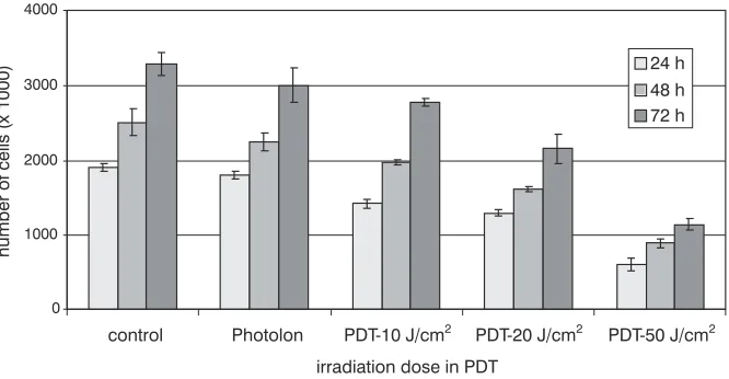
Fig. 3.Proliferation of SH−4 series cells after application of PDT−Photolon 10–50 J/cm2
was checked for normality by the Shapiro−Wilk test. Average results of two independent samples (trials) were compared using Student’sttest when the assumption of variance homogeneity was met or Student’sttest with separate estimation of vari− ance (Cochrane−Cox test) when not. The homo− geneity of variance for two samples (trials) was checked using Snedecor’sFtest. Determination of homogeneous groups was performed by the Duncan test. The assumption of homogeneity of variance was checked by the Levene test. When comparing the averages of at least three samples (trials), analysis of variance (ANOVA) was per− formed. Results for which p< 0.05 were consid− ered statistically significant.
Results
Figure 2 shows the results of the survival of SH−4 melanoma cells subjected to photodynamic therapy with Photolon irradiated at doses of 10, 20, and 50 J/cm2at 24, 48, and 72 h after irradiation.
The average values indicating the numbers of cells in the particular cultures are presented in the dia− gram as percentages of the control. A decrease in survival rate can be seen even for the lowest dose of irradiation applied (10 J/cm2). The results of the
WST−1 test, which was the basis for the diagram included in Fig. 2, confirmed the earlier observa− tions from cultures stained with trypan blue (Fig. 3) Figure 3 shows changes in the proliferation of SH−4 cells which underwent photodynamic thera− py with Photolon under the same conditions. Increased irradiation doses resulted in a drop in the cell counts of the cultures, although it did not sig− nificantly change the proliferative activity of the cells that survived the therapy.
Quantitative assessment of melanin in the cells of the examined melanoma series irradiated with the lowest dose of light applied in the experiment (10 J/cm2), was carried out with the spectrophoto−
metric measurement of absorption at λ= 410 nm.
The melanin content was determined based on the standard curve for DOPA−melanin (Table 1).
The influence of the conditions of photody− namic therapy with Photolon when irradiating the culture with doses of 10, 20, and 50 J/cm2on the
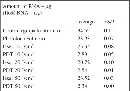
expression of genes encoding the key enzymes of the melanogenesis pathway (tyrosynase (TYR), DHICA oxidase (TYRP−1), and dopa−chrome tau− tomerase (TYRP−2)) was also analyzed. The amounts of total RNA extracted from the cultures subjected to PDT are presented in Table 2.
An attempt was made to present the expres− sions of the selected genes relative to the number of copies of basal metabolism genes, i.e. β−actin
and GAPDH [22] (Table 3). However, the analysis
of variance of the average values of β−actinand
GAPDHexpression (test probabilities (p) provid−
ed in Table 4) revealed significant differences in the number of transcripts of the genes in the applied study arrangement (p< 0.005), which does not justify making such conversions. The number of mRNA copies for the specific genes are pre− sented as recalculated into µg of total RNA in the given sample.
The expressions of the genes encoding the main enzymes of melanogenesis 3 h after comple− tion of photodynamic therapy with Photolon under
Amount of melanin in pg/cell
(Ilość melaniny wyrażona w pg/komórkę)
Series control laser 10 J/cm2 PDT−Photolon, 10 J/cm2
average ± SD average ±SD average ±SD
SH−4 16.35 0.79 17.58 0.79 21.42 1.25
Table 1.Amount of melanin expressed in pg/cell in control cultures and cultures irradiated with 10 J/cm2(λ= 662 nm), without photo−sensitizer and in the presence of Photolon (72 h after the irradiation)
Tabela 1.Ilość melaniny wyrażona w pg/komórkę w hodowlach kontrolnych oraz hodowlach naświetlanych 10 J/cm2(λ= 662 nm), bez fotouczulacza i w obecności Fotolonu (po 72 godz. od naświetlania)
Amount of RNA – µg (Ilość RNA – µg)
average ±SD Control (grupa kontrolna) 34.62 0.12
Photolon (Fotolon) 23.93 0.07
laser 10 J/cm2 23.35 0.08
PDT 10 J/cm2 2.89 0.05
laser 20 J/cm2 20.72 0.10
PDT 20 J/cm2 2.54 0.01
laser 50 J/cm2 23.52 0.03
PDT 50 J/cm2 2.34 0.00
Table 2.Obtained amounts of total RNA, after extracting from cultures subjected to PDT
Number of gene copies/µg RNA (Liczba kopii genów/µg RNA)
β−actin GAPDH
average ± SD average ± SD Control (grupa
kontrolna) 1160977 344652 2516323 354085
Photolon 79578 9550 1452919 1292443
(Fotolon)
Laser 10 J/cm2 713763 129770 1286308 891728
PDT 10 J/cm2 3808 1221 63058 13308
laser 20 J/cm2 314672 32277 6348209 464715
PDT 20 J/cm2 2610 1040 40297 18131
laser 50 J/cm2 208617 38343 2646469 435690
PDT 50 J/cm2 1146 263 11294 3525
Table 3.Number of RNA copies for β−actin andGAPDHper µg of total RNA
Tabela 3.Liczba kopii RNA dla β−aktyny iGAPDHna µg RNA całkowitego
β−actin GAPDH
Control (Grupa
kontrolna) x x
10 J/cm2 x x
20 J/cm2 x x
50 J/cm2 x x
Photolon
(Fotolon) x x
PDT 10 J/cm2 x x
PDT 20 J/cm2 x x
Table 4.Test probability (p) for the results of variance analysis (ANOVA) and homogeneous groups of actin
β−geneand GAPDHexpression, determined for cultures
without and with addition of photo−sensitizer
Tabela 4.Prawdopodobieństwa testowe (p) wyników analizy wariancji (ANOVA) oraz grupy jednorodne ekspresji β−genuaktyny iGAPDHwyznaczone dla hodowli bez i z dodatkiem fotouczulacza
p
= 0.00021
p
< 0.00001
p
< 0.00001
p
= 0.00002
1 10 100 1000 10000 100000 1000000
TYR control
Photolon 10 J/cm2
PDT-10 J/cm2
20 J/cm2
PDT-20 J/cm2
50 J/cm2
PDT-50 J/cm2
1 10 100 1000 10000 100000 1000000 10000000
TYRP-1 control
Photolon 10 J/cm2
PDT-10 J/cm2
20 J/cm2
PDT-20 J/cm2
50 J/cm2
PDT-50 J/cm2
1 10 100 1000 10000 100000
TYRP-2 control
Photolon 10 J/cm2
PDT-10 J/cm2
20 J/cm2
PDT-20 J/cm2
50 J/cm2
PDT-50 J/cm2
number of mRNA copies/
µg
RNA
number of mRNA copies/
µg
RNA
number of mRNA copies/
µg
RNA
Fig. 4.Number of RNA copies of genes TYR, TYRP−1, and TYRP−2
converted into µg of total RNA in the examined cultures
Ryc. 4.Liczba kopii RNA genów
the experimental conditions are presented in Fig. 4. Distinct changes can be seen in the gene expres− sions in the cultures subjected to photodynamic therapy, both in the number of copies of individual genes and in their profile. Tables 5 and 6 contain data of the statistical analysis of the expressions of the genes (TYR, TYRP−1, andTYRP−2). For all cul− tures, the applied therapy conditions led to signif− icant changes in their expressions (Table 5). Only in the control cultures (laser−irradiated without the addition of photo−sensitizer, Fig. 4) were no statis− tically significant differences noted regarding the number of gene transcripts compared with the con− trol cultures without photo−sensitizer and also without irradiation (Table 5).
Despite the differences, notable in Fig. 4, in
the number of RNA copies for TYR, TYRP−1, and
TYRP−2in the cultures irradiated at doses of 10,
20, and 50 J/cm2, the differences were not been sta−
tistically significant and all the changes that occurred were ranked into one group (Table 6).
Discussion
Melanoma is still one of the most serious problems and challenges in the treatment of neo− plasms. Due to its being diagnosed too late, its aggressiveness, and its resistance to commonly applied treatments, it is still considered one of the most frequent causes of death. Until recently, prac− tically no other treatment methods besides early surgical intervention and chemotherapy have been introduced for melanoma patients [2]. However, research conducted in recent years indicates a high level of differentiation concerning sensitivity to irradiation among melanoma cells, which creates new therapeutic possibilities. Cases have been described of permanent cure of melanoma patients subjected to radiotherapy. Because of the limited knowledge of the molecular effects and distant consequences of such therapy, these are mainly inoperable tumors located in places where surgical intervention is impossible or in a stage or form precluding other therapeutic actions. Most fre− quently, these are cases of melanoma of the organ of vision or lesions in the area of nose and sensi− tive parts of the face. Such information also induces research into the possibilities of introduc− ing phototherapy to melanoma treatment. Besides
in vitrostudies, first reports appeared concerning
positive completion of phototherapy in cases of melanoma of the iris [2]. The cases of 14 patients with melanoma metastases have also been described (13 of which had been treated with other methods earlier), in whom neoplastic cells were completely eliminated after one PDT−chlorin e6 procedure (in 8 cases) or after a few series of ther− apy (in 6 cases) [24]. The search is also underway for rational diagnostic indicators enabling prog−
TYR TYRP−1 TYRP−2
Photolon (–) Photolon (+) Photolon (–) Photolon (+) Photolon (–) Photolon (+) SH−4 p < 0.05 p < 0.05 p = 0.0559 p < 0.05 p = 0.0835 p < 0.05
Table 5.Test probabilities (p) of results of single factor variance analysis of data concerning expression of genes for tyrosy− nase (TYR), DHICA oxidase (TYRP−1), and dopa−chrome tautomerase (TYRP−2) in the cells subjected to photodynamic ther− apy (photo−sensitizer – „+”) and in cells irradiated without photo−sensitizer (photo−sensitizer – „–”); where for p < 0.05 the observed effects are statistically significant
Tabela 5. Prawdopodobieństwa testowe (p) wyników jednoczynnikowej analizy wariancji danych dotyczących ekspresji genów tyrozynazy (TYR), oksydazy DHICA (TYRP−1) i tautomerazy dopachromu (TYRP−2) w komórkach poddanych terapii fotodynamicznej (fotouczulacz „+”) oraz w komórkach naświetlanych bez fotouczulacza (fotouczulacz „–”); gdy p < 0,05, obserwowane rezultaty są istotne statystycznie
Gene Sample Homogenous groups
(Gen) (Próbka) (Grupy jednorodne)
TYR Photolon x
PDT 10 J/cm2 x
PDT 20 J/cm2 x
PDT 50 J/cm2 x
TYRP−1 Photolon x
PDT 10 J/cm2 x
PDT 20 J/cm2 x
PDT 50 J/cm2 x
TYRP−2 Photolon x x
PDT 10 J/cm2 x
PDT 20 J/cm2 x
PDT 50 J/cm2 x
Table 6.Homogeneous groups of cell cultures subjected to PDT−Fotolon (determined for samples with photo−sen− sitizer), in respect of the number of copies of tyrosynase (TYR), DHICA oxidase (TYRP−1), and dopa−chrome tau− tomerase (TYRP−2) genes
nostication of the sensitivity of specific neoplastic lesions to a selected type of therapy [25–27].
The research presented in this study concern− ing the consequences of applying PDT−Photolon in cultures of SH−4 melanoma reveals a drop in cell survival after the photodynamic procedure (Figs. 2 and 3). After a single photodynamic procedure the number of living cells decreased by ca. 40% (after applying a dose of 10 J/cm2) up to ca. 50% (when
the dose was 50 J/cm2) 72 h after irradiation.
However, this did not change (at doses of 10 and 20 J/cm2) the proliferative activity of the surviving
cells significantly.
An important element of the melanoma cells’ response to stress−inducing factors, which also includes the 662−nm laser radiation applied in this experiment, is, among other defense mechanisms, the ability to synthesize melanin pigment. Even at the lowest dose of radiation applied in the experi− ment (10 J/cm2) the amount of pigment increased
by ca. 30% 72 h after irradiation (Table 1). Changes were also noted at the expression levels of the genes encoding the key enzymes for melano− genesis, i.e. TYR, TYRP−1, andTYRP−2. Decreases in the expressions of the genes could be observed as well as a change in profile, i.e. the TYR/TYRP−1/
/TYRP−2ratio (Fig. 4, Tables 5 and 6).
The extraction of RNA from the cells subject− ed to photodynamic procedures indicated an extremely significant decrease, by a factor of over 10, in the amount of ribonucleic acid (Table 3), which attests to the destructive influence of the applied therapy on the SH−4 melanoma cells.
Because substantial changes in the transcrip− tional activity of numerous pro− and anti−apoptotic genes, genes of thermal−shock proteins, and genes encoding anti−oxidative enzymes can be observed simultaneously in cells (the present authors’ unpublished data), it appears difficult to state explicitly whether the reduced copy numbers of the determined genes recalculated per 1 µg of total RNA is due to an actual reduction in the expres− sions of these genes or rather the transcript num− bers constitute a proportionally reduced share in the pool of total RNA. Supportive of the latter option is at least the increased amount of the pig− ment after application of PDT−Photolon. A sub− stantial change resulting from the photodynamic procedure is undoubtedly the change in the expres− sion profile of TYR, TYRP−1, and TYRP−2. Irrespective of the aberrations in the expressions of all the remaining genes, also including those defined as ”housekeeping genes” (Tables 3 and 4) and considered stable regarding transcription level in correctly functioning cells, the TYR/TYRP−1/
/TYRP−2 ratio of gene copies was substantially
altered after PDT−Photolon. In all the variants of
PDT applied, a substantial drop in the number of
TYRtranscripts and increase in TYRP−2transcripts (Tables 5 and 6) can be noted.
The numbers of RNA copies for the specific genes are presented in this study in relation to the pool of total RNA, which results from the statisti− cally significant changes in RNA content for β−actin
and GAPDHin specific cultures (Table 4). This sit−
uation gives evidence of major disturbances in the functioning of cells subjected to PDT (changes in both basal metabolism and structural changes con− nected with the cell’s cytoskeleton) and precludes making the respective conversions [22].
It should be stressed that the change in TYR,
TYRP−1, and TYRP−2 expression profile that was
noted in the studied cultures does not depend on the irradiation dose (10–50 J/cm2) (Table 6). The differ−
ences seen in Fig. 4 between the number of RNA copies for TYRP−2at PDT 10 J/cm2, PDT 20 J/cm2,
and PDT 50 J/cm2 do not show statistical signifi−
cance and were all qualified into one group when homogeneous groups were determined (Table 6).
References
[1] Fabris C, Valduga G, Miotto G:Photosensitization with zinc (II) phthalocyanine as a switch in the decision between apoptosis and necrosis. Cancer Res 2001, 61, 7495–7500.
[2] Ruka W, Nowecki ZI, Rutkowski P:Czerniaki skóry u dorosłych. Medipage Warszawa 2005.
[3] Kumala S, Niemiec P, Wideł M, Hancock R, Rzeszowska−Wolny J:Apoptosis and clonogenic survival in three tumour cell lines exposed to gamma rays or chemical genotoxic agents. Cell Mol Biol Lett 2003, 8, 655–665. [4] Latocha M, Pilawa B, Kuśmierz D, Zielińska A, Nawrocka D:Changes in free radicals system on IMR−90 and
C−32 cells during photodynamic therapy. Pol J Environ Stud 2006, 15, 4A, 154–156.
[5] Córdoba F, Braathen LR, Weissenberger J: 5−aminolaevulinic acid photodynamic therapy in a transgenic mouse model of skin melanoma. Exp Dermatol 2005, 14, 6, 429–437.
[6] Saczko J, Kulbacka J, Chwiłkowska A: The influence of photodynamic therapy on apoptosis in human melanoma cell line. Folia Histochem Cytobiol 2005, 43, 3, 129–132.
[7] Latocha M, Pilawa B, Zdybel M, Wilczok T:Effect of Laser Radiation on Free Radicals in Human Cancer G361 Cells. Acta Physica Pol A 2005, 2, 108, 409–412.
[8] Nevřelová P, Kolářová H, Bajgar R:Measurement of reactive oxygen species after photodynamic therapy in vitro. Scripta Med 2005, 78, 5, 281–290.
[9] Lim DS, Ko SH, Lee WY:Silkworm−pheophorbide a mediated photodynamic therapy against B16F10 pigment− ed melanoma. J Photochem Photobiol B 2004, 74, 1, 1–6.
[10] Urbańska K: Uczulanie pigmentowanych komórek i guzów czerniaków zwierzęcych na promieniowanie jonizujące i światło (rozprawa habilitacyjna), Wydawnictwo Uniwersytetu Jagiellońskiego, Kraków 2000. [11] Pasewicz A, Idziak D, Koloczek J: Pair correlation function analysis of 5−(4−hexa−decyloxyphenyl)−
−10,15,20−tri(4−pyridyl)porphyrin and 5−(4−methoxycarbonylphenyl)−10,15,20−tri(4−pyridyl)porphyrin. J Mol Struct 2008, 875, 1–3, 167–172.
[12] Grinholc M, Szramka B, Olender K, Graczyk A:Bactericidal effect of photodynamic therapy against methi− cillin−resistant Staphylococcus aureusstrain with the use of various porphyrin photosensitizers. Acta Biochim Pol 2007, 54, 3, 665–670.
[13] Copley L, van der Watt P, Wirtz KW, Parker MI, Leaner VD:Photolon, a chlorin e6 derivative, triggers ROS production and light−dependent cell death via necrosis. IJBCB 2008, 40, 227–235.
[14] Ramaswamy B, Manivasager V, Chin WWL, Soo KC, Olivo M:Photodynamic diagnosis of human nosopha− ryngeal carcinoma xenograft model using the novel Chlorin e6 photosensitizer Fotolon. Int J Oncol 2005, 26, 1501–1506.
[15] Istotin YP, Laptsevich TP, Bizyuk SA, Alexandrova EN:Photodynamic efficacy of topical application of chlo− rine6 – polyvinylpyrrolidone complex in tumor−bearing rats. Exp Oncol 2006, 28, 4, 299–302.
[16] Isakau HA, Trukhacheva TV, Zhebentyaev AI, Petrov PT:HPLC study of chlorin e6 and its molecular com− plex with polyvinylpyrrolidone. Biomed Chromatogr 2007, 21, 318–325.
[17] Parkhots MV, Lapina VA, Butorina DN:Spectral and photochemical characteristics of the photosensitizers chlorin e6and photolon in the presence of melanin. Optic Spectros 2005, 98, 3, 1562–6911.
[18] http://www.genecards.org/cgi−bin/carddisp.pl?gene=TYR. [19] http://www.genecards.org/cgi−bin/carddisp.pl?gene=TYRP1. [20] http://www.genecards.org/cgi−bin/carddisp.pl?gene=TYRP2.
[21] Cieszka KA, Hill HZ, Hill GJ, Plonka PM:Growth and pigmentation in genetically related Cloudman S91 melanoma cell lines treated with 3−isobutyl−1−methyl−xanthine and β−melanocyte−stimulating hormone. Exp Dermatol 1995, 4, 4, 192–198.
[22] Romanowski T, Markiewicz A, Bednarz N, Bielawski KP:Geny metabolizmu podstawowego jako geny referen− cyjne w ilościowym oznaczaniu ekspresji genów metodą real−time PCR. Postepy Hig Med Dosw 2007, 61, 500–510. [23] Trichopoulos N, Damato B:Photodynamic therapy for recurrent hyphema after proton beam radiotherapy of iris
melanoma. Graefes Arch Clin Exp Ophthalmol 2007, 245, 10, 1573–1575.
[24] Sheleg SV, Zhavrid EA, Khodina TV:Photodynamic therapy with chlorin e6 for skin metastases of melanoma. Photodermatol Photoimmunol Photomed 2004, 20, 21–26.
[25] Konopacka M, Rzeszowska−Wolny J:The bystander effect−induced formation of micronucleated cells is inhib− ited by antioxidants, but the parallel induction of apoptosis and loss of viability are not affected. Mutat Res 2006, 593, 1–2, 32–38.
[26] Kumala S, Niemiec P, Wideł M, Hancock R, Rzeszowska−Wolny J:Apoptosis and clonogenic survival in three tumour cell lines exposed to gamma rays or chemical genotoxic agents. Cell Mol Biol Lett 2003, 8, 655–665. [27] Córdoba F, Braathen LR, Weissenberger J: 5−aminolaevulinic acid photodynamic therapy in a transgenic
mouse model of skin melanoma. Exp Dermatol 2005, 14, 6, 429–437.
cytotoxic effects of photodynamic therapy. As results of the research conducted by the present authors on survival and proliferative activity in var− ious series of melanoma cells, the presence of melanins as well as active antioxidant enzymes
[28] Monfrecola G, Procaccini EM, D’Onofrio D:Hyperpigmentation induced by topical 5−aminolaevulinic acid plus visible light. J Photochem Photobiol B 2002, 68, 2–3, 147–155.
[29] Ma LW:A new method for photodynamic therapy of melanotic melanoma – effects of depigmentation with vio− let light photodynamic therapy. J Environ Pathol Toxicol Oncol 2007, 26, 3, 167–172.
[30] Lim DS, Ko SH, Lee WY:Silkworm−pheophorbide a mediated photodynamic therapy against B16F10 pigment− ed melanoma. J Photochem Photobiol B 2004, 74, 1, 1–6.
Address for correspondence:
Małgorzata Latocha Department Cell Biology Medical University of Silesia Jedności 8
41−200 Sosnowiec Poland
Tel.: +48 32 364 12 10 E−mail: [email protected]
Conflict of interest: None declared
![Fig. 1. Complex of chlorin e6–PVP (Photolon) [16]](https://thumb-us.123doks.com/thumbv2/123dok_us/8772107.1757571/3.595.73.280.68.179/fig-complex-of-chlorin-e-pvp-photolon.webp)