Jerzy Garcarek
1, A–F, Jacek Kurcz
1, B–D, F, Maciej Guziński
1, B–D, F, Dariusz Janczak
2, A, E, F,
Marek Sąsiadek
1, A, E, FTen Years Single Center Experience in Percutaneous
Transhepatic Decompression of Biliary Tree in Patients
with Malignant Obstructive Jaundice
Przezskórne przezwątrobowe odbarczenie dróg żółciowych u pacjentów
chorych na żółtaczkę mechaniczną o etiologii nowotworowej – 10-letnie
doświadczenia własne
1 Chair of Radiology, Department of General and Interventional Radiology and Neuroradiology, Wroclaw Medical University, Poland
2 Department of Surgery, 4th Military Clinical Hospital in Wrocław, Poland
A – research concept and design; B – collection and/or assembly of data; C – data analysis and interpretation;
D – writing the article; E – critical revision of the article; F – final approval of article; G – other
Abstract
Background. Percutaneous transhepatic biliary drainage (PTBD) is a method of biliary tree decompression, applied as palliative treatment in patients with malignant biliary tree critical stenosis/obstruction, but also as a potentially curative treatment in patients with non-malignant biliary tree stenosis. Novel instrumentation dedicated to PTBD has been designed in recent years, which makes it possible to perform more advanced procedures in patients with severe extensive malignant biliary tree stenosis/obstruction.
Objectives. The first primary goal of the study was to compare both the rate and types of short- and long-term complications in patients who had undergone PTBD between 2000 and 2006 with patients treated between 2007 and 2011. The second primary goal of the study was to work out an original algorithm of efficient management in patients undergoing PTBD. An additional goal was to assess the efficacy of PTBD and the overall survival of the patients.
Material and Methods. One-hundred twenty-eight consecutive PTBD procedures performed between 2000 and 2006 in patients with malignant biliary jaundice were analyzed retrospectively. Similarly, retrospective analysis of 73 consecutive procedures in patients with malignant biliary jaundice performed between 2007 and 2011 was car-ried out. Subsequently, the results of both subsets were compared to each other. The PTBD procedure was guided fluoroscopy each time. PTBD involved external biliary drainage and/or stenting of the strictured/occluded seg-ments of extra- and intrahepatic biliary ducts.
Results. The analysis demonstrated a statistically significant decrease in the overall incidence of short- and long-term complications in patients undergoing PTBD in 2007–2011 in comparison to the subset treated in 2000–2006. Among the early complications, a significant decrease in sub- and pericapsular contrasted bile leaks was shown. The evaluation of long-term complications demonstrated lower incidence of the falling out of the draining cath-eter. The implementation of novel instrumentation made it possible to perform biliary stenting in 63.7% cases of common bile duct (CBD) obstruction (vs. 37.5% in procedures carried out in 2000–2006). However, no statistically significant difference in survival between the two analyzed subsets was demonstrated.
Conclusions. The analysis of rate and types of complications made it possible to establish authors own algorithm of management in different types of biliary obstructions and strictures. The modification of procedure technique, pos-tinterventional management and usage of the new generation of low-profile instrumentation for percutaneous access dedicated to PTBD has resulted in a significant reduction of the complication rate in the last 5 years. Higher frequency of CBD stenting improves the quality of life in this subset of patients (Adv Clin Exp Med 2012, 21, 5, 621–632).
Key words: biliary stenting, biliary drainage, biliary decompression, malignant obstructive jaundice.
Adv Clin Exp Med 2012, 21, 4, 621–632 ISSN 1899–5276
ORIGINAl PAPERS
Streszczenie
Wprowadzenie. Przezskórny przezwątrobowy drenaż dróg żółciowych (PTBD) jest metodę odbarczenia dróg żół-ciowych, znajduje zastosowanie jako leczenie paliatywne u chorych ze zwężeniem/niedrożnością dróg żółciowych o etiologii nowotworowej, również jako terapia prowadząca do wyleczenia u pacjentów ze zwężeniami dróg żółcio-wych o etiologii nienowotworowej. Wprowadzone w ostatnich latach nowoczesne instrumentarium przeznaczone do PTBD pozwala na wykonywanie bardziej zaawansowanych zabiegów u pacjentów z nasilonymi zwężeniami/ /niedrożnością dróg żółciowych o etiologii nowotworowej.
Cel pracy. Porównanie częstości oraz typów powikłań wczesnych i odległych u pacjentów poddanych PTBD w latach 2000–2006 z grupą leczoną w latach 2007–2011. Drugi główny cel pracy stanowiło wypracowanie własne-go alwłasne-gorytmu postępowania u pacjentów poddawanych PTBD.
Materiał i metody. Retrospektywnej analizie poddano 128 zabiegów PTBD wykonanych w latach 2000–2006. Otrzymane wyniki zestawiono z wynikami 73 procedur PTBD wykonanych w latach 2007–2011. Wykonywany pod kontrolą prześwietlenia PTBD obejmował drenaż zewnętrzny i/lub stentowanie zwężonych/niedrożnych odcinków dróg żółciowych wewnątrz- i zewnątrzwątrobowych.
Wyniki. Wykazano znamienne zmniejszenie częstości powikłań wczesnych i odległych w podgrupie pacjentów poddanych PTBD w latach 2007–2011 w porównaniu z pacjentami leczonymi w latach 2000–2006. Analizując powikłania wczesne, wykazano istotne zmniejszenie częstości pod- i okołotorebkowych zacieków żółci. Ocena powikłań odległych wykazała znamienne zmniejszenie częstości wypadania cewnika drenującego. Zastosowanie instrumentarium nowej generacji umożliwiło zakończenie zabiegu wszczepieniem stentów w 63,7% przypadków niedrożności przewodu żółciowego wspólnego (CBD) (vs. 37,5% w zabiegach wykonywanych w latach 2000–2006). Okres przeżycia w porównywanych podgrupach jednak nie wydłużył się istotnie statystycznie.
Wnioski. Analiza częstości i typów powikłań pozwoliła na wypracowanie własnego algorytmu postępowania w poszczególnych typach niedrożności/zwężeń dróg żółciowych. Modyfikacja techniki zabiegu, postępowania poza-biegowego oraz zastosowanie nowego rodzaju niskoprofilowego instrumentarium przeznaczonego do PTBD spo-wodowało znamienne zmniejszenie częstości powikłań obserwowanych w ostatnich 5 latach. Zwiększenie częstości stentowania CBD poprawia jakość życia chorych poddawanych PTBD (Adv Clin Exp Med 2012, 21, 5, 621–632).
Słowa kluczowe: stentowanie dróg żółciowych, drenaż dróg żółciowych, dekompresja dróg żółciowych, żółtaczka mechaniczna nowotworowa.
Pancreatic cancer and biliary tract and gall bladder malignancies constitute the most common reasons for malignant obstructive jaundice. Other causes include hepatic metastases and recurrent postsurgical pancreatic or gastric malignancy. For practical reasons, malignant obstructive jaundice should be divided into intra- and extrahepatic bili-ary obstruction. Percutaneous transhepatic bilibili-ary access is reserved for inoperable patients, in whom no endoscopic procedure can be performed because of anatomical conditions associated with previous surgery, coexisting complications or the poor gen-eral state of the patient. Nowadays, development of the techniques and devices for interventional ra-diology and implementation of elastic, low-profile equipment enables combined multilevel biliary stenting, which permits biliary tree decompression and recovers physiological bile flow, which contrib-utes to a substantially higher comfort of life. Failure of the passage through the obstruction or anatomi-cal or clinianatomi-cal backgrounds extort the implementa-tion of external or internal-external drainage, which is associated with higher risk of complications and a cause of discomfort for the patients.
Material and Methods
The results of biliary tree decompression in pa-tients with a stricture or obstruction of malignant
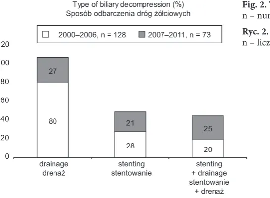
origin were analyzed retrospectively. Two hun-dred one procedures of percutaneous transhepatic biliary tree decompression in 204 patients (98.5%) with malignant biliary stricture or obstruction were performed between 2000 and 2011 (Fig. 1). One hundred twenty eight procedures were carried out between 2000 and 2006, while 73 procedures were performed between 2007 and 2011 with the application of modified methods of treatment and low-profile instrumentation. The analyzed group included 117 females (58.6%) aged 32–81 (median age 61.1) and 84 males (41%) aged 41–79 (median age 67.2). Biliary tree decompression was divided into the following categories: exclusively drain-age – percutaneous or combined (both endoscopic and percutaneous), exclusively stenting, drainage + stenting (Fig. 2).
puncture) was advanced. A dedicated catheter col-laborating with hydrophilic compatible guidewires meant for manipulation within the biliary tracts was applied. The decision regarding the method of biliary tree decompression was taken during the procedure depending mainly on anatomical relations, the patient’s clinical condition, expected survival time, and occasionally the patient’s pref-erence was taken into account. Biliary stenting was performed after insertion of a hemostatic sheath (6–8F in size) with a marker at its tip. Since 2007, biliary tree stenting and removal of the hemostatic sheath was followed each time by embolization of the approach canal using a couple of plugs made of rolled sponge (Spongostan). Typically nitinol stents were implanted, rarely balloon-expanding bare metal stents and – exceptionally – covered stents. Between 2000 and 2006, external biliary drainage was most commonly performed using 7F Kaastrup catheters which were blocked at the tip with a balloon, less frequently the authors used
Mac lock catheters (COOK) of 10.2F in diameter, and internal-external drainage was performed ex-clusively by means of Mac lock or Flexima (Boston Scientific) catheters. Since 2007, biliary drainage was carried out using only Mac lock or Flexima catheters. The catheters were fixed to the skin with the original kits and since 2007 additionally using a skin suture. The proximal visible portion of the catheter was labeled with a marker. Postint-erventionally, the patient was instructed about the necessity of careful toilet drainage of the catheter (flushing 2–3 times daily using 20 ml of saline). Since 2007, a bile sample has been taken intraint-erventionally for smear and antibiotics have been administered in the postinterventional period as standard management. The administration of an-algesics in the first 24 hours after the procedure has also been standard. In clinically severe patients with symptoms of cholangitis and/or with hepatic abscess, biliary drainage together with aggressive antibiotic administration was the first stage of
24 25
20
17
13 14
15 23
21 19
5 5
0 5 10 15 20 25 30
2000 2001 2002 2003 2004 2005 2006 2007 2008 2009 2010 2011
Type of biliary decompression (%) Sposób odbarczenia dróg żółciowych
20 40 60 80
drainage
drenaż stentowaniestenting + drainagestenting stentowanie
+ drenaż 0
100
120 2000–2006, n = 128 2007–2011, n = 73
20 27
80 21
25
28
Fig. 1. The number of procedures performed between 2000 and 2011
Ryc. 1. liczba zabiegów wykonanych w latach 2000–2011
Fig. 2. Types of biliary tract decompression. n – number of patients
management; after clinical improvement – usually 2–6 weeks postinterventionally – biliary stenting was performed. In cases of good general condition but with accompanying symptoms of cholangitis, biliary stenting was carried out, however the ex-ternal-internal drainage catheter was left in place until the symptoms subsided.
Results
The statistical analysis was performed using the chi-squared test and Fisher’s exact test by means of Statistica (StatSoft) software. Values of p < 0.05 were regarded as statistically significant.
Periinterventional mortality (up to the 7th day) amounted to 2.98% (6 out of 201 pts). Complica-tions were divided into early (the periinterven-tional period) – manifesting within the first 24 hours after the procedure (Tab. 1) and late (Tab. 2) – occurring during the postinterventional medi-cal treatment. Minor complications like intrainter-ventional shivers, intra- and periinterintrainter-ventional pain or a transient fever subsiding within 24 hours were not regarded as significant and not analyzed in the study.
The complications were isolated or – more commonly – multiple in a single patient. Total
Table 1. Early complications (manifested within 24 hours postinterventionally)
Tabela 1. Powikłania wczesne (ujawniające się w ciągu 24 godzin od zabiegu)
Type of complication
(Rodzaj powikłania) Patients treated 2000–2006 (Pacjenci leczeni w latach 2000– –2006) n = 128
Percentage
(Odsetek) Percentage (Odsetek) Patients treated 2007–2011 (Pacjenci lecze-ni w latach 2007–2011) n = 73
p value (Wartość p)
Sub- and epicapsular leakage of contrast bile
(Pod- oraz zewnątrztorebkowy zaciek żółci kontrastowej) *
41 32 15.1 11 0.02
Perforation of inferior (visceral) hepatic surface
(Perforacja dolnej (trzewnej) powierzchni wątroby)
3 2.3 0 0 0.55
Bleeding originating from intercos-tal vessels
(Krwawienie z naczyń między-żebrowych)
8 6.3 2.7 2 0.4
Bleeding from drain lasting > 24 hours
(Krwawienie z drenu utrzymujące się > 24 godzin)
18/92 19.6 20.0 8/40 0.56
Total (Suma)* 70 21 0.001
* Statistically significant (chi-squared test, Fisher’s exact test). * Istotne statystycznie (test c2, test dokładności Fishera).
analysis of the early and late complications in sub-sets of patients demonstrated that single or com-bined complications occurred in 71/128 patients treated between 2000 and 2006 (55.5% of the sub-set) and in 20/73 patients treated between 2007 and 2011 (27.4% of the subgroup).
Survival in the followed-up group of 111 pa-tients treated between 2000 and 2006 was between 1 week and 13 months (median survival 3.5 months; loss of follow-up in 17 patients) whereas in the group of 61 followed-up patients treated between 2007 and 2011 between 1 week and 14 months (median survival 3.9 months; loss of follow-up in 12 cases). The analysis of patients demonstrated no significant difference in median survival between the two subsets.
The quality of life was evaluated using a sur-vey concerning physical and psychological factors as well as the necessity of rehospitalization due to procedure-related complications. The authors observed a significant difference in quality of life between the subgroup treated 2000–2006 and the subgroup treated 2007–2011 (p = 0.01).
Examples of the different types of procedures are demonstrated in Figs. 3–8.
Discussion
Jaundice, as the first symptom of pancreatic, bile duct or gall bladder malignancies, usually is proof of an advanced neoplastic process. [1] Other reasons include hepatic metastatic lesions or re-current pancreatic or gastric malignancy after sur-gery. [1] Only approximately 10% of the patients can be qualified for tumor resection. In other cases decompression of the biliary tree can be performed endoscopically by means of plastic prostheses and recently also self-expandable stents [2, 3]. Some patients are disqualified from the endoscopic pro-cedure due to neoplasmatic infiltration (no duo-denal access), previous surgical procedures or ob-struction of earlier inserted biliary prosthesis [4]. All the patients in present analysis were disquali-fied from endoscopic intervention. It should be stressed that the patients, who usually presented with severely advanced disease, a high level of se-rum bilirubin and were in poor general condition, required particularly careful preparation and care-ful choice of interventional strategy.
The procedure of percutaneous transhepatic biliary tree drainage and stenting is characterized by a potentially high risk of serious complications. [5] All the patients included in present analysis suffered from malignant biliary tree stricture or obstruction and presented with clinical symptoms of obstructive jaundice. Typically the intervention was performed as an emergency procedure due to rapidly increasing bilirubinaemia which ranged from 6.0 to 35.5 mg/dl (median total bilirubin 18.5 mg/dl). Preinterventional diagnostic manage-ment included abdominal ultrasound (US) with a detailed assessment of the liver (evaluation of in-trahepatical biliary tract diameter, site of blockage and localization of primary or metastatic masses). Other imaging modalities included magnetic res-onance cholangiopancreatography (MRCP) or, more frequently, computed tomography (CT) of the abdomen. Histopathologically, biliary tract cancer (mainly Klatskin tumor), gall bladder ma-lignancy, pancreatic and gastric cancer metastases, direct infiltration or hilar hepatic metastases were revealed most commonly.
Table 2. late complications
Tabela 2. Powikłania odległe
Type of complication
(Rodzaj powikłania) Patients treated 2000–2006 (Pacjenci leczeni
w latach 2000–2006) n = 128
Percentage
(Odsetek) Percentage (Odsetek) Patients treated 2007–2011 (Pacjenci leczeni w latach 2007–2011) n = 73
p value (Wartość p)
Haemobilia
(Hemolibia) 4 3.1 0 0 0.3
Perihepatic abscess
(Ropień okołowątrobowy) 7 5.5 4.8 3 0.89 Hepatic abscess
(Ropień wątroby) 9 7.1 1.6 1 0.17
Sepsis
(Posocznica) 5 3.9 3.1 2 1
Cholangitis (Zapalenie dróg żółciowych)
18 14.0 6.3 4 0.12
Bile leakage along the drain
(Zaciek żółci wzdłuż drenu) 12/80 18.8 21.7 5/23 0.44 Drain falling out
(Wypadnięcie drenu)* 31/92 33.7 13.5 5/37 0.02 Stent occlusion and/or
stent migration
(Zamknięcie i/lub migracja stentu)
9/48 18.8 17.5 7/40 0.88
Total (Suma)* 95 27 0.001
A minority of patients presented with compli-cations after previous endoscopic or surgical inter-vention. The choice of endoscopy as the preferred method of biliary intervention and its improve-ment in recent years has caused a reduction in the number of transhepatical procedures [6].
The authors do not regard the presence of bile with blood in the collecting sac within 24 hours after the procedure as a complication. If prolonged or extensive bleeding is observed, it is reasonable to pull back the drain 1–2 cm and continue observa-tion. In cases of extensive bleeding, it is advisable to remove the drain, perform embolization of the access canal and repeat the procedure via another approach. Plain X-ray of the abdomen and/or ab-dominal USG should be performed as follow-up examinations every 2–4 weeks in order to check the location of the catheter, location of the stent(s) and the efficacy of the decompression. If biliary obstruction or stent migration is observed, a sec-ondary transhepatic procedure is necessary.
Basic imaging modalities include USG, MRCP and CT [7, 8]. It should be remembered that a de-tailed knowledge of previous therapeutic proce-dures and the results of microscopic examina-tions are necessary to make a precise report. USG should give answers concerning the extent of bile tree dilatation, site of the lesion causing obstruc-tion, the presence of metastases and other
patho-logical lesions in the liver including pathopatho-logical fluid collections, patency and location of the portal vein and its intrahepatical branches. MRCP allows for noninvasive evaluation of biliary tree anato-my, the site and – occasionally – the reason for obstruction [9, 10]. CT permits assessment of the local and regional staging of the malignancy, iden-tification of the type of the lesion and the detec-tion of distant metastases [7]. All the informadetec-tion mentioned above makes it possible to evaluate the patient’s condition and estimated survival, which
Fig. 3. Obstruction of common bile duct (due to infil-trating gastric carcinoma). External drainage. Distal portion of the drain inserted via the right lobe biliary ducts is seen in left lobe biliary ducts
Ryc. 3. Niedrożność przewodu wątrobowego wspólne-go i przewodu żółciowewspólne-go wspólnewspólne-go (naciek w prze-biegu raka żołądka). Drenaż zewnętrzny. Dystalny odcinek drenu wprowadzonego przez przewody żół-ciowe prawego płata wątroby widoczny w przewodach żółciowych lewego płata wątroby
Fig. 4. a)Obstruction of both left and right hepatic ducts as well as proximal segment of common bile duct (due to cholangiocarcinoma), b) simultaneous com-bined internal-external drainage of both liver lobes. Distal portions of drains are placed in duodenum
Ryc. 4. a)Niedrożność lewego i prawego przewodu wątrobowego oraz przewodu wątrobowego wspólnego (w przebiegu cholangiocarcinoma), b) jednoczasowy drenaż wewnętrzno-zewnętrzny obu płatów wątroby. Dystalne odcinki drenów są umieszczone w dwunastnicy
A
is of fundamental importance when a decision on the method of biliary tree decompression is made. Other aspects the operator has to consider before making the decision regarding the method of bil-iary tree decompression include: age of the patient, general health condition, potential additional thology within the biliary tracts and the liver, pa-tient’s expectations regarding the procedure and acceptance of discomfort related to the external biliary drain as well as the ability to assure outpa-tient care in the postinterventional period.
Percutaneous transhepatic decompression of the biliary tree can be achieved by means of ex-ternal or exex-ternal-inex-ternal drainage, plastic pros-thesis implantation or biliary stenting. Each of the methods mentioned above has its advantages and disadvantages, which is why the choice of the method is individual and depends on the clinical setting and therapeutic capabilities of particular
Fig. 5. a) Severe stricture of common bile duct (due to pancreatic carcinoma), b) stenting of common bile duct and right hepatic duct using self-expandable nitinol stent
Ryc. 5. a) Znaczne zwężenie przewodu żółciowego wspólnego (w przebiegu raka trzustki), b) stentowanie przewodu żółciowego wspólnego i prawego przewodu wątrobowego za pomocą samorozprężalnego stentu nitinolowego
Fig. 6. a) Obstruction of left and right hepatic ducts and proximal common bile duct (due to metastatic lesion in hepatic hilum), b) combined biliary stenting. One stent connects left hepatic duct with the right one. The second stent was implanted into the right hepatic duct and common bile duct
Ryc. 6. a) Niedrożność prawego i lewego przewodu wątrobowego oraz przewodu wątrobowego wspólnego (na skutek zmian przerzutowych we wnęce wątroby), b) złożone stentowanie dróg żółciowych. Jeden ze sten-tów łączy lewy przewód wątrobowy z prawym. Drugi stent łączy prawy przewód wątrobowy z przewodem żółciowym wspólnym
A
B A
patients [2, 3, 11]. The experience of the interven-tional team plays a great role in minimizing the risk of potential complications.
According to the literature, the technical suc-cess of the procedure amounts to nearly 100% but there is a large discrepancy regarding the in-cidence of serious complications [5]. According to present material, the technical success of the procedure was 98.5% and all cases of intervention failure were associated with a lack of dilated intra-hepatic bile ducts and accompanying ascites which constitute relative contraindications to the inter-vention. However, a significantly elevated level of serum bilirubin was always an indication for the procedure as a life-saving intervention.
In present material, the authors focused atten-tion on the most frequent complicaatten-tions and how to avoid them. The most common postinterven-tional complications included the falling out of the drain from the biliary ducts and subcapsular or perihepatic leakage of contrast bile (mixture of contrast medium and bile). large bile collections visualized on the USG were treated by means of percutaneous drainage. leakage of bile along the drain was usually the effect of long-term (lasting a couple of months) biliary drainage and was often the first symptom of sub-occlusion (“overgrowth”) of the lumen of the drain. The exchange of the drain for a new one, occasionally of a larger diam-eter, was the solution to this clinical problem. The
falling out of the drain from biliary ducts and the associated necessity of reintervention was often as-sociated with insufficient knowledge of the nurse staff and/or patient’s family about the proper drain care and often a direct burden of the drain with the collective sac. In the last 5 years, the authors have paid particular attention to informing the pa-tients precisely how to care for the drain and about the necessity to apply the belts to fix the collecting sacs. Moreover, the drain is – except for the appli-cation of the original kit which is typically glued to the skin – additionally sutured to the skin and the visible portion of the drain is labeled in order to detect its dislocation early enough. Follow-up im-aging modalities are also aimed at early complica-tion deteccomplica-tion because, in cases of incomplete dis-location of the drain, it is still possible to exchange it for a new one and place in a proper position. Complete dislocation of the drain out of the bili-ary tree implicates the necessity of reintervention via another approach. In these cases, the first canal remaining in the liver parenchyma – if does not occlude itself – causes, on one hand, perihepatic leakage formation and, on the other hand, the bile ducts to remain undilated, which to a great extent makes the repetitive procedure difficult and in-creases the risk of complications. The management the authors have implemented in recent years has contributed to a reduction in the incidence of the complications mentioned above as compared to the previous period from 33.7% to 13.5%.
Moss et al. [6] performed a metaanalysis com-paring the results of palliative endoscopic biliary stenting/prosthesis implantation to traditional surgery in patients with inoperable pancreatic cancer. According to their results, currently endo-scopic implantation of metal stents is the manage-ment of choice in patients with pancreatic cancer with symptomatic impairment of distal biliary duct patency. Piñol et al. [11] compared the results of the usage of percutaneously implanted metal self-expandable stents to the results of endoscopic implantation of plastic prostheses in patients with malignant biliary obstruction. The overall medi-an survival was significmedi-antly longer in the subset that had undergone percutaneous self-expandable metal stent implantation. According to the results obtained by these authors, percutaneous biliary stenting (PBS) is an important alternative to the traditional therapeutic method – endoscopic im-plantation of polyethylene prosthesis. Aljiffry et al. [12] assessed optimal palliative management in patients with inoperable cholangiocellular can-cer (CCC). According to their study, biliary duct stenting was the most efficient method of pal-liative treatment. Meller et al. [13] retrospectively analyzed patients with malignancy not originating
Fig. 7. Plastic “double mushrooms” prosthesis implant-ed percutaneously transhepatically due to common bile duct occlusion. Contrast medium seen in duodenum proves that the prosthesis is patent
primarily from the liver, biliary ducts and pancre-as but impairing biliary tree patency. According to their results, PBS is a safe and effective method of management that makes it possible to prolong the estimated survival as well as jaundice-free period. Briggs et al. [14] demonstrated the usefulness of percutaneous implantation of short metal stents prior to surgery in patients with operable pancre-atic head cancer complicated by impairment of bil-iary duct patency. Rasmussen et al. [15] analyzed the incidence of stent-associated complications in 48 patients with papilla of Vater malignancy who had undergone percutaneous stent implantation due to biliary duct obstruction. The fracture of the stent was shown in 8% of the patients. According to these authors, stent fracture should be regard-ed as risk factor of recurrent biliary obstruction. Chiou et al. [16] compared the efficacy of bili-ary stenting in 102 consecutive patients who had undergone PBS at the level of the hepatic hilum and in the common bile duct (CBD). The aver-age stent patency period was significantly longer in the subset who had undergone stenting at the level of the hepatic hilum. Neal et al. [17] present-ed the short- and long-term results of combinpresent-ed
percutaneous-endoscopical biliary stenting in 106 patients with malignant biliary obstruction. The procedure was technically successful in 96.2% of the patients. 16.7% of the patients died during the hospitalization, the main cause of death being biliary tract sepsis. Mean survival was 100 days. In 13.7% of cases, impatency of the implanted stent was observed. Yee et al. [18] compared the inci-dence of serious percutaneous transhepatic biliary drainage (PTBD) complications in patients with benign biliary diseases and in patients with malig-nant biliary obstruction. The study demonstrated a significantly lower incidence of serious periin-terventional complications and deaths in patients with benign biliary diseases. The main periinter-ventional complications in the subset with malig-nancies included: cholangitis, migration or falling out of the drain and drain impatency. The more frequent occurrence of cholangitis in patients with malignancies can be associated with impaired im-munity due to a background disease, medication or malnutrition but can also result from multilevel biliary duct stricture, because bile congestion in undrained segments of the liver predisposes the development of cholangitis. Hamlin et al. [19]
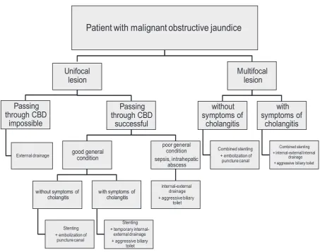
Patient with malignant obstructive jaundice
Unifocal
lesion
Passing
through CBD
impossible
External drainage
Passing
through CBD
successful
good general
condition
without symptoms of cholangitis
Stenting + embolization of
puncture canal
with symptoms of cholangitis
Stenting + temporary
internal-external drainage + aggressive biliary
toilet
poor general condition sepsis, intrahepatic
abscess
internal-external drainage + aggressive biliary
toilet
Multifocal
lesion
without
symptoms of
cholangitis
Combined stenting + embolization of puncture canal
with
symptoms of
cholangitis
Combined stenting + internal-external/internal
drainage + aggressive biliary toilet
Fig. 8. Algorithm of management in patients with obstructive malignant jaundice
analyzed complications in 109 patients who had undergone PTBD. A positive technical effect of the procedure was reached in 97% of their patients. Serious complications, including septic shock, hemorrhage, subphrenical abscess and artero-ve-nous fistula, were manifested in 5 patients (4.2% of patients who had undergone PTBD); in 3 patients (2.5%), the complication resulted in a fatal out-come. Mueller et al. [20] performed a study among 188 patients who had undergone PTBD; the most numerous subgroup (54% of the patients) were pa-tients with pancreatic cancer. A positive technical effect of the procedure was achieved in 94% of cas-es. Serious complications in the periinterventional period occurred in 8% of the patients – 7 patients developed sepsis, in 6 cases serious bleeding oc-curred, 3 patients died.
It should be remembered that a puncture of blood vessels (portal and/or intrahepatic) in pa-tients with cholangitis undergoing PTBD can result in bilemia and subsequent bacteriemia as well as the development of sepsis. The puncture of intra-hepatic blood vessels is particularly dangerous in patients with blood coagulation disorders. Due to the reasons mentioned above, the technique of one-wall puncture seems to be useful in a daily routine and safer in comparison to the traditionally-used double-wall puncture followed by pulling back the needle. USG-guided biliary tree puncture seems to be an optimal solution, particularly in cases of only mild dilatation of the intrahepatic bile ducts. Kloek et al. [21] performed a study comparing the out-comes of endoscopic biliary drainage (EBD) prior to surgery to PTBD in patients with operable hilar CCC. The researchers demonstrated a significant-ly higher incidence of infectious complications in the subgroup after EBD. Additionally, a necessity of multiple procedures in a single patient as well as a prolongation of drainage duration were shown in the EBD subgroup, therefore PTBD seems to be more advantageous in the analyzed group of pa-tients. Paik et al. [4] compared the results of pal-liative percutaneous biliary self-expandable metal stenting (SEMS) to endoscopic stenting in patients with advanced hilar CCC. The rate of successful biliary tree decompression was significantly high-er in patients undhigh-ergoing phigh-ercutaneous stenting. Jeong et al. [22] analyzed the risk of development of pancreatitis in patients with malignant biliary obstruction who had undergone PTDB to the risk of pancreatitis after transampullary or retroam-pullary stent implantation. Pancreatitis occurred more often in patients with the transampullarily implanted stent, however the correlation was not statistically significant. Sut et al. [23] presented a report on the long-term results of percutaneous transhepatic cholangiography (PTC) combined
with biliary stenting as a method of palliative treat-ment in patients with malignant biliary obstruc-tion. Major complications of the procedure were cholangitis and acute pancreatitis. A high 30-day mortality (43%) was noted. Van Delden et al. [5] also pointed out a high incidence of complica-tions after PTBD combined with biliary stenting and significant 30-day mortality. However, these researchers associate the high mortality with the natural course of background disease rather than with postinterventional complications. In 10–30% of patients after PTBD, a secondary procedure is necessary because of recurrent jaundice. Saluja et al. [24] compared the results of percutaneous and endoscopic biliary drainage to patients with gall bladder malignancy. The authors concluded that implementation of PTBD permits better bile drainage and is associated with a lower compli-cation rate (in particular cholangitis) when com-pared to the EBS subset. Tsuyuguchi et al. [25], in their report, presented types of stents used for endoscopic biliary drainage. Metal stents provide patency longer as compared to the plastic stents, however their disadvantage is high cost. There-fore, according to this report, implantation of metal stents is advisable in patients with estimated survival exceeding 6 months. The implementation of plastic stents in patients with estimated survival less than 6 months is not regarded as a mistake. On the other hand, the implementation of covered stents is associated with a higher incidence of acute pancreatitis and stent migration.
References
[1] Björnsson E, Gustafsson J, Borkman J, Kilander A: Fate of patients with obstructive jaundice. J Hosp Med 2008, 3(2), 117–123.
[2] Gerges C, Schumacher B, Terheggen G, Neuhaus H: Expandable metal stents for malignant hilar biliary obstruc-tion. Gastrointest Endosc Clin N Am 2011, 21(3), 481–497.
[3] Huibregtse I, Fockens P: Plastic biliary stents for malignant biliary diseases. Gastrointest Endosc Clin N Am 2011, 21(3), 435–445.
[4] Paik WH, Park YS, Hwang JH, Lee SH, Yoon CJ, Kang SG, Lee JK, Ryu JK, Kim YT, Yoon YB: Palliative treatment with self-expandable metallic stents in patients with advanced type III or IV hilar cholangiocarcinoma: a percutaneous versus endoscopic approach. Gastrointest Endosc 2009, 69(1), 55–62.
[5] Van Delden OM, Laméris JS: Percutaneous drainage and stenting for palliation of malignant bile duct obstruc-tion. Eur Radiol 2008, 18(3), 448–456.
[6] Moss AC, Morris E, Mac Mathuna P: Palliative biliary stents for obstructing pancreatic carcinoma. Cochrane Database Syst Rev 2006(1), CD004200.
[7] Maurea S, Caleo O, Mollica C, Imbriaco M, Mainenti PP, Palumbo C, Mancini M, Camera L, Salvatore M:
Comparative diagnostic evaluation with MR cholangiopancreatography, ultrasonography and CT in patients with pancreatobiliary disease. Radiol Med 2009, 114(3), 390–402.
[8] Domagk D, Wessling J, Reimer P, Hertel L, Poremba C, Senninger N, Heinecke A, Domschke W, Menzel J:
Endoscopic retrograde cholangiopancreatography, intraductal ultrasonography, and magnetic resonance cholan-giopancreatography in bile duct strictures: a prospective comparison of imaging diagnostics with histopathologi-cal correlation. Am J Gastroenterol 2004, 99(9), 1684–1689.
[9] Sozański T, Sokolska V, Moroń K: Magnetic resonance cholangiopancreatography after failed or incomplete endoscopic retrograde cholangiopancreatography. Adv Clin Exp Med 2009, 18, 3, 297–302.
[10] Sozański T, Badowski R, Sokolska V, Moroń K: Postępy w diagnostyce dróg żółciowych i przewodu trzust-kowego. Adv Clin Exp Med 2002, 11, 1, 134–140.
[11] Piñol V, Castells A, Bordas JM, Real MI, Llach J, Montañà X, Feu F, Navarro S: Percutaneous self-expanding metal stents versus endoscopic polyethylene endoprostheses for treating malignant biliary obstruction: random-ized clinical trial. Radiology 2002, 225(1), 27–34.
[12] Aljiffry M, Walsh MJ, Molinari M: Advances in diagnosis, treatment and palliation of cholangiocarcinoma: 1990–2009. World J Gastroenterol 2009, 15(34), 4240–4262.
[13] Meller MT, Arts GR, Dean JR: Outcomes in percutaneous stenting of non-hepato-biliary/pancreatic malignant jaundice. Eur J Cancer Care (Engl) 2010, 19(5), 664–668.
[14] Briggs CD, Irving GR, Cresswell A, Peck R, Lee F, Peterson M, Cameron IC: Percutaneous transhepatic inser-tion of self-expanding short metal stents for biliary obstrucinser-tion before resecinser-tion of pancreatic or duodenal malig-nancy proves to be safe and effective. Surg Endosc 2010, 24(3), 567–571.
[15] Rasmussen IC, Dahlstrand U, Sandblom G, Eriksson LG, Nyman R: Fractures of self-expanding metallic stents in periampullary malignant biliary obstruction. Acta Radiol 2009, 50(7), 730–737.
[16] Chiou YY, Tseng HS, Chiang JH, Hwang JI, Chou YH, Chang CY: Percutaneous placement of metallic stents in the management of malignant biliary obstruction. J Formos Med Assoc 2005, 104(10), 738–743.
[17] Neal CP, Thomasset SC, Bools D, Sutton CD, Garcea G, Mann CD, Rees Y, Newland C, Robinson RJ, Dennison AR, Berry DP: Combined percutaneous-endoscopic stenting of malignant biliary obstruction: results from 106 consecutive procedures and identification of factors associated with adverse outcome. Surg Endosc 2010, 24(2), 423–431.
[18] Yee AC, Ho CS: Complications of percutaneous biliary drainage: benign vs malignant diseases. AJR 1987, 148(6), 1207–1209.
[19] Hamlin JA, Friedman M, Stein MG, Bray JF: Percutaneous biliary drainage: complications of 118 consecutive catheterizations. Radiology 1986, 158, 199–202.
[20] Mueller PR, van Sonnenberg E, Ferrucci JT Jr: Percutaneous biliary drainage: technical and catheter-related problems in 200 procedures. AJR 1982, 138(1), 17–23.
[21] Kloek JJ, van der Gaag NA, Aziz Y, Rauws E, Van Delden O, Lameris J, Busch O, Gouma D, Van Gulik T:
Endoscopic and percutaneous preoperative biliary drainage in patients with suspected hilar cholangiocarcinoma. J Gastrointest Surg 2010, 14(1), 119–125.
[22] Jeong YW, Shin KD, Kim SH, Kim IH, Kim SW, Lee KA, Jeon BJ, Lee SO: The safety assessment of percutane-ous transhepatic transpapillary stent insertion inmalignant obstructive jaundice: regarding the risk of pancreatitis and the effect of preliminary endoscopic sphincterotomy. Korean J Gastroenterol 2009, 54(6), 390–394.
[23] Sut M, Kennedy R, McNamee J, Collins A, Clements B: long-term results of percutaneous transhepatic cholan-giographic drainage for palliation of malignant biliary obstruction. J Palliat Med 2010, 13(11), 1311–1313.
[24] Saluja SS, Gulati M, Garg PK, Pal H, Pal S, Sahni P, Chattopadhyay TK: Endoscopic or percutaneous biliary drainage for gallbladder cancer: a randomized trial and quality of life assessment. Clin Gastroenterol Hepatol 2008, 6(8), 944–950.
Address for correspondence:
Jacek Kurcz Chair of Radiology
Department of General and Interventional Radiology and Neuroradiology Wroclaw Medical University
Borowska 213 50-556 Wrocław Poland
Tel.: +48 71 733 16 60 E-mail: [email protected]
Conflict of interest: None declared